. 2013 Oct 14;27(1):5-18.
doi: 10.1016/j.devcel.2013.09.003.

WT1 maintains adrenal-gonadal primordium identity and marks a population of AGP-like progenitors within the adrenal gland

Affiliations

WT1 maintains adrenal-gonadal primordium identity and marks a population of AGP-like progenitors within the adrenal gland

Roberto Bandiera et al. Dev Cell. .

Abstract

Adrenal glands and gonads share a common primordium (AGP), but the molecular events driving differentiation are poorly understood. Here we demonstrate that the Wilms tumor suppressor WT1 is a key factor defining AGP identity by inhibiting the steroidogenic differentiation process. Indeed, ectopic expression of WT1 precludes differentiation into adrenocortical steroidogenic cells by locking them into a progenitor state. Chromatin immunoprecipitation experiments identify Tcf21 and Gli1 as direct targets of WT1. Moreover, cell lineage tracing analyses identify a long-living progenitor population within the adrenal gland, characterized by the expression of WT1, GATA4, GLI1, and TCF21, that can generate steroidogenic cells in vivo. Strikingly, gonadectomy dramatically activates these WT1(+) cells and leads to their differentiation into gonadal steroidogenic tissue. Thus, our data describe a mechanism of response to organ loss by recreating hormone-producing cells at a heterotopic site.


Figures

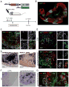
Fig. 1
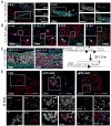
Fig. 1. Repression of WT1 is required to allow adrenocortical differentiation
(A) Immunostaining against WT1 (green), SF1 (red) and GATA4 (blue) in early (E9.5) and late (E9.75) AGP. WT1 and GATA4 characterize AGP cells from the earliest stages, whereas SF1 can only be detected in late AGP cells. (B) WT1+ cells (green) are detected in the mesenchyme surrounding the adrenal primordium prior encapsulation (E11.5 and E12.5) and in the adrenal capsule at later developmental stages. Few WT1+, SF1- cells are also located within the adrenal cortex after encapsulation (arrow). (C) Coexpression of WT1 and SF1 persists in E11.5 and E12.5 gonads until later stages. (D) Schematic representation of the genetic approach used to ectopically express WT1−KTS isoforms in steroidogenic cells. Black arrow-heads indicate LoxP sites. An equivalent strategy was used to knock in and ectopically express Wt1+KTS isoforms. (E) WT1 (blue) SF1 (red) and GATA4 (green) immunofluorescence on adrenal sections from CTR, − and +KTSGOF E15.5 embryos. Two cell populations can be distinguished in −KTSGOF adrenal cortex (insets): WT1high, which have strong GATA4 and low SF1 expression (arrows); and WT1low, which instead have reduced or absent GATA4 and strong SF1 expression (arrowheads). Please note that the cytoplasmic WT1 staining in control adrenals represents background noise. me, mesonephros; ce, coelomic epithelium; ap, adrenal primordium, Co, adrenal cortex; Ca, adrenal capsule. See also Fig. S1.
Fig. 2
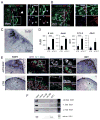
Fig. 2. Cells ectopically expressing WT1-KTS are blocked in an AGP-like state throughout life
(A) Haematoxylin and eosin staining of adrenals from wild type, + and −KTSGOF adult mice. Arrows indicate capsular-like cells within the adrenal cortex of −KTSGOF animals. (B) Immunostaining against WT1 (red) and SF1 (green) on adrenals from wild type, + and −KTSGOF adult mice shows the persistence of WT1+ expressing cells within the adrenal cortex of −KTSGOF and +KTSGOF mice. (C) Immunostaining for WT1 (green) and SF1 (red) on adult adrenal glands reveals the presence of WT1+ cells within the adrenal capsule (left panel) and in rare patches located in the subcapsular cortex (right panel). (D) WT1 positive capsular and cortical cells (red) found in adult wildtype animals also express GATA4 (green). Note the cytoplasmic GATA4 signal in steroidogenic cells represents background (asterisk). (E) Quantification of Wt1 and Gata4 messenger RNAs reveals increased expression of both genes in −KTSGOF adrenals. E 14.5: Wt1, CTR: 100 ± 61.85, n=6; −KTSGOF: 522.28 ± 89.27, n=6; Gata4, CTR: 100 ± 35.38, n=6; −KTSGOF: 407.20 ± 109.67, n=6. Adult: Wt1, CTR: 100 ± 48.20, n=9; −KTSGOF: 10578.96 ± 12509.51, n=3; Gata4, CTR: 100 ± 69.51, n=6; −KTSGOF: 569.70 ± 309.06, n=4. *** P< 0.001 using student T test. Ca, adrenal capsule; Co, adrenal cortex; m, adrenal medulla. See also Fig. S2.
Fig. 3
Fig. 3. WT1-KTS ectopic expression directly upregulates Tcf21 and Gli1
(A-B) LacZ (green) and WT1 (red) immunofluorescence on adrenal sections from adult Gli1:LacZ mice. (A) The adrenal capsule is composed of at least three cells types that can be distinguished by the presence or absence of WT1 and GLI1 (WT1+ GLI1-, arrowhead; WT1- GLI1+ asterisk; WT1+ GLI1+, arrows in insets). (B) WT1+ adrenocortical patches are also positive for GLI1 expression. (C) Tcf21 RNA in situ hybridization showing expression of this marker in the same cells. (D) Tcf21 and Gli1 in adrenal glands of E14.5 embryos or adult −KTSGOF mice show increased expression. E14.5: Tcf21, CTR: 100 ± 33.29, n=4; −KTSGOF: 461.52 ± 102.07, n=6; Gli1, CTR: 100 ± 42.58, n=4; −KTSGOF: 317.34 ± 63.02, n=6. Adult: Tcf21 CTR: 100 ± 110.91, n=20; −KTSGOF: 298.77 ± 123.12, n=8; Gli1, CTR: 100 ± 147.46, n=10; −KTSGOF: 334.15 ± 305.52, n=4. *** P< 0.001, * P< 0.05 using student T test. The data presented were normalized for Hprt1 expression. (E) WT1 (red) and SF1 (green) immunofluorescence on consecutive sections to Tcf21 (left panel) or Gli1 (right panel) RNA in situ hybridization on E18.5 adrenals, shows overlap between WT1 and the RNA for the two markers in both CTR and −KTSGOF. Please note that the cytoplasmic WT1 staining in control adrenals is background. (F) ChIP analysis on −KTSGOF adrenals shows binding of WT1-KTS to the proximal promoter of Tcf21 and to an intronic region of Gli1. A region located -2.7kb upstream of Tcf21 with background amplification is shown as a negative control. Ca, adrenal capsule; Co, adrenal cortex. See also Fig. S3.
Fig. 4
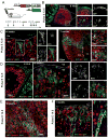
Fig. 4. WT1 expressing cells are progenitors able to differentiate into steroidogenic cells
(A) Schematic representation of the cell lineage tracing experiments performed on Wt1:Cre-ERT2; mTmG mice or embryos indicating the time point when tamoxifen was administered. (B-F) WT1 or DNA (blue), SF1, 3βHSD2 or AKR1b7 (red) and GFP (green) immunofluorescence on samples of adrenal glands from Wt1:Cre-ERT2; mTmG mice of different ages treated with tamoxifen at E12.5 (B and C) or E14.5 (D to F). During development the majority of GFP+ cells are localized within the capsule (B and D E18.5). Postnatal patches of WT1+, GFP+ cells expand under the adrenal capsule and few cells acquire a steroidogenic phenotype. F) Quantification of the number of GFP+ patches of cells within the adrenal cortex of E18.5 embryos and 3 weeks old Wt1:Cre-ERT2; mTmG mice treated with tamoxifen at E14.5. The overall number of GFP+ clusters increases from 4.9 ± 2.42 (2.6 ± 1.84 WT1+, 2.3 ± 2.45 WT1-) (N=10) to 16.33 ± 4.78 (1.25 ± 0.84 WT1+, 14.0 ± 4.55 WT1-) (N=5). Co, adrenal cortex; Ca, adrenal capsule; m, adrenal medulla. See also Fig. S4.
Fig. 5
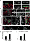
Fig. 5. WT1+ cells maintain the ability to generate subcapsular patches and to differentiate into steroidogenic cells throughout life
(A-D) Immunostaining against WT1 (blue), SF1 (red) and GFP (green) on adrenals from Wt1:Cre-ERT2; mTmG mice treated with 4 consecutive doses of tamoxifen between 10 and 12 weeks of age and collected after 10 days (A), 1 month (B) and 7 months (C and D) after the last administration. Almost all GFP+ cells also express WT1 and SF1 (A-C). GFP+ SF1+ WT1- cells can be detected in a subset of patches (B-C). GFP+ WT1+ SF1- capsular cells can be detected within the adrenal capsule up to 7 month after force-feeding (D). (E-F) Quantification of the number of GFP+ patches within the adrenal cortex of Wt1:Cre-ERT2; mTmG mice treated with tamoxifen at 3 weeks (E) or 11.5 weeks (F) of age. The number of GFP+ clusters increases significantly from 3.56 ± 2.70 (N=9) to 6.00 ± 2.35 (N=9), 3 days and 9 weeks after force-feeding at 3 weeks of age, respectively. No significant increase in the number of GFP+ clusters is detected in animals force-fed at 11.5 weeks, from 3.38 ± 2.00 (N=8) to 5.75 ± 1.98 (N=8). Co, adrenal cortex; Ca, adrenal capsule. See also Fig. S5.
Fig. 6
Fig. 6. WT1+ cells generate cells with gonadal characteristics in the adrenal cortex upon gonadectomy
(A) Schematic representation of the experimental design. (B-E) Adrenal glands from mice sacrificed 10 weeks after gonadectomy. Gonadectomy induces massive invasion of the adrenal cortex by GFP+ cells, derived from WT1+ cells (B). Higher magnification shows that the majority of GFP+ cells have lost WT1 expression, whereas SF1 is present (C). (D) RNA in situ hybridization shows that cells expressing cytochrome P450scc, do not express the adrenocortical specific marker 21-hydroxylase. Please note that the three stainings in panels C to D were performed on consecutive sections, thus representing the same area. (E) CYP17 (red), a marker of cells producing sex hormones (gonadal cells) appears in some cells within the adrenal cortex of gonadectomised mice, but is never found in control animals. Coexpression of CYP17 and GFP in the same cells indicates that they are directly derived from WT1 expressing cells. (F-G) LH receptor RNA in situ hybridization shows expression of this gene in gonadectomised animals (F), where it colocalises with GFP+ cells (G). Co, adrenal cortex; Ca, adrenal capsule; m, adrenal medulla. See also Fig. S6.
Fig. 7
Fig. 7. Schematic representation of the fate of WT1+ progenitors during development and adulthood
Our data expose clear parallels between mechanisms that drive adrenogonadal development and the differentiation of AGP-like cells in the adult adrenal cortex. Progenitor cells are characterized by WT1 and GATA4 expression. WT1 prevents differentiation by regulating expression of key genes such as GLI1 and TCF21. Suppression of WT1 is thus a key step to allow steroidogenic differentiation both during development and or adult AGP-like cells. Upon gonadectomy AGP-like cells respond by differentiating into gonadal steroidogenic cells.

Comment in

Similar articles

Cited by

References

    1. Anderson DJ, Axel R. A bipotential neuroendocrine precursor whose choice of cell fate is determined by NGF and glucocorticoids. Cell. 1986;47:1079–1090. - PubMed
    1. Bickmore WA, Oghene K, Little MH, Seawright A, van Heyningen V, Hastie ND. Modulation of DNA binding specificity by alternative splicing of the Wilms tumor wt1 gene transcript. Science. 1992;257:235–237. - PubMed
    1. Bielinska M, Genova E, Boime I, Parviainen H, Kiiveri S, Leppaluoto J, Rahman N, Heikinheimo M, Wilson DB. Gonadotropin-induced adrenocortical neoplasia in NU/J nude mice. Endocrinology. 2005;146:3975–3984. - PubMed
    1. Bielinska M, Parviainen H, Porter-Tinge SB, Kiiveri S, Genova E, Rahman N, Huhtaniemi IT, Muglia LJ, Heikinheimo M, Wilson DB. Mouse strain susceptibility to gonadectomy-induced adrenocortical tumor formation correlates with the expression of GATA-4 and luteinizing hormone receptor. Endocrinology. 2003;144:4123–4133. - PubMed
    1. Bingham NC, Verma-Kurvari S, Parada LF, Parker KL. Development of a steroidogenic factor 1/Cre transgenic mouse line. Genesis. 2006;44:419–424. - PubMed

Publication types

MeSH terms

LinkOut - more resources