. 2013 Oct 16;33(42):16490-501.
doi: 10.1523/JNEUROSCI.2131-13.2013.

Opposing action of nuclear factor κB and Polo-like kinases determines a homeostatic end point for excitatory synaptic adaptation

Affiliations

Opposing action of nuclear factor κB and Polo-like kinases determines a homeostatic end point for excitatory synaptic adaptation

Anca B Mihalas et al. J Neurosci. .

Abstract

Homeostatic responses critically adjust synaptic strengths to maintain stability in neuronal networks. Compensatory adaptations to prolonged excitation include induction of Polo-like kinases (Plks) and degradation of spine-associated Rap GTPase-activating protein (SPAR) to reduce synaptic excitation, but mechanisms that limit overshooting and allow refinement of homeostatic adjustments remain poorly understood. We report that Plks produce canonical pathway-mediated activation of the nuclear factor κB (NF-κB) transcription factor in a process that requires the kinase activity of Plks. Chronic elevated activity, which induces Plk expression, also produces Plk-dependent activation of NF-κB. Deficiency of NF-κB, in the context of exogenous Plk2 expression or chronic elevated neuronal excitation, produces exaggerated homeostatic reductions in the size and density of dendritic spines, synaptic AMPA glutamate receptor levels, and excitatory synaptic currents. During the homeostatic response to chronic elevated activity, NF-κB activation by Plks subsequently opposes Plk-mediated SPAR degradation by transcriptionally upregulating SPAR in mouse hippocampal neurons in vitro and in vivo. Exogenous SPAR expression can rescue the overshooting of homeostatic reductions at excitatory synapses in NF-κB-deficient neurons responding to elevated activity. Our data establish an integral feedback loop involving NF-κB, Plks, and SPAR that regulates the end point of homeostatic synaptic adaptation to elevated activity and are the first to implicate a transcription factor in the regulation of homeostatic synaptic responses.


Figures
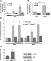
Figure 1.
Figure 1.
NF-κB is activated through the canonical pathway by Plks requiring intact kinase activity. A, Plk2 specifically activates NF-κB in heterologous HEK 293T cells. Luciferase assay in cells transfected with reporters containing wild-type or mutant NF-κB binding sites and increasing amounts (10, 100, 300 ng) of wild-type Plk2 [*p ≤ 0.0003 relative to Plk2 negative control (Ctrl); n = 6]. B, Constitutively active Plk2T236E activates NF-κB more robustly than wild-type Plk2 by luciferase assay in HEK 293T cells. Cells were transfected with wtκB or mtκB reporters and 10 ng of wild-type Plk2 or constitutively kinase active Plk2T236E (*p ≤ 7.5 × 10⁻⁴ relative to Ctrl; #p = 1.55 × 10⁻⁵; n = 6). C, Neuronally expressed Plks activate NF-κB. NF-κB luciferase reporter assay in hippocampal neurons transfected with increasing amounts (0.5–1.0–2.0 μg) of wild-type or kinase-dead Plk mutants (Plk2K108M and Plk3K91R) is shown (*p ≤ 0.05 relative to Ctrl; n = 6). D, Plks activate NF-κB through the canonical pathway. IKK kinase assay in HEK 293T cells expressing GFP or GFP-Plk2T236E from a lentivirus using GST-IκBα as a substrate. Phosphorylated IκBα is normalized to total immunoprecipitated IKK (*p ≤ 0.0001; n ≥ 6). All statistics are reported from at least three independent experiments. n, Number of independent biological replicates. Error bars indicate SEM.
Figure 2.
Figure 2.
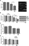
NF-κB and Plk interaction determines neuronal spine density and excitatory neurotransmitter receptor composition. A, Neuronal NF-κB opposes and limits Plk2-induced spine loss. Average spine densities (left) quantitated from confocal projections of dendrites (representative segments; right) from DIV21 RelAF/F murine cultured hippocampal neurons transduced with CreERT2 by lentivirus infection and treated with vehicle or OHT to induce p65-deficiency. Plk2 (either wild-type or kinase-active with data pooled) was expressed for 24 h (*p ≤ 1.2 × 10⁻⁵ relative to Ctrl; **p = 1.83 × 10⁻⁶, ANOVA; n ≥ 44 dendritic segments from ≥10 independent experiments). Scale bar, 10 μm. B, NF-κB limits Plk2-induced loss of surface GluA1 AMPA receptor subunits in hippocampal neurons. Averages of normalized total and surface GluA1 quantitated by densitometry (left) from immunoblots (representative blots; right) after surface biotinylation from DIV21 neurons in the presence (Ctrl) or absence of p65 (OHT) are shown, and wild-type Plk2 was transduced by lentiviral infection. β-Actin was used as the loading control (*p ≤ 0.012 relative to Ctrl; **p = 0.004; #p = 0.009, ANOVA; n ≥ 7 independent experiments). C, Surface to total GluA1 ratio does not significantly change with Plk2 overexpression in wild-type or p65 deficient neurons (data from same experiments as in B). D, NF-κB and Plk2 do not regulate inhibitory GABAA receptors in neurons (biotinylation assay as is Fig. 1B). Densitometric quantification of average surface and total GABAARs. All error bars indicate SEM.
Figure 3.
Figure 3.
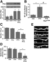
Hyperactivity induces Plk2 which is required for hyperactivity induction of NF-κB. A, Representative immunoblot (inset) and quantification of Plk2 levels in lysates from chronic PTX (PTX, 100 μm, 24 h) or mock-treated DIV21 hippocampal cultures in the presence (Ctrl or PTX) or absence of p65 (OHT or OHT/PTX). HSC70 was used as the loading control (*p ≤ 4.54 × 10⁻⁴, ANOVA; n = 5 independent experiments). B, NF-κB luciferase reporter in DIV19 hippocampal neurons. NF-κB transcriptional activity is significantly increased by 12 h PTX treatment (50–100 μm; *p = 0.003), but this is prevented by expression of the kinase-dead Plk2K108M (#p = 0.01; n = 3 independent experiments). C, NF-κB limits decreases in spine density by chronic PTX. Hippocampal neurons (DIV21, RelAF/F mice) in the presence or absence of p65 (Cre expression 48 h) were vehicle or PTX treated (100 μm for 24 h; *p ≤ 1.62 × 10⁻⁴, ANOVA; n ≥ 15 dendritic segments from 4 independent experiments). D, NF-κB limits decreases in spine volumes by chronic PTX. Spine head volumes were measured using Imaris software from the same dendritic segments as in A (*p ≤ 5.42 × 10⁻⁴, ANOVA). E, Confocal projections of representative dendrite segments for data in C and D. Scale bar, 10 μm. All error bars indicate SEM.
Figure 4.
Figure 4.
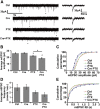
NF-κB opposes the homeostatic downregulation of synaptic strength by elevated activity. A, Representative traces of mEPSCs recorded from p65-wild-type or p65-deficient (Cre) DIV21 RelAF/F hippocampal cultures after treatment with vehicle or PTX (100 μm for 20–28 h). Right, Enlarged single events (marked by asterisks) from each condition. B, NF-κB limits the homeostatic decrease in AMPAR-dependent mEPSC amplitudes in response to elevated activity in hippocampal neurons. Average AMPAR-dependent mEPSC amplitudes under the indicated conditions (*p ≤ 0.023, ANOVA). C, Cumulative percentage plot of mEPSC amplitudes from all recorded neurons (nCtrl = 12, nCre = 10, nPTX = 10, nCre+PTX = 11, where n represents individual neurons recorded from 10 independent experiments). K–S statistical analysis shows a significant left shift in the cumulative distribution of mEPSC amplitudes from Ctrl after chronic PTX both in the presence (PTX; p = 0.014) and absence of p65 (Cre/PTX; p = 9.67 × 10⁻⁴). There is also a statistically significant left shift in the cumulative distribution of mEPSC amplitudes in neurons treated with PTX in the absence of p65 (Cre/PTX) compared to neurons with p65 (PTX; p = 8.65 × 10⁻³). D, Average AMPAR-dependent mEPSC frequency under the indicated conditions. E, Cumulative percentage plot of mEPSC IEIs from all recorded neurons. K–S test shows statistically significant right shifts from Ctrl in the cumulative distribution of mEPSC IEIs after chronic PTX in the absence of p65 (Cre/PTX; p = 0.018), and between chronic PTX in the presence (PTX) and absence of p65 (Cre/PTX; p = 0.035), but there is no statistical difference from Ctrl in the presence of PTX (PTX; K–S test). All error bars indicate SEM.
Figure 5.
Figure 5.
NF-κB positively regulates SPAR protein in vitro and in vivo. A, NF-κB limits Plk2-induced SPAR protein loss in neurons. Representative immunoblot (inset) and quantification of SPAR levels in lysates from mock or Plk2 infected (lentivirus, 2 d) DIV21 hippocampal cultures in the presence (Ctrl) or absence of p65 (OHT). HSC70 was used as the loading control (*p ≤ 2.87 × 10⁻³, ANOVA; n = 5 independent experiments). B, C, Levels of neuronal SPAR positively correlate with NF-κB activity. B, NF-κB reporter assay shows that NF-κB activity is higher in developing (DIV16) compared to mature (DIV21) hippocampal cultures. C, Representative immunoblot and quantitated levels of SPAR protein in DIV16 RelAF/F hippocampal cultures in the presence (Ctrl) or absence (OHT) of p65, or following rescue of p65-deficiency by expression of exogenous p65 (OHT/p65; using lentiviral expression of Myr-GFP-F2A-p65). Average data are plotted normalized to Ctrl. HSC70 was used for the loading control (*p ≤ 0.04; n ≥ 3 independent experiments). D, Representative immunoblot (inset) and normalized quantification of SPAR protein levels in RelAF/F P14 mice transduced with control GFP or GFP-Cre by AAV injections at P0. SPAR levels are significantly reduced (p = 2.07 × 10⁻⁴; nGFP = 3, nGFP-Cre = 6 mice) by in vivo NF-κB deficiency. E, Immunoblot showing expression of GFP and GFP-Cre from RelAF/F hippocampi transduced with AAV injected at P0 and harvested at P14. F, Confocal Z-stack projections of a P12 mouse hippocampus showing viral expression of GFP-Cre following P0 lateral ventricle AAV injections. Hippocampi were cryosectioned after harvest at P12 with Hoechst dye used to counterstain nuclei. We observe >50% infection efficacy of neurons in the hippocampal pyramidal layer (CA1 shown in image) and in the dentate gyrus (DG). All error bars indicate SEM.
Figure 6.
Figure 6.
SPAR is transcriptionally upregulated by NF-κB and rescues the decrease in synaptic strength due to loss of p65 in neurons exposed to hyperactivity. A, NF-κB induces a reporter driven by the SPAR promoter assayed in HEK 293T cells. Diagram (top inset) of SPAR promoter luciferase reporter, with ∼1.2 kb flanking the mouse SPAR transcriptional start site, containing four putative κB DNA binding sites. Plot of reporter assay in cells transduced with 0 to 10 ng of p65 subunit of NF-κB (*p ≤ 0.01; n = 3 independent experiments). B, NF-κB induction by Plk2 leads to an increased association of p65 with SIPA1L1 promoter in hippocampal neurons. ChIP assays were performed with either anti-p65 antibody or isotype-matched IgG, in DIV21 hippocampal dissociated cultures expressing either GFP (Ctrl) or GFP-tagged constitutively active Plk2T236E; using either primers within the SIPA1L1 promoter (left) or upstream control primers (right; *p = 0.008; n = 5 independent experiments). The relative abundance of specific amplicons in the IP compared to the input DNA is shown on the y-axis. C, NF-κB does not regulate SPAR protein levels from a construct lacking the endogenous SPAR promoter in neurons. Inset, Diagram of construct containing the SPAR coding sequence fused directly to luciferase to allow sensitive quantification, and driven by a constitutive CMV promoter. SPAR protein level is quantified by luciferase activity from RelAF/F DIV16 dissociated hippocampal cultures transfected with a dose titration of the fusion construct in the presence or absence of p65. D, Expression of exogenous SPAR rescues the overshoot of spine head volume loss in p65-deficient (Cre) neurons exposed to elevated activity. Average spine head volumes after chronic PTX are plotted as a percentage of control neurons (100%) not exposed to PTX to illustrate that low levels of SPAR expression rescue the exaggerated loss of spine volume in p65-deficient neurons exposed to elevated activity (*p = 1.87 × 10⁻⁸, ANOVA; Cre/PTX/SPAR; n ≥ 10 dendritic segments from 4 independent experiments), but do not alter average spine volumes without heightened activity (Cre/SPAR). E, Exogenous SPAR expression opposes excessive loss of surface synaptic GluA1 in response to elevated activity (PTX) in p65-deficient (Cre) neurons. Plot of average surface synaptic GluA1 density for the indicated conditions. Synaptic GluA1 was quantified (Imaris, Bitplane) from confocal images as surface GluA1 puncta colocalized with transfected PSD95 puncta in DIV21 RelAF/F hippocampal pyramidal neurons (*p ≤ 3.39 × 10⁻³, #p = 2.16 × 10⁻³, ANOVA; n ≥ 10 dendritic segments from 4 independent experiments). F, Representative confocal projections of dendrites from pyramidal neurons for the indicated conditions, as quantified in D and E. Right panels are masked (Imaris) for ubiquitously expressed mCherry fluorophore (left) to allow confirmation by visual inspection that illustrated puncta are within the presented dendritic segments. Scale bar, 10 μm. All error bars indicate SEM.
Figure 7.
Figure 7.
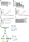
NF-κB, Plk2, and SPAR kinetics during PTX-induced hyperactivity. A, B, NF-κB limits the homeostatic SPAR loss in response to elevated activity in neurons. Representative immunoblot (A) and quantification (B) of SPAR levels in lysates from chronic PTX or mock-treated DIV21 hippocampal cultures in the presence (Ctrl or PTX) or absence of p65 (OHT or OHT/PTX). HSC70 serves as the loading control (*p ≤ 3.39 × 10⁻³, ANOVA; n = 5 independent experiments). C–F, Evaluation of neuronal Plk2, NF-κB, and SPAR kinetics during a PTX (100 μm) time course (0, 3, 7, 12, 24 h) reveals an initial excessive loss of SPAR during Plk2 dominance (3 h; C, E, F), followed by NF-κB activation (D) and subsequent partial recovery of SPAR levels (E, F). F, Representative Plk2, SPAR, and GAPDH (loading control) immunoblot of hippocampal lysates during PTX time course (quantified in C, E). Plots of NF-κB luciferase reporter assay (D) and immunoblot quantification for Plk2 (C) and SPAR (E) in the same neurons from DIV21 dissociated hippocampal cultures (*p ≤ 0.035, #p = 0.012; n ≥ 3 independent experiments). All error bars indicate SEM. G, Summary diagram for the function of the NF-κB transcription factor as an integral feedback sensor in the homeostatic response to chronic elevated activity. Activity-dependent induction of Plk is sufficient to mediate both SPAR protein degradation as well as activation of NF-κB with resulting delayed feedback through NF-κB-dependent upregulation of SPAR expression. These countervailing forces act to tune the outcome of the homeostatic synaptic response, as is revealed by the much more drastic loss of dendritic spine number, size, synaptic receptor content, and function in NF-κB-deficient neurons. Importantly, in mature neurons, these changes do not occur with NF-κB loss under basal conditions, but only in NF-κB-deficient neurons undergoing a homeostatic response to elevated synaptic activity.

Similar articles

Cited by

References

    1. Ahn HJ, Hernandez CM, Levenson JM, Lubin FD, Liou HC, Sweatt JD. c-Rel, an NF-kappaB family transcription factor, is required for hippocampal long-term synaptic plasticity and memory formation. Learn Mem. 2008;15:539–549. doi: 10.1101/lm.866408. - DOI - PMC - PubMed
    1. Ang XL, Seeburg DP, Sheng M, Harper JW. Regulation of postsynaptic RapGAP SPAR by Polo-like kinase 2 and the SCFbeta-TRCP ubiquitin ligase in hippocampal neurons. J Biol Chem. 2008;283:29424–29432. doi: 10.1074/jbc.M802475200. - DOI - PMC - PubMed
    1. Boersma MC, Dresselhaus EC, De Biase LM, Mihalas AB, Bergles DE, Meffert MK. A requirement for nuclear factor-kappaB in developmental and plasticity-associated synaptogenesis. J Neurosci. 2011;31:5414–5425. doi: 10.1523/JNEUROSCI.2456-10.2011. - DOI - PMC - PubMed
    1. Burrone J, O'Byrne M, Murthy VN. Multiple forms of synaptic plasticity triggered by selective suppression of activity in individual neurons. Nature. 2002;420:414–418. doi: 10.1038/nature01242. - DOI - PubMed
    1. Chakraborty T, Chowdhury D, Keyes A, Jani A, Subrahmanyam R, Ivanova I, Sen R. Repeat organization and epigenetic regulation of the DH-Cmu domain of the immunoglobulin heavy-chain gene locus. Molecular Cell. 2007;27:842–850. doi: 10.1016/j.molcel.2007.07.010. - DOI - PubMed

Publication types

MeSH terms