. 2013 Oct 21;210(11):2477-91.
doi: 10.1084/jem.20121999. Epub 2013 Oct 7.

IL-4 directly signals tissue-resident macrophages to proliferate beyond homeostatic levels controlled by CSF-1

Affiliations

IL-4 directly signals tissue-resident macrophages to proliferate beyond homeostatic levels controlled by CSF-1

Stephen J Jenkins et al. J Exp Med. .

Abstract

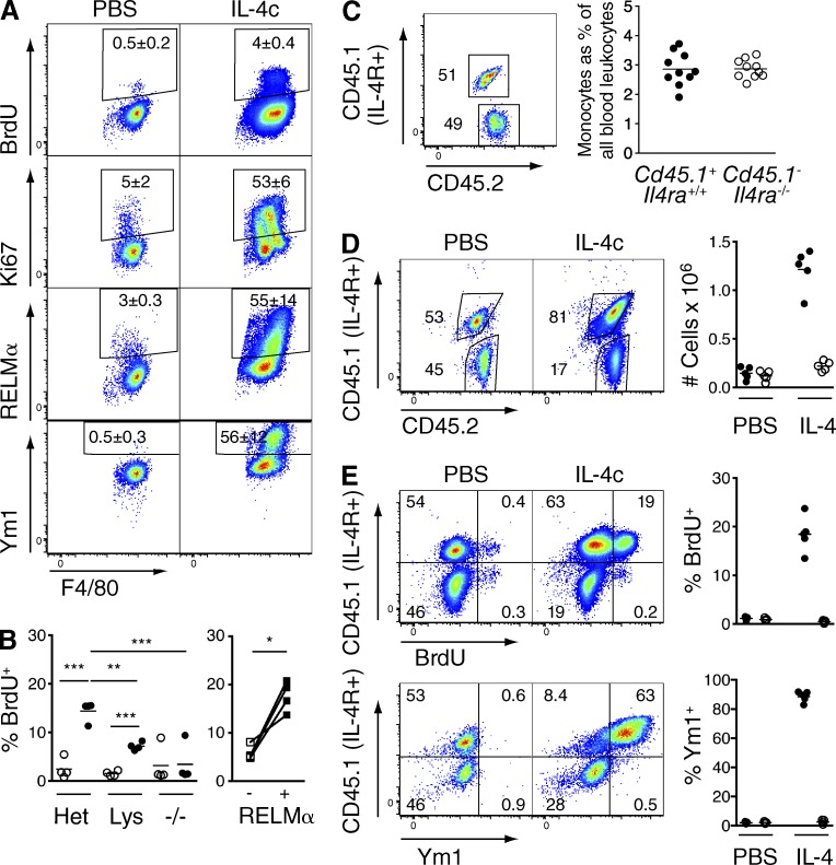
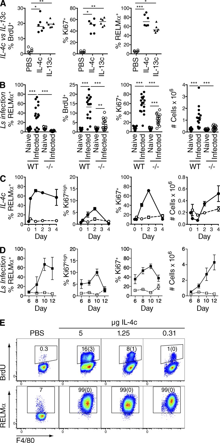
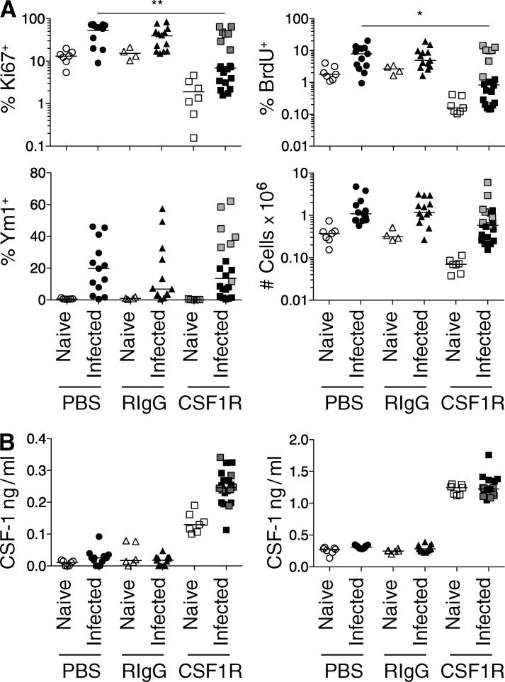
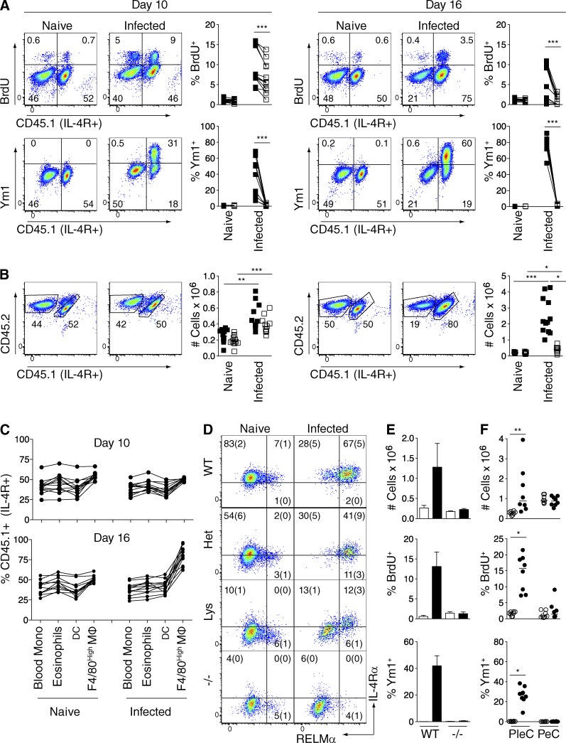
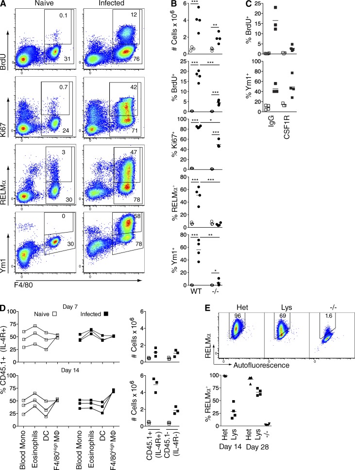
Macrophages (MΦs) colonize tissues during inflammation in two distinct ways: recruitment of monocyte precursors and proliferation of resident cells. We recently revealed a major role for IL-4 in the proliferative expansion of resident MΦs during a Th2-biased tissue nematode infection. We now show that proliferation of MΦs during intestinal as well as tissue nematode infection is restricted to sites of IL-4 production and requires MΦ-intrinsic IL-4R signaling. However, both IL-4Rα-dependent and -independent mechanisms contributed to MΦ proliferation during nematode infections. IL-4R-independent proliferation was controlled by a rise in local CSF-1 levels, but IL-4Rα expression conferred a competitive advantage with higher and more sustained proliferation and increased accumulation of IL-4Rα(+) compared with IL-4Rα(-) cells. Mechanistically, this occurred by conversion of IL-4Rα(+) MΦs from a CSF-1-dependent to -independent program of proliferation. Thus, IL-4 increases the relative density of tissue MΦs by overcoming the constraints mediated by the availability of CSF-1. Finally, although both elevated CSF1R and IL-4Rα signaling triggered proliferation above homeostatic levels, only CSF-1 led to the recruitment of monocytes and neutrophils. Thus, the IL-4 pathway of proliferation may have developed as an alternative to CSF-1 to increase resident MΦ numbers without coincident monocyte recruitment.


Figures

Figure 1.
Figure 1.
IL-4 drives CSF-1–independent MΦ proliferation. (A) BL/6 mice were injected i.p. with IL-4c or PBS plus anti-CSF1R mAb (CSF1R), rat IgG (RIgG), or PBS on days 0 and 2. The proportion of F4/80High pleural MΦs positive for Ki67, Ki67High, or RELMα and total MΦ numbers were determined by flow cytometry on day 2 after the last injection. Graphs depict individual data for four mice/group. (B) As in A, but on day 3 after daily oral gavage with vehicle control or GW2580 on days 0–3. (C) Mice were injected with a single dose of PBS or IL-4c, and the expression of CSF1R mRNA was determined 24 h later in FACS-purified F4/80High peritoneal MΦs. ***, P < 0.001 determined by two-tailed Student’s t test. Individual data for five mice/group are shown. (D) CSF-1 levels in pleural lavage fluid and serum from mice in A determined by ELISA. All data are representative of two to three separate experiments, with the same results observed in the peritoneal cavity. (A–D) Horizontal bars indicate mean values.
Figure 2.
Figure 2.
MΦ-intrinsic IL-4Rα signaling is essential for proliferation and alternative activation triggered by IL-4c. (A) LysMcreIl4ra−/lox BALB/c mice were injected i.p. on days 0 and 2 with PBS or IL-4c, and peritoneal lavage cells were analyzed on day 4 by flow cytometry for BrdU incorporation or Ki67, RELMα, and Ym1 versus F4/80 expression. Representative flow cytograms gated on F4/80High peritoneal MΦs with frequencies depicting the mean ± SEM of four mice per group. (B) Il4ra−/lox (Het), LysMcreIl4ra−/lox (Lys), or Il4ra−/− (−/−) BALB/c were treated with PBS (open) or IL-4c (closed) as in A, and BrdU incorporation by F4/80High pleural MΦs was determined on day 3 together with the frequency of BrdU+ cells in RELMα-positive (+) or negative (−) F4/80High MΦs from the IL-4c–treated LysMcreIl4ra−/lox group. *, P < 0.05; **, P < 0.01; and ***, P < 0.001 determined by ANOVA (left) or paired Student’s t test (right). Data are representative of four independent experiments inclusive of data in A, with three to four mice/group. (C) BL/6 Il4ra+/+Cd45.1+Cd45.2+ mice were lethally irradiated and reconstituted with a 50:50 mix of Il4ra+/+Cd45.1+Cd45.2+ and Il4ra−/−Cd45.1nullCd45.2+/+ congenic BM cells. The frequency of blood monocytes (gated as side scatter [SSC]lowCD11b+CD115+) derived from each BM was determined by analysis of CD45.1 and CD45.2 expression 8 wk later. A representative flow cytogram of CD45.1 and CD45.2 expression on blood monocytes is shown together with a graph depicting the frequency of CD45.1+ and CD45.1 monocytes in all blood leukocytes from 10 individual mice. (D) Mice from C were subsequently injected i.p. twice, 2 d apart, with PBS or IL-4c, and pleural lavage cells were analyzed 2 d after the last injection for CD45.1 and CD45.2 expression. Representative flow cytograms gated on all live F4/80High MΦs are shown together with a graph depicting the total number of Il4ra+/+Cd45.1+ (closed circles) and Il4ra−/−Cd45.1null (open circles) pleural F4/80High MΦs in each group, with individual data for five mice/group presented. (E) Representative flow cytograms depicting CD45.1 expression versus BrdU incorporation or Ym1 expression gated on single F4/80HighCD19 pleural MΦs from mice in D, and graphs showing the proportion of Il4ra+/+Cd45.1+ (closed circles) and Il4ra−/−Cd45.1null (open circles) F4/80HighCD19 MΦs positive for BrdU or Ym1 for individual mice. (C–E) Data are representative of two experiments. (B–E) Horizontal bars indicate mean values.
Figure 3.
Figure 3.
IL-4Rα–dependent and –independent mechanisms of proliferation during nematode infection. (A) BL/6 mice were given a single i.p. injection of IL-13c, IL-4c, or PBS, and the proportion of BrdU+, Ki67+, and RELMα+ F4/80High peritoneal MΦs was determined 36 h later. Graphs depict individual data for six mice per group and are representative of two independent experiments. Horizontal bars indicate median values. Equivalent results were obtained in the pleural cavity. *, P < 0.05; **, P < 0.01; and ***, P < 0.001 determined by Kruskal-Wallis test. (B) C57BL/6 (WT) and Il4ra−/− (−/−) mice were infected with Ls, and the total number of F4/80High pleural MΦs and the proportion positive for BrdU, Ki67, and RELMα were determined on day 10. Results are pooled from two experiments with 15–16 mice/group shown. Horizontal bars indicate mean values. **, P < 0.01; and ***, P < 0.001 determined by ANOVA. (C) BL/6 mice were given a single i.p. dose of IL-4c (solid line) or PBS (dashed line), after which the total number of pleural cavity F4/80High MΦs and the proportion positive for RELMα, Ki67High, or Ki67 were determined over a 4-d time course. Data are mean ± SEM of four mice/group and representative of two experiments, with the same results obtained for peritoneal MΦs. Day 0 represents naive. (D) As C but from Ls-infected (solid line) or naive (dashed line) mice. Data are mean ± SEM from four to five mice per group and representative of three experiments. (E) Representative flow cytograms of F4/80High peritoneal MΦs 4 d after i.p. injection of PBS or IL-4c containing 5, 1.25, or 0.31 µg IL-4 on days 0 and 2. Frequencies are means with SEM in parentheses of three to six IL-4c–treated animals per group or a single value for pooled cells from three PBS-treated mice. A repeat using a single injection regimen verified dose effect for both of IL-4c and IL-13c.
Figure 4.
Figure 4.
Elevated CSF-1 production contributes to MΦ proliferation during tissue nematode infection. (A) BL/6 mice were infected with Ls and injected i.p. on day 8 with anti-CSF1R mAb (CSF1R), rat IgG (RIgG), or PBS. Total number of F4/80High pleural MΦs and the proportion positive for BrdU, Ki67, or Ym1 were determined on day 10. Data are pooled from two experiments with 7, 13, and 20 mice for naive and PBS-, RIgG-, and anti-CSF1R–treated infected, groups, respectively. *, P < 0.05; and **, P < 0.01 determined by Kruskal-Wallis test. (B) CSF-1 levels in pleural lavage (left) or serum (right) from mice in A determined by ELISA. (A and B) Horizontal bars indicate median.
Figure 5.
Figure 5.
IL-4Rα signaling to MΦs provides a competitive advantage during tissue nematode infection. (A) BL/6 Il4ra+/+Cd45.1+Cd45.2+ mice were lethally irradiated and reconstituted with a 50:50 mix of Il4ra+/+Cd45.1+Cd45.2+ and Il4ra−/−Cd45.1nullCd45.2+/+ congenic BM cells over 8 wk. Mice were infected with Ls, after which BrdU incorporation or Ym1 expression versus expression of CD45.1 was determined on single F4/80HighCD19 pleural MΦs at days 10 and 16 after infection. Representative flow cytograms are shown, whereas graphs depict the proportion of IL-4Rα+CD45.1+ (closed squares) and IL-4RαCD45.1 (open squares) cells positive for BrdU or Ym1, with each line illustrating paired measurements from individual mice. Data are pooled from two experiments, with 11–12 mice/group. ***, P < 0.001 determined by paired Student’s t test. (B) CD45.1 and CD45.2 expression gated on all F4/80High pleural MΦs from mice in A and graphical presentation of the total number of IL-4Rα+CD45.1+ (closed squares) and IL-4RαCD45.2+/+ (open squares) subsets of these cells. *, P < 0.05; **, P < 0.01; and ***, P < 0.001 determined by Kruskal-Wallis test. (C) Frequency of IL-4Rα+CD45.1+ cells within CD115+CD11b+SSCLow blood monocytes and pleural cavity eosinophils (SSCHigh, F4/80Low, MHCII), DCs (CD11cHighMHCIIHigh), and F4/80High MΦs at days 10 and 16 after infection of mice in A, with lines joining cells of individual mice. (D) Il4ra+/+ (WT), Il4ra−/lox (Het), LysMcreIl4ra−/lox (Lys), or Il4ra−/− (−/−) mice were infected with Ls or left naive, and RELMα and IL-4Rα staining on F4/80High pleural MΦs was determined at day 17. Frequencies are means with SEM in parentheses of five to seven mice/group. The emergence of RELMα+ MΦs in LysMcreIl4ra−/lox mice was confirmed in a further three independent experiments between days 14 and 60 after infection. (E) C57BL/6 (WT) or Rag1−/− (−/−) mice were infected with Ls, and the total number of pleural cavity F4/80High MΦs and the proportion incorporating BrdU or expressing Ym1 were determined on day 10, with mean and SEM of five mice/group shown. (F) As in E, but both peritoneal (PeC) and pleural (PleC) cavity F4/80High MΦs from naive (open circles) or Ls-infected C57BL/6 mice were analyzed at 10 d after infection. Data are representative of three experiments with eight mice/group. *, P < 0.05; and **, P < 0.01 determined by Kruskal-Wallis test. (B and F) Horizontal bars indicate median values.
Figure 6.
Figure 6.
IL-4–dependent proliferation occurs during GI nematode infection and provides a competitive advantage to IL-4Rα+ MΦs. (A) BALB/c mice were infected orally with Hp, and peritoneal lavage cells were assessed at day 7. Representative flow cytograms of all peritoneal cells showing gates and frequencies for all or BrdU+, Ki67+, RELMα+ or Ym1+, F4/80High MΦs. Data are representative of five experiments. (B) BALB/c (WT) or Il4−/− (−/−) mice were infected with Hp (closed symbols) or left naive (open symbols), and the total peritoneal F4/80High MΦs and the proportion positive for BrdU, Ki67, Ym1, and RELMα+ was determined on day 7. Data are representative of three experiments with three to four mice per group. *, P < 0.05; **, P < 0.01; and ***, P < 0.001 determined by ANOVA. (C) As in B but after treatment of BALB/c mice with anti-CSF1R mAb (CSF1R) or rat IgG (IgG) on day 6. (D) Il4ra+/+Cd45.1+Cd45.2+ mice were lethally irradiated and reconstituted for 8 wk with a 50:50 mix of Il4ra+/+Cd45.1+Cd45.2+ and Il4ra−/−Cd45.1nullCd45.2+ congenic BM cells before infection with Hp. The frequency of CD45.1+ cells in different leukocyte subsets in the peritoneal cavity in naive (open symbols) and Hp-infected mice (closed symbols) was determined on days 7 and 14 after infection. The graphs show total numbers of Il4ra+/+Cd45.1+ and Il4ra−/−Cd45.1null peritoneal F4/80High MΦs obtained in naive (open) and infected (closed) mice at each time point. (E) Il4ra−/lox (Het), LysMcreIl4ra−/lox (Lys), or Il4ra−/− (−/−) mice were infected with Hp, and RELMα expression by F4/80High peritoneal MΦs was determined on days 14 and 28. (C–E) Data are from independent experiments performed once and are confirmatory of data from the Ls model. (B–E) Horizontal bars indicate mean values.
Figure 7.
Figure 7.
CSF-1 but not IL-4 stimulates neutrophil and monocyte recruitment alongside elevated proliferation of resident tissue MΦs. (A) BL/6 mice were injected i.p. with PBS, Fc–CSF-1, or IL-4c, and Ki67 and RELMα expression by F4/80High peritoneal MΦs was determined 24 h later. Representative flow cytograms show all live peritoneal lavage cells. Data are representative of two independent experiments. (B) Total Ly-6C+CD115+ monocytes and Ly-6CIntermediateSSCIntermediate neutrophils in peritoneal lavage cells from mice in A and representative flow cytograms depicting gating strategies. Top flow cytograms also depict CD115+Ly6C monocytes/MΦs (left gate), and bottom flow cytograms show the SSCHighLy6CLow eosinophil gate, both of which exhibited no change in total number. (A and B) Horizontal bars indicate mean values.

Similar articles

Cited by

References

    1. Ajami B., Bennett J.L., Krieger C., Tetzlaff W., Rossi F.M. 2007. Local self-renewal can sustain CNS microglia maintenance and function throughout adult life. Nat. Neurosci. 10:1538–1543 10.1038/nn2014 - DOI - PubMed
    1. Ajami B., Bennett J.L., Krieger C., McNagny K.M., Rossi F.M. 2011. Infiltrating monocytes trigger EAE progression, but do not contribute to the resident microglia pool. Nat. Neurosci. 14:1142–1149 10.1038/nn.2887 - DOI - PubMed
    1. Alikhan M.A., Jones C.V., Williams T.M., Beckhouse A.G., Fletcher A.L., Kett M.M., Sakkal S., Samuel C.S., Ramsay R.G., Deane J.A., et al. 2011. Colony-stimulating factor-1 promotes kidney growth and repair via alteration of macrophage responses. Am. J. Pathol. 179:1243–1256 10.1016/j.ajpath.2011.05.037 - DOI - PMC - PubMed
    1. Aziz A., Soucie E., Sarrazin S., Sieweke M.H. 2009. MafB/c-Maf deficiency enables self-renewal of differentiated functional macrophages. Science. 326:867–871 10.1126/science.1176056 - DOI - PubMed
    1. Behnke J.M., Wakelin D. 1977. Nematospiroides dubius: stimulation of acquired immunity in inbred strains of mice. J. Helminthol. 51:167–176 10.1017/S0022149X0000746X - DOI - PubMed

Publication types

MeSH terms

Substances