. 2013 Oct 5;12(1):115.
doi: 10.1186/1476-4598-12-115.

CD164 regulates the tumorigenesis of ovarian surface epithelial cells through the SDF-1α/CXCR4 axis

Affiliations

CD164 regulates the tumorigenesis of ovarian surface epithelial cells through the SDF-1α/CXCR4 axis

Ai-Fang Huang et al. Mol Cancer. .

Abstract

Background: CD164 (endolyn), a sialomucin, has been reported to play a role in the proliferation, adhesion, and differentiation of hematopoietic stem cells. The potential association of CD164 with tumorigenicity remains unclear.

Methods: The clinicopathological correlation of ovarian cancer with CD164 was assessed in a 97-patient tumor tissue microarray. Overexpression or silence CD164 was to analyze the effect of CD164 on the proliferation, colony formation and apoptosis via a mouse xenograft and western blotting analysis. The subcellular localization of CD164 was collected in the immunohistochemical and confocal analysis.

Results: Our data demonstrated that higher expression levels of CD164 were identified in malignant ovarian cancer cell lines, such as SKOV3 and HeyA8. The clinicopathological correlation analysis showed that the upregulation of CD164 protein was significantly associated with tumor grade and metastasis. The overexpression of CD164 in human ovarian epithelial surface cells promoted cellular proliferation and colony formation and suppressed apoptosis. These tumorigenicity effects of CD164 were reconfirmed in a mouse xenograft model. We also found that the overexpression of CD164 proteins increased the amounts of CXCR4 and SDF-1α and activated the SDF-1α/CXCR4 axis, inducing colony and sphere formation. Finally, we identified the subcellular localization of CD164 in the nucleus and cytosol and found that nuclear CD164 might be involved in the regulation of the activity of the CXCR4 promoter.

Conclusions: Our findings suggest that the increased expression of CD164 is involved in ovarian cancer progression via the SDF-1α/CXCR4 axis, which promotes tumorigenicity. Thus, targeting CD164 may serve as a potential ovarian cancer biomarker, and targeting CD164 may serve as a therapeutic modality in the management of high-grade ovarian tumors.


Figures
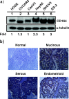
Figure 1
Figure 1
Quantification of the expression of CD164 in ovarian cancer cell lines and an ovarian tissue array. a) Cell lysates from indicated ovarian cells were subjected to immunoblot analysis with an antibody against CD164 protein. α-tubulin was used as a loading control. b) Immunohistochemical staining of malignant and normal ovarian tissue section stained with rabbit anti-CD164 antibodies and CD164 levels in representative ovarian tumor tissue. Scale bars = 50 μm.
Figure 2
Figure 2
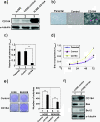
CD164 overexpression alters cell morphology and induces malignant transformation/anchorage independent growth in hOSE. a) Cell lysates from hOSE-CD164 cells were subjected to an immunoblot analysis with an antibody against the CD164 protein. α-tubulin was used as a loading control. b) The morphology of hOSE-CD164 cells compared with hOSE parental and hOSE-vector control cells by phase-contrast microscopy. (×100 magnification and Scale bars = 50 μm). c) The adhesive ability of hOSE-CD164 and control cells was detected by an adhesion assay. **p < 0.01 (three independent experiments were performed and the data are expressed as the means ± s.e.m.). d) Proliferation of hOSE-CD164 cells and control cells were analyzed using a BrdU proliferative assay (three independent experiments and data were mean ± s.e.m.). e) Effect of CD164 overexpression on the colony formation ability of hOSE cells. Soft agar colony formation of hOSE-CD164 and BEAS2B-CD164 cells paired with control cells was calculated after 28 days of culture. The graph showed the number of colonies (means ± s.e.m.) after 4 weeks of culture for three independent experiments. The p values (determined by Student’s t test) were relative to control cells. **p < 0.01 (three independent experiments and data were means ± s.e.m.). f) Cell lysates from hOSE-CD164 cells were subjected to immunoblot analysis for antibodies against anti-apoptotic Bcl-2 and apoptotic Bax protein. α-tubulin was used as a loading control.
Figure 3
Figure 3
CD164 overexpression promotes tumor formation in nude mice. a) The tumor growth of hOSE-CD164 cells, control cells and SKOV3 cells subcutaneously injected into female athymic nude mice (n = 8, each group) was assessed every 5 days for 45 days by measuring two perpendicular diameters and calculating the tumor volume in mm3. Tumor size was measured during 7–8 weeks and calculated as follows: volume = length × width2 × 1/2. ** p < 0.01. Data represent the means ± SEM. b) Macroscopic appearance of hOSE-CD164 (top and bottom, red), hOSE control (top, yellow) and SKOV3 (bottom, yellow) tumors formed subcutaneous tumors. c) Quantification of the tumor weight of hOSE-vector and hOSE-CD164 cells (4 × 106) after subcutaneous injection in the flanks of nude mice (n = 8, each group). Tumor weight was measured in the hOSE-CD164 cells and compared with hOSE-vector cells (*** p < 0.001) and SKOV3 (** p < 0.01) nude mice. d) Macroscopic appearance of hOSE-CD164 tumors (upper) spread and disseminated in the peritoneal cavity (bottom, arrows). e) hOSE-CD164 cells, control cells and SKOV3 cells (4 × 106) were injected into the peritoneal cavity of nude mice. Tumor weight was measured in the hOSE-CD164 cells and compared with hOSE-vector cells (*** p < 0.001) or SKOV3 (** p < 0.01) nude mice. f) At autopsy, tumors were excised and ascite fluid was collected and measured. *** p < 0.001.
Figure 4
Figure 4
CD164 downregulation rescues the effects of CD164 overexpression on tumorigenicity in nude mice. a) Total RNAs were extracted from SKOV3-shCD164, HeyA8-shCD164 and respective control cells for the qRT-PCR analysis. Quantitative analysis of CD164 transcript levels relative to GAPDH was calculated and compared with SKOV3 or HeyA8 control cells. b) Doxycycline-inducible SKOV3-shCD164 and HeyA8-shCD164 cells were treated with 5 μg/ml doxycycline. After 48 hours, the cells were harvested and subjected to immunoblot analysis with antibody against CD164 protein, cleaved PARP and caspase 3 and its cleaved fragment. α-tubulin was used as a loading control. c and d) SKOV3-shCD164 cells and SKOV3-control cells were subcutaneously injected in the flanks of nude mice (n = 8). Nude mice were treated continuously with Dox and harvested at 8 weeks after Dox treatment. Tumor sizes and tumor weight c) were measured and analyzed, and Kaplan-Meier survival curves were recorded d).
Figure 5
Figure 5
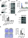
CD164 overexpression upregulates the SDF1α /CXCR4 axis to activate downstream PI3K/Akt signaling pathway. a-b) Cell lysates from hOSE-CD164 and hOSE-vector cells were subjected to a) immunoprecipitation with antibody against CXCR4 and immunoblot analysis with antibody against CD164 and a control antibody (upper) or immunoprecipitation with antibody against CD164 and immunoblot analysis with antibodies against CXCR4 and CXCR7 and a control antibody (bottom); b) immunoblot analysis with antibodies against CD164, CXCR4, pPDK1, pAkt and its phosphorylated form pAktSer-473, CXCR7, p53 and p21. α-tubulin was used as a loading control. c) Quantitative mRNA analysis of CD164, CXCR4 and CXCR7 in hOSE-CD164 cells and indicated ovarian cells was performed by qRT-PCR, and mRNA levels were normalized with individual GAPDH mRNA. Fold changes of specific mRNA expression were compared with that of hOSE control cells. d) The amount of SDF1α in culture medium from hOSE-CD164 cells and indicated ovarian cells was measured using an ELISA analysis. e) The hOSE-CD164 cells showed downregulated CXCR4 expression and were subjected to immunoblot analysis with antibodies against CXCR4, pPDK1 and pAktSer-473. α-tubulin was used as a loading control. f) For the colony formation analysis, the hOSE-CD164 cells were treated with the CXCR4 antagonist AMD3100 or CXCR4 was downregulated using shRNA. Quantitation of anchorage-independent growth of indicated conditioned cells was performed using anchorage independent assay. The presented data are the means of three experiments (means ± S.D.; n = 3).
Figure 6
Figure 6
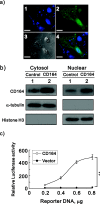
Nuclear CD164 upregulates CXCR4 promoter activity. a) Representative hOSE cells transiently transfected with EGFP–tagged CD164 constructs are shown. Approximately 100 transfected cells were examined to evaluate the intracellular distribution of each of the EGFP fusion proteins under a confocal microscope. Nuclei were stained with TOTO-3. Bar = 20 μm. b) Fractionated cytosolic and nuclear lysates from hOSE-CD164 and hOSE-vector cells were subjected to immunoblot analysis with antibody against CD164. α-tubulin was used as a loading control. c) Indicated amount of CXCR4 reporter DNAs were transiently transfected into hOSE-CD164 and hOSE-vector cells. After 36 hours, the luciferase activity in the transfected cell extracts was determined and the fold induction of the activity was estimated relative to that of cells transfected with the control vector in hOSE-vector cells. The presented data are the means of three experiments (means ± S.D.; n = 3).
Figure 7
Figure 7
The effects of CD164 on stem cell marker and sphere formation. a) A quantitative mRNA analysis of ALDH-1, CD44, Nanog, Nestin, Oct4 and Sox2 in hOSE and hOSE-CD164 cells was performed by qRT-PCR, and mRNA levels are expressed relative to individual GAPDH mRNA. The presented data are the means of three experiments (means ± S.D.; n = 3). b) Spheroid formation of hOSE and hOSE-CD164 and shCXCR4 cells after culture in ultralow plates for 7 days. **p < 0.01. c) Western blot analysis of hOSE and hOSE.CD164 cell lysates were subject to against antibodies for CSC markers, including Nanog, Oct4 and Sox-2. α-tubulin was used as a loading control.

Similar articles

Cited by

References

    1. Naora H, Montell DJ. Ovarian cancer metastasis: integrating insights from disparate model organisms. Nat Rev Cancer. 2005;12:355–366. - PubMed
    1. Lengyel E. Ovarian cancer development and metastasis. Am J Pathol. 2010;12:1053–1064. doi: 10.2353/ajpath.2010.100105. - DOI - PMC - PubMed
    1. Tingulstad S, Skjeldestad FE, Halvorsen TB, Hagen B. Survival and prognostic factors in patients with ovarian cancer. Obstet Gynecol. 2003;12:885–891. doi: 10.1016/S0029-7844(03)00123-6. - DOI - PubMed
    1. Dubeau L. The cell of origin of ovarian epithelial tumours. Lancet Oncol. 2008;12:1191–1197. doi: 10.1016/S1470-2045(08)70308-5. - DOI - PMC - PubMed
    1. Bowtell DD. The genesis and evolution of high-grade serous ovarian cancer. Nat Rev Cancer. 2010;12:803–808. doi: 10.1038/nrc2946. - DOI - PubMed

Publication types

MeSH terms

LinkOut - more resources