. 2014 Jan;28(1):382-94.
doi: 10.1096/fj.13-230037. Epub 2013 Sep 30.

Role of mitofusin 2 (Mfn2) in controlling cellular proliferation

Affiliations

Role of mitofusin 2 (Mfn2) in controlling cellular proliferation

Kuang-Hueih Chen et al. FASEB J. 2014 Jan.

Abstract

It has been reported that Mitofusin2 (Mfn2) inhibits cell proliferation when overexpressed. We wanted to study the role of endogenous Mfn2 in cell proliferation, along with the structural features of Mfn2 that influence its mitochondrial localization and control of cell proliferation. Mfn2-knockdown clones of a B-cell lymphoma cell line BJAB exhibited an increased rate of cell proliferation. A 2-fold increase in cell proliferation was also observed in Mfn2-knockout mouse embryonic fibroblast (MEF) cells as compared with the control wild-type cells, and the proliferative advantage of the knockout MEF cells was blocked on reintroduction of the Mfn2 gene. Mfn2 exerts its antiproliferative effect by acting as an effector molecule of Ras, resulting in the inhibition of the Ras-Raf-ERK signaling pathway. Furthermore, both the N-terminal (aa 1-264) and the C-terminal (aa 265-757) fragments of Mfn2 blocked cell proliferation through distinct mechanisms: the N-terminal-mediated inhibition was due to its interaction with Raf-1, whereas the C-terminal fragment of Mfn2 inhibited cell proliferation by interacting with Ras. The inhibition of proliferation by the N-terminal fragment was independent of its mitochondrial localization. Collectively, our data provide new insights regarding the role of Mfn2 in controlling cellular proliferation.

Keywords: ERK; HSG; Raf; Ras.


Figures

Figure 1.
Figure 1.
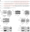
Overexpression of Mfn2 inhibits proliferation of B lymphoma cell lines BJAB (A, B) and RL (C, D). Overexpression of Mfn2 by adenovirally mediated gene transfer in BJAB cells (A) and RL (C) cells, shown by WB (25 μg cell lysates) with an anti-Mfn2 antibody. β-Tubulin was used as a loading control. Inhibitory effect of Mfn2 on BJAB growth (B) and RL growth (D) was examined by WST-1 assay (n=3, performed in quadruplicate). *P < 0.01.
Figure 2.
Figure 2.
Mfn2-knockdown BJAB cells grow faster than the control cells. A) BJAB cells were transduced with lentiviral particles expressing Mfn2-specific or nonsilencing shRNAmirs. Top panel: residual expression levels of Mfn2 in 3 stable Mfn2-silencing clones relative to the nonsilencing control. Bottom panel: quantitative analysis of Mfn2 protein abundance in these stable cell lines (n=3 independent experiments). *P < 0.01 vs. control group. B) Growth curve of the 3 stable Mfn2 silencing clones and the nonsilencing control, assayed by the cleavage of the tetrazolium salt WST-1 (n=3). *P < 0.01 vs. control group.
Figure 3.
Figure 3.
Mfn2 involved in controlling cell proliferation. A) WB of total cell lysates (30 μg) from Mfn2−/− and WT MEFs probed with an anti-Mfn2 antibody. B) Growth curve of MEFs from WT or Mfn2−/− mice determined by cell counting (n=3, performed in triplicate). *P < 0.01. C) Level of Mfn2 protein in Mfn2−/− MEFs infected with either Adv-LacZ or Adv-Mfn2 at 100 pfu/cell. D) Inhibitory effect of Mfn2 on serum-induced Mfn2−/− MEFs proliferation (n=3, performed in quadruplicate). *P < 0.01.
Figure 4.
Figure 4.
Introduction of Mfn2 into Mfn2−/− MEF cells inhibits serum-induced phosphorylation of ERK1/2 and Raf-1. A) WB of total cell lysates (25 μg) from Mfn2−/− and WT MEFs for phosphorylated (p-ERK1 and p-ERK2) and total ERK1/2. B) Total and phosphorylated ERK1/2 in Mfn2−/− MEFs infected with different titers (pfu) of Adv-LacZ or Adv-Mfn2. C) WB of total cell lysates (25 μg) from Mfn2−/− and WT MEFs for phosphorylated (p-Raf-1) and total Raf-1. D) Total and phosphorylated Raf-1in Mfn2−/− MEFs infected with different titers of Adv-LacZ or Adv-Mfn2.
Figure 5.
Figure 5.
Mfn2 associates with Ras protein. A) Cell lysates (500 μg) from Mfn2−/− MEFs transfected with Flag-Mfn2 construct were immunoprecipitated with anti-Ras monoclonal antibody (2 μg) and immunoblotted with either anti-Flag antibody (top panel) or anti-Pan Ras polyclonal antibody (bottom panel). Total cell extracts (25 μg) were used as positive control for expression of Flag-tagged Mfn2 and Ras. B) Whole-cell lysates (500 μg) from WT or Mfn2−/− MEFs were immunoprecipitated with anti-Ras antibody and immunoblotted with either anti-Mfn2 or anti-Ras antibody. Total extracts (25 μg) were used as the control for the expression of Mfn2 and Ras.
Figure 6.
Figure 6.
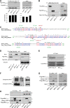
Mfn2 is a Ras effector molecule. A) Sequence similarity between the C-terminal region of human Mfn2 (aa 490–757) and the C-terminal region of NORE1A (aa 191–413) containing RA and SARAH domains. Red box indicates the RA domain of NORE1A (aa 191–363); black box indicates the SARAH domain of NORE1A (aa 366–413). Asterisks indicate identity; colons indicate strong conservation; periods indicate weak conservation. B) Lysates (500 μg) from Mfn2−/− MEFs cotransfected with flag-tagged Mfn21–756 along with different HA-tagged K-Ras plasmids, WT-K-Ras, CA-K-Ras and CA-K-Ras-E37G, were immunoprecipitated with anti-HA monoclonal antibody (2 μg) and immunoblotted with either anti-Flag antibody (top panel) or anti-HA antibody (bottom panel). Total cell extracts (25 μg) were used to monitor the expression of flag-tagged Mfn2 and HA-tagged Ras. Similar results were obtained in 3 independent experiments. C) Expression levels of individual Ras constructs (WT-K-Ras, CA-K-Ras, and CA-K-Ras-E37G) were comparable. Mfn2−/− MEFs were cotransfected with empty vector and indicated HA-tagged Ras constructs, and 25 μg of total cell lysates was analyzed by WB analysis. D) Lysates (25 μg) from Mfn2−/− MEFs cotransfected with a flag-tagged Mfn2 plasmid together with H-Ras construct were analyzed by immunoblotting analysis. β-Tubulin was used as loading control. E) Total cell lysates (25 μg) from Mfn2−/− MEFs cotransfected with WT-Raf-1 together with indicated HA-tagged K-Ras constructs were analyzed by immunoblotting using anti-Raf-1 and anti-HA antibodies. β-Tubulin was used as loading control. F) Flag-tagged Mfn21-757 plasmid and indicated flag-tagged K-Ras constructs were expressed separately using in vitro transcription and translation system. Mfn2 protein was allowed to interact separately with the indicated Ras proteins first and then immunoprecipitated with anti-Ras antibody and immunoblotted with anti-Flag antibody. Similar results were obtained in 3 independent experiments. G, H) Mfn1 associates with Ras. Flag-tagged Mfn1 plasmid and HA-tagged CA-K-Ras plasmid were coexpressed in Mfn2−/− MEFs. Cell lysates (500 μg) were immunoprecipitated with either α-Flag monoclonal antibody (G) or α-HA monoclonal antibody (H), and the immunocomplexes were analyzed by WB analysis; 50 μg of total cell extracts was used as input.
Figure 7.
Figure 7.
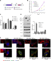
Both N- and C-terminal fragments of Mfn2 inhibit cell proliferation. A) Schematic representation of Mfn2 protein showing different domains. B) Mfn2−/− MEFs were infected with either nothing (control) or 100 pfu/cell of Adv-LacZ, adenovirus containing a full-length Mfn2 (Adv-Mfn21–757), N-terminal fragment of Mfn2 (Adv-Mfn21–264), or C-terminal fragment of Mfn2 (Adv-Mfn2265–757). Cell proliferation assay was performed in quadruplicate by WST-1 assay (n=3 independent experiments). *P < 0.01 vs. control groups. C) Cell cycle distribution of Mfn2−/− MEFs infected with recombinant adenoviruses (30 pfu) carrying different fragments of Mfn2 gene (n=3). D) Expression of different mitochondrial proteins after overexpression of different fragments of Mfn2. Mfn2−/− MEFs were transfected with either control vector or Flag-tagged Mfn21–757, Flag-tagged Mfn21–264, or Flag-tagged Mfn2265–757 plasmids for 48 h. Total cell extracts (35 μg) were used for WB analysis to check the expression of the indicated mitochondrial proteins. Right panel shows quantitation of the bands normalized to γ-tubulin. E) Antiproliferative capacity of Mfn2 protein is partially independent of its mitochondrial localization. WT MEFs (i), Mfn2−/− MEFs (ii), and Mfn2−/− MEfs transfected with flag-tagged Mfn21–757 (iii, iv), Mfn21–264 (v, vi), or Mfn2265–757 (vii, viii) were incubated with 200 nM MitoTracker Red CMXRos and then incubated with mouse anti-Flag antibody, subsequently incubated with anti-mouse IgG Alexa Fluor 350. Cells were then examined with a confocal microscope.
Figure 8.
Figure 8.
While the N-terminal fragment of Mfn2 interacts with Raf-1, the C-terminal fragment interacts with Ras. A) Lysates (500 μg) from Mfn2−/− MEFs transfected with either control vector or plasmids containing Flag-tagged Mfn21–264 or Flag-tagged Mfn2265–757 fragments were immunoprecipitated with α-Ras monoclonal antibody and immunoblotted with anti-Flag antibody (top panel) and an anti-Pan Ras polyclonal antibody (bottom panel). B) Flag-tagged Mfn21–757 and Mfn2265–757 plasmids and Flag-tagged CA-K-Ras plasmid were expressed separately using an in vitro transcription and translation system. Mfn2 proteins were allowed to interact separately with the CA-K-Ras protein first and then immunoprecipitated with α-Mfn2 polyclonal antibody and immunoblotted with α -Flag monoclonal antibody. C) Sequence comparison between Mfn2 N-terminal protein (aa 97–325) and Ras like GTPase superfamily. Asterisks indicate identity; colons indicate strong conservation; periods indicate weak conservation. D) Mfn2−/− MEFs were cotransfected with WT-Raf-1 and indicated Flag-tagged Mfn2 plasmids. Cell lysates (500 μg) were immunoprecipitated with either α-Flag antibody or normal mouse IgG and immunoblotted with either α-Raf-1 antibody (top panel) or α-Flag antibody (bottom panel). Total extracts (25 μg) were used to monitor the expression of Flag-tagged Mfn21–264, Flag-tagged Mfn21–757, and Raf-1. E) Status of phosphorylated and total ERK1/2 in cells cotransfected with WT-Raf-1/empty vector or WT-Raf-1/Flag-tagged Mfn21–264 plasmids. F) Mfn2 associates with endogenous c-Raf protein. Cell lysates (500 μg) of cell lysates from 293A were immunoprecipitated with anti-Mfn2 polyclonal antibody and immunoblotted with either anti-Raf-1 antibody (top panel) or anti-Mfn2 monoclonal antibody (bottom panel); 50 μg of total extract was used as an input. G) Flag-tagged Mfn21–757, Flag-tagged Mfn21–264, and Raf-1 constructs were expressed separately using an in vitro transcription and translation system. Mfn2 proteins were allowed to interact separately with the Raf-1 protein first and then immunoprecipitated with α-Raf-1 monoclonal antibody and immunoblotted with α -Flag monoclonal antibody (top panel) and α Raf-1 polyclonal antibody (bottom panel). H) Mfn2 does not interact with B-Raf; 500 μg of lysates from Mfn2−/− MEFs transfected with either control vector or plasmids containing Flag-tagged Mfn21–264 or Flag-tagged Mfn21–757 were immunoprecipitated with α-Flag antibody and immunoblotted with α-B-Raf antibody (top panel) and an α-Flag antibody (bottom panel); 50 μg of total cell extracts was used to monitor the expression of Flag-Mfn21–264, Flag-Mfn21–757, and B-Raf.

Similar articles

Cited by

References

    1. De Brito O. M., Scorrano L. (2008) Mitofusin 2 tethers endoplasmic reticulum to mitochondria. Nature 456, 605–610 - PubMed
    1. De Brito O. M., Scorrano L. (2009) Mitofusin-2 regulates mitochondrial and endoplasmic reticulum morphology and tethering: the role of Ras. Mitochondrion 9, 222–226 - PubMed
    1. Zuchner S., Mersiyanova I. V., Muglia M., Bissar-Tadmouri N., Rochelle J., Dadali E. L., Zappia M., Nelis E., Patitucci A., Senderek J., Parman Y., Evgrafov O., Jonghe P. D., Takahashi Y., Tsuji S., Pericak-Vance M. A., Quattrone A., Battaloglu E., Polyakov A. V., Timmerman V., Schroder J. M., Vance J. M. (2004) Mutations in the mitochondrial GTPase mitofusin 2 cause Charcot-Marie-Tooth neuropathy type 2A. Nat. Genet. 36, 449–451 - PubMed
    1. Misko A. L., Sasaki Y., Tuck E., Milbrandt J., Baloh R. H. (2012) Mitofusin2 mutations disrupt axonal mitochondrial positioning and promote axon degeneration. J. Neurosci. 32, 4145–4155 - PMC - PubMed
    1. Hernandez-Alvarez M. I., Thabit H., Burns N., Shah S., Brema I., Hatunic M., Finucane F., Liesa M., Chiellini C., Naon D., Zorzano A., Nolan J. J. (2010) Subjects with early-onset type 2 diabetes show defective activation of the skeletal muscle PGC-1α/mitofusin-2 regulatory pathway in response to physical activity. Diabetes Care 33, 645–651 - PMC - PubMed

Publication types

LinkOut - more resources