Diffusion tensor imaging of the spinal cord: insights from animal and human studies
- PMID: 24064483
- PMCID: PMC4986512
- DOI: 10.1227/NEU.0000000000000171
Diffusion tensor imaging of the spinal cord: insights from animal and human studies
Abstract
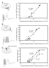
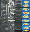
Diffusion tensor imaging (DTI) provides a measure of the directional diffusion of water molecules in tissues. The measurement of DTI indexes within the spinal cord provides a quantitative assessment of neural damage in various spinal cord pathologies. DTI studies in animal models of spinal cord injury indicate that DTI is a reliable imaging technique with important histological and functional correlates. These studies demonstrate that DTI is a noninvasive marker of microstructural change within the spinal cord. In human studies, spinal cord DTI shows definite changes in subjects with acute and chronic spinal cord injury, as well as cervical spondylotic myelopathy. Interestingly, changes in DTI indexes are visualized in regions of the cord, which appear normal on conventional magnetic resonance imaging and are remote from the site of cord compression. Spinal cord DTI provides data that can help us understand underlying microstructural changes within the cord and assist in prognostication and planning of therapies. In this article, we review the use of DTI to investigate spinal cord pathology in animals and humans and describe advances in this technique that establish DTI as a promising biomarker for spinal cord disorders.
Figures


Similar articles
-
Reproducibility, temporal stability, and functional correlation of diffusion MR measurements within the spinal cord in patients with asymptomatic cervical stenosis or cervical myelopathy.J Neurosurg Spine. 2018 May;28(5):472-480. doi: 10.3171/2017.7.SPINE176. Epub 2018 Feb 9. J Neurosurg Spine. 2018. PMID: 29424671 Free PMC article.
-
Application of diffusion tensor imaging (DTI) in pathological changes of the spinal cord.Med Sci Monit. 2012 Jun;18(6):RA73-9. doi: 10.12659/msm.882891. Med Sci Monit. 2012. PMID: 22648262 Free PMC article. Review.
-
Correlation of magnetic resonance diffusion tensor imaging and clinical findings of cervical myelopathy.Spine J. 2013 Aug;13(8):867-76. doi: 10.1016/j.spinee.2013.02.005. Epub 2013 Mar 21. Spine J. 2013. PMID: 23523441
-
Magnetic resonance diffusion tensor imaging in patients with cervical spondylotic spinal cord compression: correlations between clinical and electrophysiological findings.Spine (Phila Pa 1976). 2012 Jan 1;37(1):48-56. doi: 10.1097/BRS.0b013e31820e6c35. Spine (Phila Pa 1976). 2012. PMID: 21228747
-
The role of diffusion tensor imaging in spinal pathology: A review.Neurol India. 2017 Sep-Oct;65(5):982-992. doi: 10.4103/neuroindia.NI_198_17. Neurol India. 2017. PMID: 28879883 Review.
Cited by
-
Feasibility of 3.0 T diffusion-weighted nuclear magnetic resonance imaging in the evaluation of functional recovery of rats with complete spinal cord injury.Neural Regen Res. 2015 Mar;10(3):412-8. doi: 10.4103/1673-5374.153689. Neural Regen Res. 2015. PMID: 25878589 Free PMC article.
-
Longitudinal assessment of recovery after spinal cord injury with behavioral measures and diffusion, quantitative magnetization transfer and functional magnetic resonance imaging.NMR Biomed. 2020 Apr;33(4):e4216. doi: 10.1002/nbm.4216. Epub 2020 Jan 13. NMR Biomed. 2020. PMID: 31943383 Free PMC article.
-
Diffusion weighted imaging as a biomarker of retinoic acid induced myelomeningocele.PLoS One. 2021 Jun 30;16(6):e0253583. doi: 10.1371/journal.pone.0253583. eCollection 2021. PLoS One. 2021. PMID: 34191842 Free PMC article.
-
Impact of Deep Learning Denoising Algorithm on Diffusion Tensor Imaging of the Growth Plate on Different Spatial Resolutions.Tomography. 2024 Apr 2;10(4):504-519. doi: 10.3390/tomography10040039. Tomography. 2024. PMID: 38668397 Free PMC article.
-
Diffusion Assessment of Cortical Changes, Induced by Traumatic Spinal Cord Injury.Brain Sci. 2017 Feb 17;7(2):21. doi: 10.3390/brainsci7020021. Brain Sci. 2017. PMID: 28218643 Free PMC article.
References
-
- Le Bihan D, Breton E, Lallemand D, Grenier P, Cabanis E, Laval-Jeantet M. MR imaging of intravoxel incoherent motions: application to diffusion and perfusion in neurologic disorders. Radiology. 1986 Nov;161(2):401–407. - PubMed
-
- Clark CA, Werring DJ. Diffusion tensor imaging in spinal cord: methods and applications - a review. NMR Biomed. 2002 Nov-Dec;15(7–8):578–586. - PubMed
-
- Barker GJ. Diffusion-weighted imaging of the spinal cord and optic nerve. J Neurol Sci. 2001 May 1;186(Suppl 1):S45–49. - PubMed
-
- Demir A, Ries M, Moonen CT, et al. Diffusion-weighted MR imaging with apparent diffusion coefficient and apparent diffusion tensor maps in cervical spondylotic myelopathy. Radiology. 2003 Oct;229(1):37–43. - PubMed
Publication types
MeSH terms
Grants and funding
LinkOut - more resources
Full Text Sources
Other Literature Sources
Medical

