. 2013 Oct 24;122(17):3062-73.
doi: 10.1182/blood-2013-05-500801. Epub 2013 Sep 12.

Host programmed death ligand 1 is dominant over programmed death ligand 2 expression in regulating graft-versus-host disease lethality

Affiliations

Host programmed death ligand 1 is dominant over programmed death ligand 2 expression in regulating graft-versus-host disease lethality

Asim Saha et al. Blood. .

Abstract

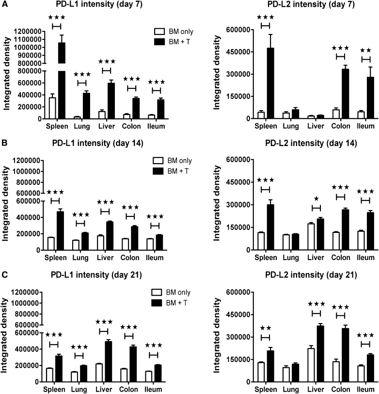
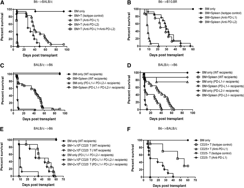
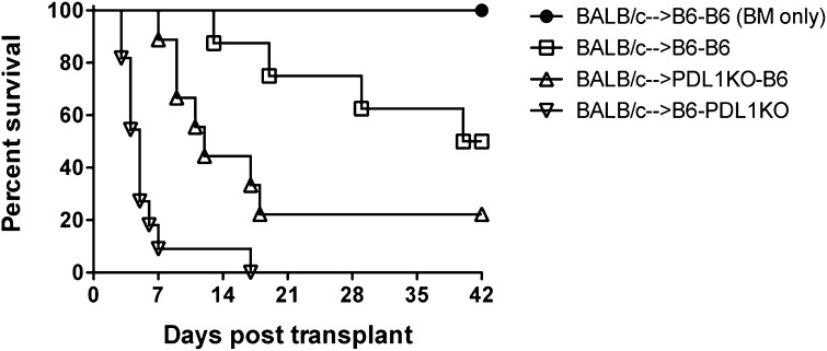
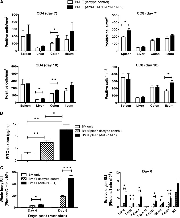
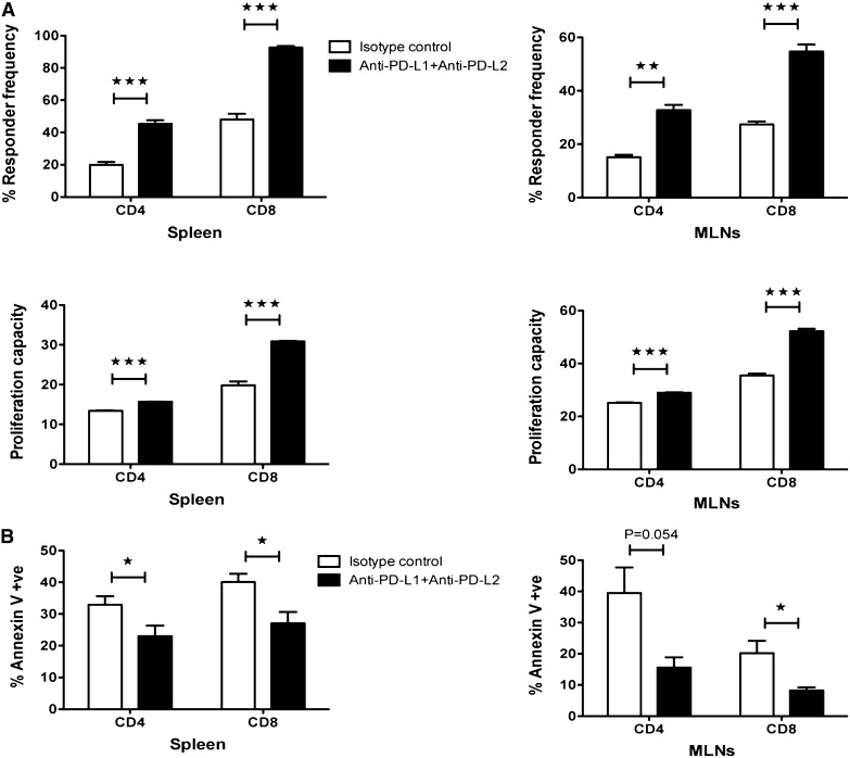
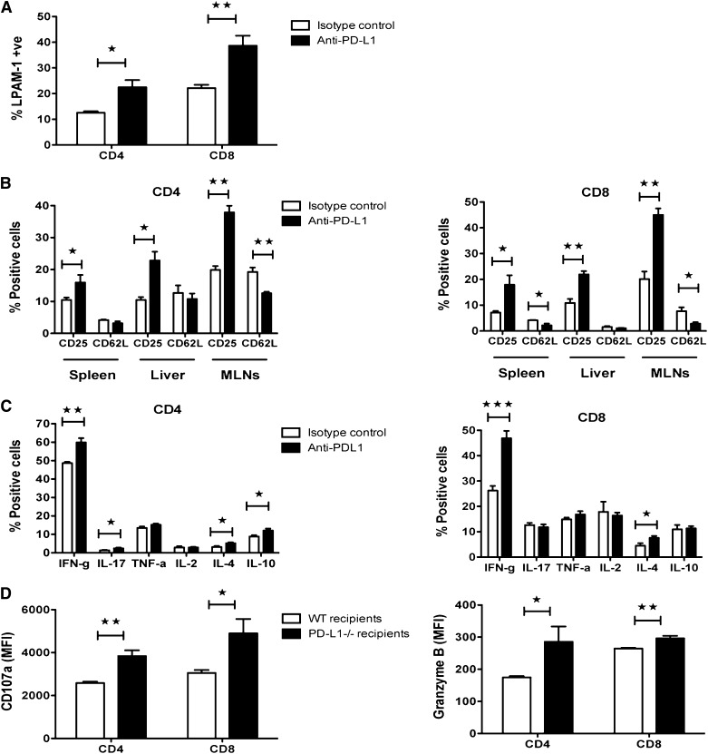
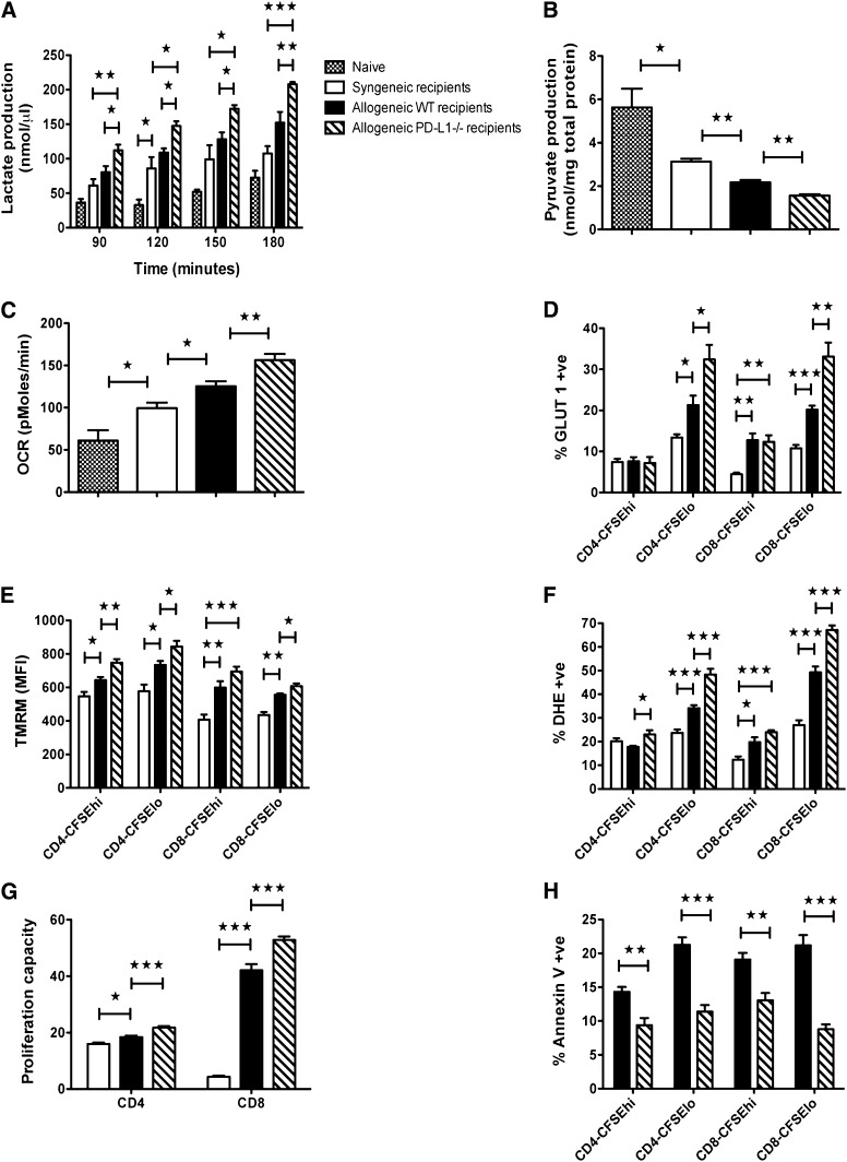
Programmed death 1 (PD-1) and its ligands, PD-L1 and PD-L2, play an important role in the maintenance of peripheral tolerance. We explored the role of PD-1 ligands in regulating graft-versus-host disease (GVHD). Both PD-L1 and PD-L2 expression were upregulated in the spleen, liver, colon, and ileum of GVHD mice. Whereas PD-L2 expression was limited to hematopoietic cells, hematopoietic and endothelial cells expressed PD-L1. PD-1/PD-L1, but not PD-1/PD-L2, blockade markedly accelerated GVHD-induced lethality. Chimera studies suggest that PD-L1 expression on host parenchymal cells is more critical than hematopoietic cells in regulating acute GVHD. Rapid mortality onset in PD-L1-deficient hosts was associated with increased gut T-cell homing and loss of intestinal epithelial integrity, along with increased donor T-cell proliferation, activation, Th1 cytokine production, and reduced apoptosis. Bioenergetics profile analysis of proliferating alloreactive donor T-cells demonstrated increased aerobic glycolysis and oxidative phosphorylation in PD-L1-deficient hosts. Donor T-cells exhibited a hyperpolarized mitochondrial membrane potential, increased superoxide production, and increased expression of a glucose transporter in PD-L1-deficient hosts. Taken together, these data provide new insight into the differential roles of host PD-L1 and PD-L2 and their associated cellular and metabolic mechanisms controlling acute GVHD.


Figures

Figure 1
Figure 1
Enhanced expression of PD-1 ligands in acute GVHD. Lethally irradiated BALB/c recipients were given 107 B6 BM cells alone or with 2 × 10⁶ B6 T-cells. Immunofluorescence staining was performed on day 7 (A), day 14 (B), and day 21 (C) for PD-L1 and PD-L2 (4 mice/group). Images were captured at ×200 magnification, using an Olympus FluoView 500 or Olympus FluoView 1000 BX2 confocal laser scanning microscope and analyzed with Adobe Photoshop CS3 (version 10) for determination of the relative fluorescence staining intensity. Data are representative of 2 independent experiments. *P < .05; **P < .01; ***P < .001.
Figure 2
Figure 2
PD-1/PD-L1, but not PD-1/PD-L2, blockade exacerbates acute GVHD. (A) Lethally irradiated BALB/c recipients were given 107 B6 BM cells alone or with 2 × 10⁶ B6 T-cells and treated with isotype-matched control antibody, anti-PD-L1, anti-PD-L2, or anti-PD-L1 and anti-PD-L2 mAbs (n = 10–18/group). Kaplan–Meier survival curve of transplanted mice (isotype control vs anti-PD-L1 [P < .0001]; isotype control vs anti-PD-L2 [P = .055]; isotype control vs anti-PD-L1 and anti-PD-L2 [P = .0001]). (B) Lethally irradiated B10.BR recipients were given 107 B6 BM cells alone or with 5 × 10⁶ B6 splenocytes and treated with isotype-matched control antibody, anti-PD-L1, or anti-PD-L2 mAb (n = 8–14/group). Kaplan–Meier survival curve of transplanted mice (isotype control vs anti-PD-L1 [P < .0001]; isotype control vs anti-PD-L2 [P = .356]). (C) Lethally irradiated wt B6 recipients or PD-L1−/−PD-L2−/− recipients were given 107 BALB/c BM cells alone or with 5 × 10⁶ BALB/c splenocytes (n = 14–34/group). Kaplan–Meier survival curve of transplanted mice (BM + splenocytes: wt vs PD-L1−/−PD-L2−/− recipients; P < .0001). (D) Lethally irradiated wt B6 recipients, PD-L1−/− recipients, or PD-L2−/− recipients were given 107 BALB/c BM cells alone or with 5 × 10⁶ BALB/c splenocytes (n = 8–17/group). Kaplan–Meier survival curve of transplanted mice (BM + splenocytes: wt vs PD-L1−/− recipients [P = .0001]; BM + splenocytes: wt vs PD-L2−/− recipients [P = .003]; BM + splenocytes: PD-L1−/− vs PD-L2−/− recipients [P = .029]). (E) Lethally irradiated wt B6 recipients or PD-L1−/−PD-L2−/− recipients were given 107 BALB/c BM cells alone or with 1 × 10⁶ BALB/c CD25-depleted T-cells or with 2 × 10⁶ BALB/c CD25-depleted T-cells (n = 8/group). Kaplan–Meier survival curve of transplanted mice (BM + 1 × 10⁶ CD25 T: wt vs PD-L1−/−PD-L2−/− recipients [P = .0006]; BM + 2 × 10⁶ CD25 T: wt vs PD-L1−/−PD-L2−/− recipients [P = .0006]). (F) Lethally irradiated BALB/c recipients were given 107 B6 BM cells alone or with 0.75 × 10⁶ B6 T-cells or with 0.75 × 10⁶ B6 CD25-depleted T-cells and treated with isotype-matched control antibody or anti-PD-L1 mAbs (n = 8–10/group). Kaplan–Meier survival curve of transplanted mice (BM + T cells: isotype control vs anti-PD-L1 [P < .0001]; BM + CD25-depleted T cells: isotype control vs anti-PD-L1 [P = .074]; BM + T cells [isotype control] vs BM + CD25-depleted T cells [isotype control] [P = .0008]).
Figure 3
Figure 3
PD-L1 expression on parenchymal cells is critical for suppression of acute GVHD. Lethally irradiated wt B6 or PD-L1−/− recipients were given BM cells from PD-L1−/− or wt B6 mice, respectively, to create chimeras. We also created control chimeras (wt→wt). After 3 months, these chimeras were reirradiated and infused with allogeneic BALB/c BM cells with 10 × 106 BALB/c splenocytes. Kaplan–Meier survival curve of transplanted mice (n = 8–11/group). wt→ wt vs PD-L1−/−→wt chimeras [P = .06]; wt→wt vs wt→PD-L1−/− chimeras [P < .0001]; PD-L1−/−→wt vs wt→ PD-L1−/− chimeras [P = .0009]). Data are representative of 2 independent experiments.
Figure 4
Figure 4
Blockade of PD-1/PD-1 ligand interactions induced preferential tissue damage by donor T-cells. (A) Lethally irradiated B10.BR recipients were given 107 B6 BM cells with 2 × 10⁶ B6 T-cells and treated with isotype-matched control antibody or anti-PD-L1 and anti-PD-L2 mAbs. Immunohistochemistry staining for CD4 (clone RM4-5) and CD8 (clone 53-6.7) T-cells on day 7 and day 10 after BMT. Cell numbers in spleen, liver, colon, and ileum are shown. Cells were quantified by counting the number of antibody-binding-positive cells in a 100 mm2 field of view under the microscope and obtaining an average of counts from 4 representative fields (n = 4 mice/group). (B) Lethally irradiated B10.BR recipients were given 107 B6 BM cells alone or with 10 × 106 B6 splenocytes and treated with isotype-matched control antibody or anti-PD-L1 mAb. Plasma FITC-dextran concentration was measured on day 7 after BMT (n = 5 mice/group). (C) Lethally irradiated BALB/c recipients were given 107 B6 BM cells alone or with 1.5 × 10⁶ B6 luciferase transgenic T-cells and treated with isotype-matched control antibody or anti-PD-L1 mAb. On day 4 and day 6 after BMT, mice were injected intraperitoneally with luciferin, and after 5 minutes, mice were imaged using a Xenogen IVIS imaging system for 2 minutes (n = 7–9 mice/group). On day 6 after BMT, mice were killed, and isolated organs were imaged for 1 minute in the presence of luciferin (n = 7 mice/group). (A−C) Data are representative of 2 independent experiments. *P < .05; **P < .01; ***P < .001.
Figure 5
Figure 5
Host PD-1 ligand expression affect proliferation and survival of allogeneic donor T-cells. Lethally irradiated B10.BR recipients were given 107 B6 BM cells with 50 × 106 CFSE-labeled B6 splenocytes and treated with isotype-matched control antibody or anti-PD-L1 and anti-PD-L2 mAbs. (A) Mice were killed on day 5 after BMT (n = 5 mice/group), and splenocytes and MLNs were analyzed by flow cytometry for CFSE dilution. Cells were gated on H-2Kb+ and analyzed for CD4+ or CD8+ events. (B) Spleen and MLNs were harvested (n = 5 mice/group) on day 5 and analyzed by flow cytometry for annexin V-positive donor CD4 and CD8 T-cells. (A-B) Data are representative of 2 independent experiments. *P < .05; **P < .01; ***P < .001.
Figure 6
Figure 6
GVHD acceleration induced by PD-1/PD-1 ligand blockade resulted in increased activation and effector function of donor T-cells. (A-C) Lethally irradiated B10.BR recipients were given 107 B6 BM cells with 5 × 10⁶ B6 splenocytes and treated with isotype-matched control antibody or anti-PD-L1 mAb. (A) Mice were killed on day 7 after BMT, and splenocytes (n = 5 mice/group) were analyzed by flow cytometry for LPAM-1 (α4β7) expression on donor CD4 and CD8 T-cells. (B) Common activation markers (CD25 and CD62L) on donor CD4 and CD8 T-cells were analyzed on day 7 (n = 4–5 mice/group) by flow cytometry. (C) Intracellular cytokine staining was performed on day 7 (n = 5 mice/group) and analyzed by flow cytometry to detect the percentage of donor CD4 and CD8 T-cells producing IFN-γ, IL-17, TNF-α, IL-2, IL-4, and IL-10 in spleen. (D) Lethally irradiated wt B6 recipients or PD-L1−/− recipients were given 107 BALB/c BM cells with 5 × 10⁶ BALB/c splenocytes. Mice were killed on day 7 after BMT (n = 5 mice/group), and splenocytes were analyzed by flow cytometry for intracellular expression of CD107a or granzyme B on donor CD4 and CD8 T-cells. Data are presented as mean fluorescence intensity (MFI). (A−D) Data are representative of 2 independent experiments. *P < .05; **P < .01; ***P < .001.
Figure 7
Figure 7
GVHD acceleration induced by PD-1/PD-L1 blockade resulted in increased metabolic stress in donor T-cells. Lethally irradiated wt B6, PD-L1−/−, or Thy1.2+ BALB/c recipients were given 107 Thy1.2+ BALB/c BM cells with 30 × 106 CFSE-labeled Thy1.1+ BALB/c splenocytes. (A-H) Mice were killed on day 5 after BMT, and experiments were performed as described. Thy1.1+ donor T-cells were purified, and lactate (A) and pyruvate (B) production and oxygen consumption (C) by donor T-cells were measured. Naive Thy1.1+ BALB/c mice (n = 4) were included as control. Mice splenocytes were analyzed by flow cytometry for GLUT1 (D), TMRM (E), dihydroethidium (F), and annexin V (H) expression in undivided (CFSEhi) and divided (CFSElo) donor T-cells. MFI; mean fluorescence intensity. (G) Splenocytes were also analyzed by flow cytometry for CFSE dilution. Cells were gated on Thy1.1+ and analyzed for CD4+ or CD8+ events. (A-H) Data are representative of 5–9 mice/group from 2-3 independent experiments. *P < .05; **P < .01; ***P < .001.

Similar articles

Cited by

References

    1. Socié G, Blazar BR. Acute graft-versus-host disease: from the bench to the bedside. Blood. 2009;114(20):4327–4336. - PMC - PubMed
    1. Okazaki T, Maeda A, Nishimura H, Kurosaki T, Honjo T. PD-1 immunoreceptor inhibits B cell receptor-mediated signaling by recruiting src homology 2-domain-containing tyrosine phosphatase 2 to phosphotyrosine. Proc Natl Acad Sci USA. 2001;98(24):13866–13871. - PMC - PubMed
    1. Sheppard KA, Fitz LJ, Lee JM, et al. PD-1 inhibits T-cell receptor induced phosphorylation of the ZAP70/CD3zeta signalosome and downstream signaling to PKCtheta. FEBS Lett. 2004;574(1-3):37–41. - PubMed
    1. Freeman GJ, Long AJ, Iwai Y, et al. Engagement of the PD-1 immunoinhibitory receptor by a novel B7 family member leads to negative regulation of lymphocyte activation. J Exp Med. 2000;192(7):1027–1034. - PMC - PubMed
    1. Latchman Y, Wood CR, Chernova T, et al. PD-L2 is a second ligand for PD-1 and inhibits T cell activation. Nat Immunol. 2001;2(3):261–268. - PubMed

Publication types

MeSH terms

Substances