. 2013 Sep 11;33(37):14816-24.
doi: 10.1523/JNEUROSCI.5511-12.2013.

Neutrophils express oncomodulin and promote optic nerve regeneration

Affiliations

Neutrophils express oncomodulin and promote optic nerve regeneration

Takuji Kurimoto et al. J Neurosci. .

Abstract

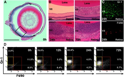
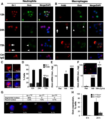
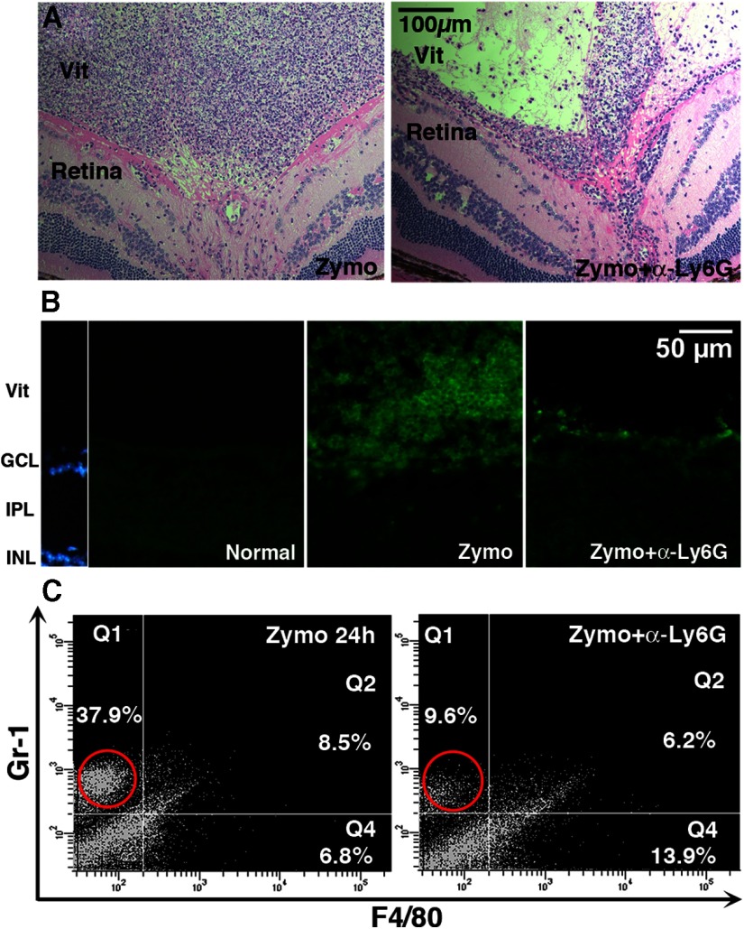
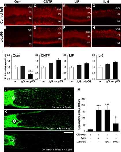
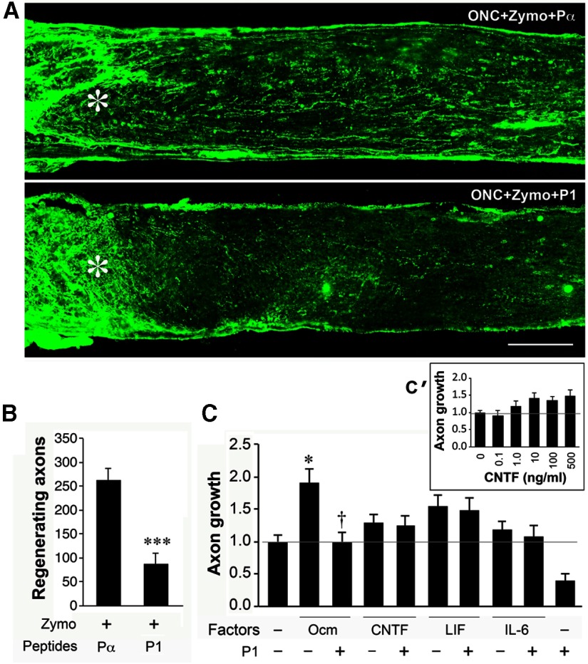
Although neurons are normally unable to regenerate their axons after injury to the CNS, this situation can be partially reversed by activating the innate immune system. In a widely studied instance of this phenomenon, proinflammatory agents have been shown to cause retinal ganglion cells, the projection neurons of the eye, to regenerate lengthy axons through the injured optic nerve. However, the role of different molecules and cell populations in mediating this phenomenon remains unclear. We show here that neutrophils, the first responders of the innate immune system, play a central role in inflammation-induced regeneration. Numerous neutrophils enter the mouse eye within a few hours of inducing an inflammatory reaction and express high levels of the atypical growth factor oncomodulin (Ocm). Immunodepletion of neutrophils diminished Ocm levels in the eye without altering levels of CNTF, leukemia inhibitory factor, or IL-6, and suppressed the proregenerative effects of inflammation. A peptide antagonist of Ocm suppressed regeneration as effectively as neutrophil depletion. Macrophages enter the eye later in the inflammatory process but appear to be insufficient to stimulate extensive regeneration in the absence of neutrophils. These data provide the first evidence that neutrophils are a major source of Ocm and can promote axon regeneration in the CNS.


Figures

Figure 1.
Figure 1.
Characterization of inflammatory cells following zymosan injection. A, Low-magnification image of the normal mouse eye. Rectangle indicates area shown in next panels. B, Higher-magnification images show cells in the vitreous 12 h after intraocular injection of zymosan and greater numbers at 24 and 72 h. C, Immunostaining for F4/80, a macrophage-specific marker, and Gr-1, a cell-surface marker expressed predominantly in neutrophils, 24 h after zymosan injection. D, Representative analyses of inflammatory cells by flow cytometry. Few Gr-1+ or F4/80+ cells are seen in the normal eye; 12 and 24 h after zymosan injection, there are large numbers of Gr-1+/F4/80 neutrophils (yellow frames) and fewer F4/80+ macrophages (red frames). At 72 h, the relative number of F4/80+ macrophages increases. Scale bars: A, 500 μm; B, 200 μm; C, 50 μm.
Figure 2.
Figure 2.
Ocm is expressed in inflammatory cells. A, Double-immunostaining for Ocm and Gr-1 in cells extracted from the vitreous. Gr-1+ cells strongly express Ocm 12 h after zymosan injection, but by 72 h, Gr-1+ cells (arrows) show little Ocm expression (arrowheads). B, Double-immunostaining for Ocm and F4/80. The number of F4/80+ cells (arrows) increases slowly, and they, too, express Ocm (arrowheads). Scale bar, 10 μm. C, Morphology of cells extracted from the vitreous at 24 h. Cells that are strongly Gr-1+ have lobulated nuclei, which is characteristic of neutrophils, whereas F4/80+ macrophages have large, round nuclei. D, Abundance of cells exhibiting strong staining for Gr-1 or F4/80 after intraocular zymosan injections. Counts are based on direct visualization of cells extracted from the eye (N ≥ 4 biologically independent samples). E, Ocm mRNA expression in FACS-sorted Gr-1+/F4/80 cells (neutrophils) and Gr-1+/F4/80+ cells (macrophages) 24 h after zymosan injection. Data are normalized to 18S RNA and then to Ocm mRNA levels in the normal retina, and are represented on a logarithmic scale (means ± SEM, N = 4 independent sortings). Significance relative to the retina: *p < 0.05; ***p < 0.001. F, F', Ocm in peripheral neutrophils isolated from normal mouse blood. Untreated neutrophils express Ocm (***p < 0.001 compared with retina) and levels increase further following a 4 h exposure to zymosan (†p < 0.05). Results are normalized as above. G, Gr-1 expression and morphology of cells isolated from the eye 24 h after zymosan injection. Table shows percentage of cells exhibiting different levels of Gr-1 staining with either polymorphic nuclei, a hallmark of neutrophils, or round nuclei, a characteristic of macrophages (based on 185 cells counted). Ninety-six percent of cells with high Gr-1 staining are neutrophils. Panels below show cells with different characteristics. g1, High Gr-1, polymorphic nuclei (neutrophils); g2–g4, variable levels of Gr-1 and round nuclei (macrophages). H, Ocm expression. Over 90% of cells with Gr-1 staining express Ocm at 6 h. The number of Gr-1-positive cells that show appreciable Ocm immunostaining declines by 24 h, as Gr-1-negative cells that express Ocm start to appear (based on 200–400 cells in 3–4 independent samples for each time point).
Figure 3.
Figure 3.
Immunodepletion of neutrophils. An antibody to Ly6G, the neutrophil-specific form of Gr-1, was administered both retro-orbitally (100 μg) and intraperitoneally (20 μg) immediately before optic nerve crush. A, Hemotoxylin-eosin staining of whole eyes shows diminished numbers of inflammatory cells in the vitreous following anti-Ly6G treatment. B, Gr-1 immunostaining reveals that immunodepletion decreases the number of Gr-1+ cells in the vitreous at 24 h. C, Flow cytometry shows a loss of Gr-1+ cells 24 h after intraocular zymosan plus anti-Ly6G antibody compared with zymosan alone. Note that the percentage of Grhigh cells shown in C is lower than in Figure 1D, perhaps due to the larger number of cells appearing in the low Gr1/low F4/80 quadrant in this experiment.
Figure 4.
Figure 4.
Neutrophil depletion decreases Ocm levels in the retina and suppresses optic nerve regeneration. A–H, Immunostaining for the indicated growth factors 1 d after intraocular injection of zymosan and systemic treatment with control IgG (A, C, E, G) or anti-Ly6G (B, D, F, H). I, Quantitation of immunoreactivity. Control IgG did not alter immunostaining for any of the factors, whereas anti-Ly6G selectively diminished immunostaining for Ocm (†p < 0.05; n ≥ 4 for each condition). J–M, Effect of neutrophil depletion on optic nerve regeneration. GAP-43+ axons are visualized by immunostaining in longitudinal sections through the mouse optic nerve 2 weeks after nerve injury and intraocular zymosan injections. Asterisks denote the injury site. Whereas treatment with control IgG had no effect (compare K, J), immune depletion of neutrophils with the anti-Ly6G antibody suppressed regeneration (L). M, Quantitation. ***Increase relative to negative controls (optic nerve crush alone) significant at p < 0.001. †Decrease relative to controls treated with normal IgG significant at p < 0.05. Results are based on N ≥ 4 cases per condition. Scale bar: A–H, 50 μm.
Figure 5.
Figure 5.
A peptide antagonist of Ocm diminishes axon regeneration. A, B, Axons regenerating through the optic nerve visualized 2 weeks after nerve injury by GAP-43 immunostaining. A, Intraocular inflammation induced by zymosan is unaffected by intraocular injection of the control peptide Pα but is suppressed by P1, a 22 aa peptide that competes with Ocm for receptor occupancy (Yin et al., 2009; compare levels of regeneration to Fig. 4, positive and negative controls). ***Decrease significant at p < 0.001 compared with cases treated with zymosan and control peptide (n = 6 mice for each). C, Specificity of the P1 peptide. Growth factors were tested in the presence of mannose (250 μm) and forskolin (15 μm), essential cofactors for Ocm. Ocm (200 ng/ml) nearly doubled the level of growth (*p < 0.05), and this effect was eliminated by a 500-fold molar excess of P1 (†p < 0.05; ANOVA, Bonferroni correction). CNTF (200 ng/ml) and LIF (400 ng/ml) had smaller effects that did not achieve statistical significance, and P1 did not alter these (samples tested in quadruplicate). IL-6 (400 ng/ml) was inactive. C', Inset, Dose–response for CNTF. Maximal effect is achieved at 10 ng/ml.

Comment in

Similar articles

Cited by

References

    1. Austyn JM, Gordon S. F4/80, a monoclonal antibody directed specifically against the mouse macrophage. Eur J Immunol. 1981;11:805–815. doi: 10.1002/eji.1830111013. - DOI - PubMed
    1. Barrette B, Hébert MA, Filali M, Lafortune K, Vallières N, Gowing G, Julien JP, Lacroix S. Requirement of myeloid cells for axon regeneration. J Neurosci. 2008;28:9363–9376. doi: 10.1523/JNEUROSCI.1447-08.2008. - DOI - PMC - PubMed
    1. Benowitz LI, Popovich PG. Inflammation and axon regeneration. Curr Opin Neurol. 2011;24:577–583. doi: 10.1097/WCO.0b013e32834c208d. - DOI - PubMed
    1. Brinkmann V, Zychlinsky A. Neutrophil extracellular traps: is immunity the second function of chromatin? J Cell Biol. 2012;198:773–783. doi: 10.1083/jcb.201203170. - DOI - PMC - PubMed
    1. Carlson SL, Parrish ME, Springer JE, Doty K, Dossett L. Acute inflammatory response in spinal cord following impact injury. Exp Neurol. 1998;151:77–88. doi: 10.1006/exnr.1998.6785. - DOI - PubMed

Publication types

MeSH terms