. 2013 Nov 1;288(44):31715-27.
doi: 10.1074/jbc.M113.501122. Epub 2013 Sep 11.

Interleukin-1 and tumor necrosis factor-α trigger restriction of hepatitis B virus infection via a cytidine deaminase activation-induced cytidine deaminase (AID)

Affiliations

Interleukin-1 and tumor necrosis factor-α trigger restriction of hepatitis B virus infection via a cytidine deaminase activation-induced cytidine deaminase (AID)

Koichi Watashi et al. J Biol Chem. .

Abstract

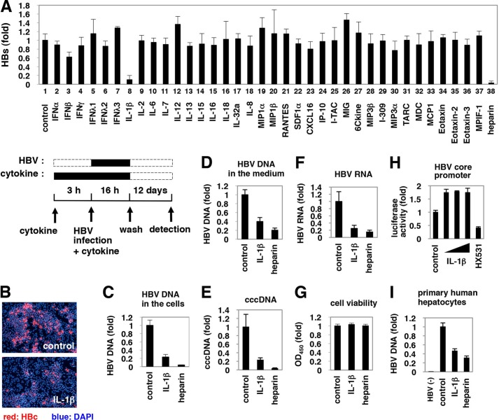
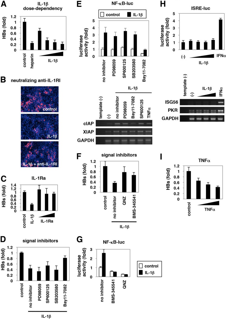
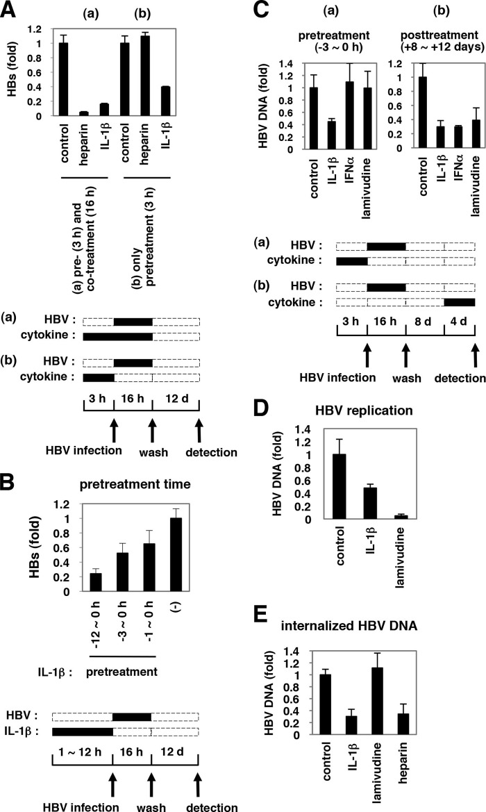
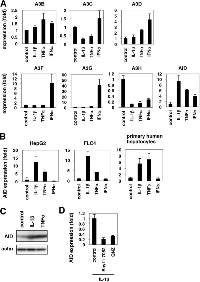
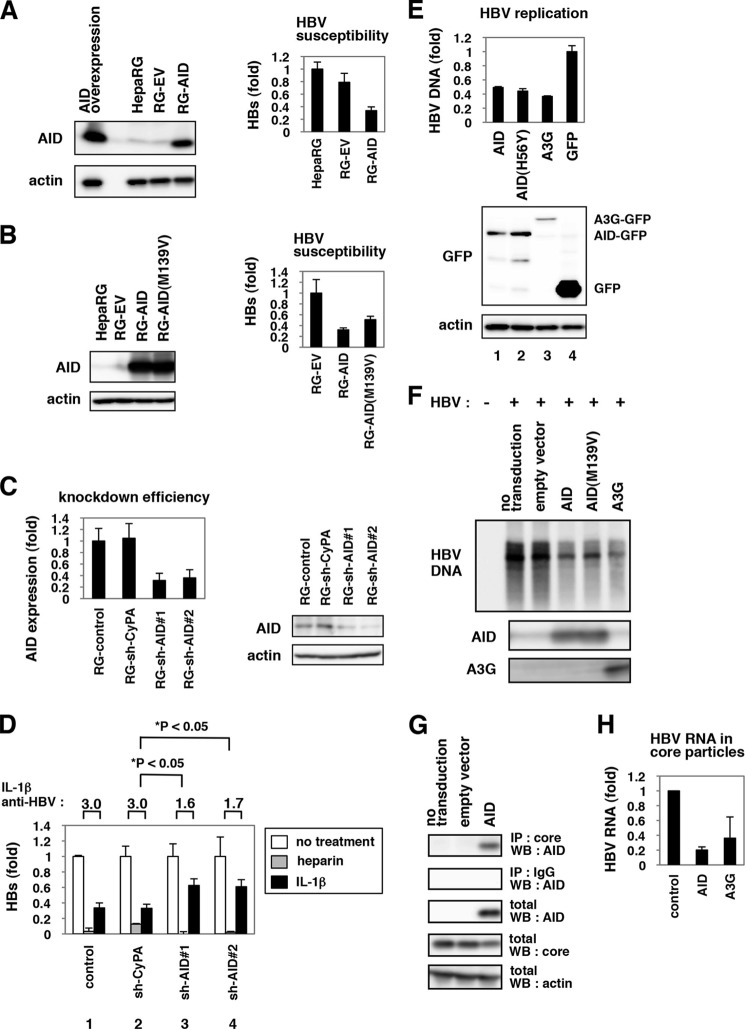
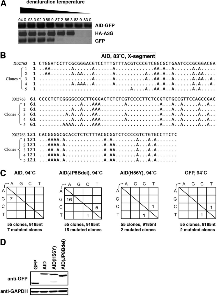
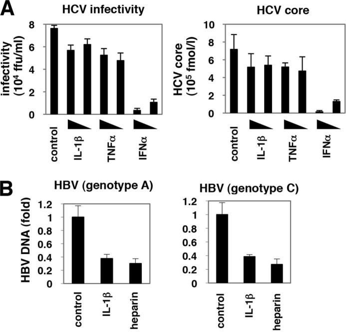
Virus infection is restricted by intracellular immune responses in host cells, and this is typically modulated by stimulation of cytokines. The cytokines and host factors that determine the host cell restriction against hepatitis B virus (HBV) infection are not well understood. We screened 36 cytokines and chemokines to determine which were able to reduce the susceptibility of HepaRG cells to HBV infection. Here, we found that pretreatment with IL-1β and TNFα remarkably reduced the host cell susceptibility to HBV infection. This effect was mediated by activation of the NF-κB signaling pathway. A cytidine deaminase, activation-induced cytidine deaminase (AID), was up-regulated by both IL-1β and TNFα in a variety of hepatocyte cell lines and primary human hepatocytes. Another deaminase APOBEC3G was not induced by these proinflammatory cytokines. Knockdown of AID expression impaired the anti-HBV effect of IL-1β, and overexpression of AID antagonized HBV infection, suggesting that AID was one of the responsible factors for the anti-HBV activity of IL-1/TNFα. Although AID induced hypermutation of HBV DNA, this activity was dispensable for the anti-HBV activity. The antiviral effect of IL-1/TNFα was also observed on different HBV genotypes but not on hepatitis C virus. These results demonstrate that proinflammatory cytokines IL-1/TNFα trigger a novel antiviral mechanism involving AID to regulate host cell permissiveness to HBV infection.

Keywords: AID; APOBEC3G; Deaminase; HBV; HepaRG; Innate Immunity; Interferon; Interleukin; Tumor Necrosis Factor (TNF); Virus.


Figures

FIGURE 1.
FIGURE 1.
Suppression of HBV infection by IL-1β. A, upper graph, HepaRG cells were pretreated with cytokines at 100 ng/ml (except for IFNα and IFNβ at 100 IU/ml) or heparin at 25 units/ml as a positive control or were left untreated (control) for 3 h and then infected with HBV in the presence of each stimuli for 16 h. After washing, cells were cultured in normal growth medium for 12 days. HBs protein secreted into the medium was quantified by ELISA. Lower scheme indicates the treatment procedure for HepaRG cells. Black and dashed line boxes indicate the periods with and without treatment, respectively. B–G and I, HepaRG cells (B–G) or PHH (I) were treated as shown in A with or without 100 ng/ml IL-1β or 25 units/ml heparin as a positive control. HBc protein in the cells (red) was detected by indirect immunofluorescence analysis, and the nucleus was stained with DAPI (blue) at 12 days post-infection (B). HBV DNA (C and I), cccDNA (E), and HBV RNA (F) in the cells as well as HBV DNA in the medium (D) were detected. Cell viability was quantified by MTT assay (G). HBV(−) in I indicates uninfected cells. All of the data, except in I, are based on the average of three independent experiments. I shows the average results from one representative experiment, but the reproducibility of the data were confirmed in three independent experiments. H, reporter plasmid carrying the HBV core promoter was transfected with HepG2 cells and then treated with or without IL-1β (1, 10, and 100 ng/ml) and an retinoid X receptor antagonist HX531 as a positive control for 6 h. Luciferase activity was measured.
FIGURE 2.
FIGURE 2.
NF-κB activation triggered by IL-1 and TNFα was critical for anti-HBV activity. A–D, F, and I, HepaRG cells were left untreated (control) or treated with varying concentrations of IL-1β (1, 10, 30, and 100 ng/ml) or 25 units/ml heparin (A), with 30 ng/ml IL-1β together with or without a neutralizing anti-IL-1RI antibody at 20 μg/ml (B), with 10 ng/ml IL-1β or varying concentrations of IL-1Ra (10, 30, and 100 ng/ml) (C), with 3 ng/ml IL-1β together with or without PD98059, SP600125, SB203580, or Bay11-7082 (D), or QNZ or BMS-345541 (F), or with TNFα (10, 100, and 300 ng/ml) (I) according to the treatment schedule shown in Fig. 1A. HBV infection was monitored by HBs protein secretion into the medium in A, C, D, F, and I and with HBc protein in the cells in B. E, G, and H, NF-κB (E and G) and ISRE activity (H) were measured by reporter assay in the cells transfected with the reporter plasmid expressing luciferase driven from five tandem repeats of NF-κB elements (E, upper graph, and G) or ISRE (H, upper graph) or by RT-PCR in the cells (E and H, lower panels) upon signaling inhibitors used in D and F together with or without IL-1β (E and G), or upon IL-1β (10, 30, and 100 ng/ml) or IFNα 100 IU/ml as a positive control (H) for 6 h. The white and black bars in the upper graph of E and G show the data in the absence or presence of IL-1β, respectively. Bands for mRNA for cIAP, XIAP, and GAPDH (E) or ISG56, PKR, or GAPDH (H) are presented in the lower panels. All of the data are based on averages of three independent experiments.
FIGURE 3.
FIGURE 3.
Defining the steps of the HBV life cycle targeted by IL-1β. A, HepaRG cells were pretreated with IL-1β or heparin for 3 h and then infected with HBV in the presence (A, panel a) or absence (A, panel b) of IL-1β or heparin for 16 h. HBV infection was monitored with HBs protein secretion from the infected cells. Only pretreatment with IL-1β and not heparin could inhibit HBV infectivity. d, day. B, HepaRG cells were pretreated with IL-1β or left untreated (−) for the indicated time (h) and infected with HBV without IL-1β. Anti-HBV activity was amplified by a prolonged treatment time. C, panel a, HepaRG cells were pretreated with 10 ng/ml IL-1β, 100 IU/ml IFNα, or 1 μm lamivudine for 3 h, followed by infection with HBV for 16 h in the absence of cytokines (pretreatment). C, panel b, HepaRG cells were infected with HBV for 16 h without pretreatment. After washing out the input virus, cells were cultured in normal medium for the first 8 days and then cultured with IL-1β, IFNα, or lamivudine for the following 4 days (post-treatment). HBV DNA in the cells was measured by real time PCR. IL-1β showed an anti-HBV activity in both pretreatment and post-treatment, although an anti-HBV effect of IFNα was seen only with post-treatment. D, HepAD38 cells were treated with 100 ng/ml IL-1β or 1 μm lamivudine, or left untreated for 6 days in the absence of tetracycline. HBV replication was evaluated by measurement of HBV DNA in the medium. E, HepaRG cells were pretreated with IL-1β, lamivudine, or heparin for 3 h or left untreated and infected with HBV for 16 h in the presence or absence of each compound. After trypsinization and extensive washing of the cells, cellular DNA was immediately recovered to detect HBV DNA. HBV DNA at 16 h post-infection was decreased by treatment with IL-1β but not lamivudine.
FIGURE 4.
FIGURE 4.
AID expression was induced by IL-1β and TNFα. A, mRNAs for A3B, -C, -D, -F, -G, -H and AID were quantified by real time RT-PCR analysis in HepaRG cells treated with 100 ng/ml IL-1β, 100 ng/ml TNFα, or 100 IU/ml IFNα for 12 h or left untreated. Graphs show the relative expression levels compared with the controls set at 1. B, AID mRNA was detected in HepG2, FLC4 cells, and PHH treated with IL-1β, TNFα, or IFNα or left untreated. Induction of AID by IL-1β and TNFα was observed in HepG2 and FLC4 cells and primary human hepatocytes. C, AID protein (upper panel) and actin levels as an internal control (lower panel) were examined by immunoblot of primary human hepatocytes treated with IL-1β or TNFα or left untreated. D, AID mRNA was detected in PHH treated with 100 ng/ml IL-1β in the presence or absence of NF-κB inhibitors, Bay11-7082, or QNZ for 12 h.
FIGURE 5.
FIGURE 5.
AID played a significant role in IL-1-mediated anti-HBV activity. A and B, left panels, HepaRG cells were transduced with a lentiviral vector carrying the expression plasmid for AID (RG-AID), AID(M139V) mutant (RG-AID(M139V)) (B), or the control vector (RG-EV). Protein expression for AID (upper panel) and actin (lower panel) in these cells, the parental HepaRG cells (HepaRG), and those transiently transfected with AID expression plasmid (AID overexpression) (A) was examined by immunoblot. Right panels, these cells were infected with HBV followed by detection of secreted HBs protein as Fig. 1A. AID-transduced cells were less susceptible to HBV infection. C, HepaRG cells were transduced with lentiviral vector carrying shRNAs for AID (RG-shAID#1 and RG-shAID#2) or for cyclophilin A (RG-shCyPA) as a control. AID mRNA (left panel) and protein (right panel) were quantified by real time RT-PCR and immunoblot analysis. D, cells produced in C were infected with HBV in the absence or presence of IL-1β or heparin, and HBs was detected in the medium as in Fig. 1A to examine the anti-HBV effect of IL-1β and heparin. The fold reduction of HBV infection by IL-1β treatment is shown as IL-1β anti-HBV above the graph. The white, gray, and black bars indicate HBs value of the cells without treatment and with heparin and IL-1β treatment, respectively. The anti-HBV activity of IL-1β but not heparin was reduced in the AID-knockdown cells. E, AID and its mutant suppressed HBV replication. HepG2 cells were cotransfected with GFP-tagged AID, AID(H56Y), A3G, and GFP itself along with an HBV-encoding plasmid. Following 3 days, cytoplasmic nucleocapsid HBV DNA was quantified (upper graph), and the overexpressed proteins as well as actin were detected (lower panels). F, lentiviral vectors carrying AID, AID(M139V) mutant, A3G, or an empty vector (empty vector) were transduced or left untransduced (no transduction) into HepG2.2.15 cells. Nucleocapsid associated HBV DNA in these cells or in HepG2 cells (HBV−) was detected by Southern blot (upper panel). AID (middle panel) and A3G protein (lower panel) were also detected by immunoblot. G, HBV core interacted with AID. HepAD38 cells transduced without (no transduction) or with AID-expressing vector or the empty vector (empty vector) were lysed and treated with anti-core antibody (1st panel) or control normal IgG (2nd panel) for immunoprecipitation (IP). Total fraction without immunoprecipitation (3rd to 5th panels) was also recovered to detect AID (1st to 3rd panels), HBV core (5th panel), and actin (5th panel) by immunoblot. WB, Western blot. H, HBV RNA in core particles was extracted as shown under “Experimental Procedures” in HepG2 cells overexpressing HBV DNA together with or without AID or A3G.
FIGURE 6.
FIGURE 6.
AID could induce hypermutation of HBV DNA. A and B, HepG2 cells were cotransfected with an expression vector for GFP-tagged AID, HA-tagged A3G, or GFP along with an HBV-encoding plasmid. 3 days after transfection, nucleocapsid-associated HBV DNA was extracted, and differential DNA denaturation PCR was performed to amplify the X gene segments. The numbers above the panels in A show denaturing temperatures. The X gene fragment amplified at 83 °C in the AID sample was cloned in to a T vector and sequenced in B. Alignment of independent five clones with reference sequence (X02763) is indicated. C, AID and its mutant (JP8Bdel) induced G-to-A and C-to-T hypermutations in HBV DNA. HepG2 cells were transfected with expression vectors of GFP-tagged AID, AID(H56Y), AID(JP8Bdel), or GFP itself together with HBV encoding plasmid. Three days after transfection, cells were harvested, and nucleocapsid-associated HBV DNA was extracted. X gene fragments were amplified at 94 °C and cloned in T vector. 55 clones were sequenced as described under “Experimental Procedures.” The numbers indicate the clone numbers carrying the mutation. D, expression of GFP, GFP-tagged AID, AID(H56Y), and AID(JP8Bdel) is shown by immunoblot.
FIGURE 7.
FIGURE 7.
Antiviral activity of AID was specific to HBV. A, Huh-7.5.1 cells were pretreated with IL-1β, TNFα, or IFNα for 3 h or left untreated and then coincubated with HCV for 4 h. After washing HCV and cytokines and culturing the cells with normal medium for 72 h, the infectivity of HCV (left panel) as well as HCV core protein (right panel) in the medium was quantified. B, HepaRG cells were treated with IL-1β or heparin or left untreated for 3 h prior to and 16 h during infection of HBV genotype A (left graph) or C (right graph) as shown in Fig. 1A. HBV infection was monitored with cellular HBV DNA at 12 days after the infection as Fig. 1C.

Similar articles

Cited by

References

    1. St Gelais C., Wu L. (2011) SAMHD1: a new insight into HIV-1 restriction in myeloid cells. Retrovirology 8, 55. - PMC - PubMed
    1. Strebel K., Luban J., Jeang K. T. (2009) Human cellular restriction factors that target HIV-1 replication. BMC Med. 7, 48. - PMC - PubMed
    1. Bertoletti A., Ferrari C. (2003) Kinetics of the immune response during HBV and HCV infection. Hepatology 38, 4–13 - PubMed
    1. Lemon S. M. (2010) Induction and evasion of innate antiviral responses by hepatitis C virus. J. Biol. Chem. 285, 22741–22747 - PMC - PubMed
    1. Saito T., Gale M., Jr. (2007) Principles of intracellular viral recognition. Curr. Opin. Immunol. 19, 17–23 - PubMed

Publication types

MeSH terms