Review
. 2013 Oct 2;32(19):2535-47.
doi: 10.1038/emboj.2013.201. Epub 2013 Sep 10.

Haematopoietic stem cell niches: new insights inspire new questions

Affiliations
Review

Haematopoietic stem cell niches: new insights inspire new questions

Fernando Ugarte et al. EMBO J. .

Abstract

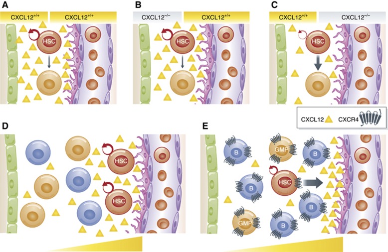
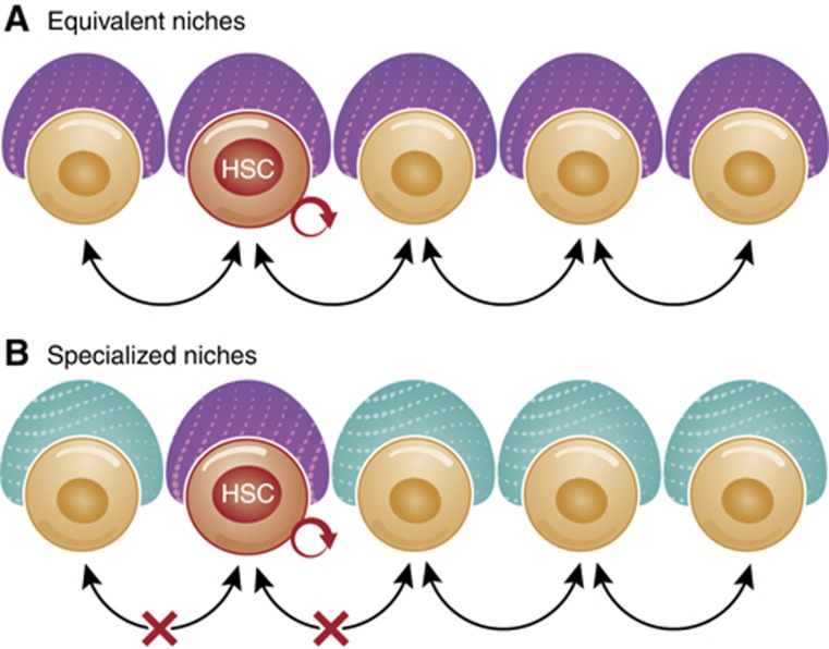
Haematopoietic stem cell (HSC) niches provide an environment essential for life-long HSC function. Intense investigation of HSC niches both feed off and drive technology development to increase our capability to assay functionally defined cells with high resolution. A major driving force behind the desire to understand the basic biology of HSC niches is the clear implications for clinical therapies. Here, with particular emphasis on cell type-specific deletion of SCL and CXCL12, we focus on unresolved issues on HSC niches, framed around some very recent advances and novel discoveries on the extrinsic regulation of HSC maintenance. We also provide ideas for possible paths forward, some of which are clearly within reach while others will require both novel tools and vision.


Conflict of interest statement

The authors declare that they have no conflict of interest.

Figures

Figure 1
Figure 1
Visualization of HSC niches at increasing resolution. Recent findings have contributed to our knowledge of structural, cellular, and molecular features important for HSC localization and maintenance. (A) Although HSCs can, at least temporarily, circulate and take up residence in organs such as spleen and liver, the majority of functional adult HSCs reside in the bone marrow. (B) Within the bone marrow, HSCs can be located in either perivascular or endosteal regions, where they interact directly or indirectly with different types of cells that comprise HSC niches (C). (D) These niche cells secrete factors that maintain haematopoietic homeostasis by favouring HSC quiescence, while allowing self-renewal and differentiation upon demand. This figure illustrates how the definition of HSC location is becoming increasingly more detailed, from organ to region, cellular, and molecular environment, by the use of novel tools and increasingly sophisticated approaches.
Figure 2
Figure 2
The hemosphere niche model. A novel view of the perivascular niche was proposed by Wang et al, where HSCs and haematopoietic progenitors are contained in defined areas termed hemospheres (Wang et al, 2012), delimited by SECs and perivascular stromal cells. This area may be highly enriched for molecular cues that promote HSC maintenance and self-renewal, such as SCF and CXCL12. Broad acceptance of this model will likely require validation by alternative genetic model and imaging techniques.
Figure 3
Figure 3
Putative lineage relationships of some of the BM cells that have been implicated in HSC function. Lineage hierarchy of the distinct candidate niche cells. Perivascular stromal cells described using different genetic models significantly overlap in terms of function and molecular profile. Some of these cells have mesenchymal stem/progenitor properties, giving rise to osteo-, adipo-, and chondrocyte lineages. Other BM cells implicated in HSC function include haematopoietic-derived osteoclasts and macrophages, sinusoidal and non-SECs, adipocytes and non-myelinating Schwann cells.
Figure 4
Figure 4
CXCL12 may affect HSC function by both direct and indirect mechanisms. In (A), the primary function of CXCL12 (yellow triangles) is to support HSC function by acting directly on HSCs. In (B), CXCL12 also affects HSC function by indirect mechanisms, by acting on CXCR4-expressing niche cells (effect number 1). This action of CXCL12 may function to reinforce an HSC-supportive environment by either cellular mechanisms, such as niche cell function or organization, and/or by molecular mechanisms, by triggering release of additional HSC-supporting factors, such as SCF or angiopoietin (blue circles), from niche cells (effect number 2).
Figure 5
Figure 5
Sequential effect of gene deletion in stem and progenitor cells. This cartoon depicts a hypothetical scenario where a secreted factor is normally expressed by both a stem/progenitor cell and at least one of its more differentiated progeny, and the effects of stem/progenitor-specific genetic deletion of the gene for this factor. (A) Prior to gene deletion, the factor is secreted by both stem/progenitor and progeny cells. (B) Soon after gene deletion, depicted here as activation of Cre recombinase specifically in stem/progenitor cells, progeny, but not stem/progenitors, will produce the factor. (C) Over time, as factor-deficient stem/progenitor cells differentiate and replenish the pool of progeny, the factor will also be missing from an increasing proportion of progeny. For example, Cre-mediated deletion of CXCL12 specifically from mesenchymal stem cells capable of differentiating into osteoblasts will first result in reduced CXCL12 secretion from MSC themselves (B), but later also in reduced CXCL12 production by their osteoblast descendants (C). This highlights the importance of the time frame of gene deletion relative to analysis of effects on HSCs. Inducible Cre models are capable of temporally more cell-specific deletion than constitutive deletion models. However, the effects of factor deletion depend on the expression pattern and levels of the factor in question, the relative cell number and expression levels in stem/progenitors versus more mature cells, and the in vivo turnover and replacement rate of progeny.
Figure 6
Figure 6
Gradients and sources of soluble factors for HSC maintenance. (AC) Effect of factor deletion on different cell types. In (A), HSC (red cell) maintenance is supported by CXCL12 expression by both osteoblasts (green cells) and (peri)vascular cells (PVCs; pink and purple cells). (B) CXCL12 deletion in OBCs fails to affect HSC numbers, as threshold levels of CXCL12 are maintained by the high CXCL12 expression by PVCs. (C) Deletion of CXCL12 in PVCs leads to overall lower levels of CXCL12. The relatively low CXCL12 expression by OBCs is insufficient to maintain threshold levels of CXCL12. The net result is a decrease in HSC numbers, either by reduced self-renewing and increased differentiating divisions (indicated by changes in arrow thickness) or by mobilization to the periphery. Thus, differential effects of cell type-specific factor deletion may reflect differences in expression levels or abundance of the expressing cell type, and does not, in itself, indicate the location of HSCs relative to a specific niche cell type. (D) Factor gradient established locally around high-expressing stromal cells. In this model, the high Cxcl12 expression in PVCs results in a high local concentration of Cxcl12 (yellow triangles), and therefore HSCs, close to PVCs. (E) Competition for CXCL12-abundant niches between HSCs and other CXCR4-expressing cells. In this model, HSCs are actively migrating towards a CXCL12 gradient, established by CXCL12-expressing BM niche cells. It is unclear how HSCs outcompete more abundant cells that express higher levels of CXCR4 and migrate with greater efficiency towards CXCL12, such as B cells (Smith-Berdan et al, 2011), for access to limited numbers of niches.
Figure 7
Figure 7
Models of niche competition. (A) In the ‘equivalent niche’ model, there are more niches capable of supporting HSCs than there are HSCs. However, HSC access to these niches, and therefore HSC expansion, is hampered by competition with other, more numerous cells. (B) In the ‘specialized niche’ model, the number of niches capable of supporting HSC self-renewal is limited. Progenitors have their own specialized niches that are not capable of supporting HSC self-renewal.

Similar articles

Cited by

References

    1. Abkowitz JL, Robinson AE, Kale S, Long MW, Chen J (2003) Mobilization of hematopoietic stem cells during homeostasis and after cytokine exposure. Blood 102: 1249–1253 - PubMed
    1. Anderson DM, Williams DE, Tushinski R, Gimpel S, Eisenman J, Cannizzaro LA, Aronson M, Croce CM, Huebner K, Cosman D (1991) Alternate splicing of mRNAs encoding human mast cell growth factor and localization of the gene to chromosome 12q22-q24. Cell Growth Differ 2: 373–378 - PubMed
    1. Bhattacharya D, Czechowicz A, Ooi AL, Rossi DJ, Bryder D, Weissman IL (2009) Niche recycling through division-independent egress of hematopoietic stem cells. J Exp Med 206: 2837–2850 - PMC - PubMed
    1. Broudy VC (1997) Stem cell factor and hematopoiesis. Blood 90: 1345–1364 - PubMed
    1. Broxmeyer HE, Orschell CM, Clapp DW, Hangoc G, Cooper S, Plett PA, Liles WC, Li X, Graham-Evans B, Campbell TB, Calandra G, Bridger G, Dale DC, Srour EF (2005) Rapid mobilization of murine and human hematopoietic stem and progenitor cells with AMD3100, a CXCR4 antagonist. J Exp Med 201: 1307–1318 - PMC - PubMed

Publication types