. 2013 Nov;24(21):3406-19.
doi: 10.1091/mbc.E13-04-0185. Epub 2013 Sep 4.

Sec16 influences transitional ER sites by regulating rather than organizing COPII

Affiliations

Sec16 influences transitional ER sites by regulating rather than organizing COPII

Nike Bharucha et al. Mol Biol Cell. 2013 Nov.

Abstract

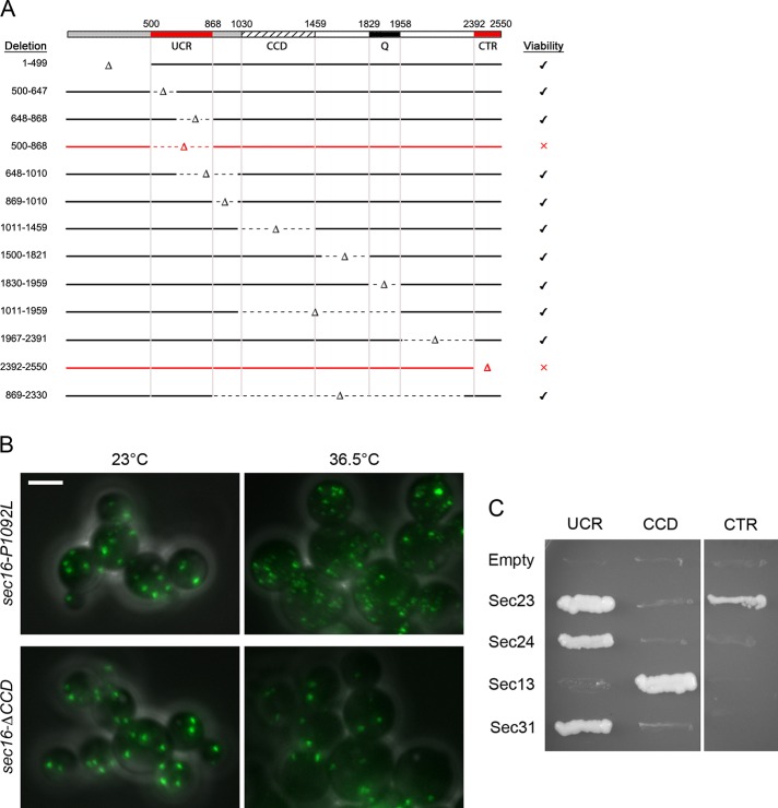
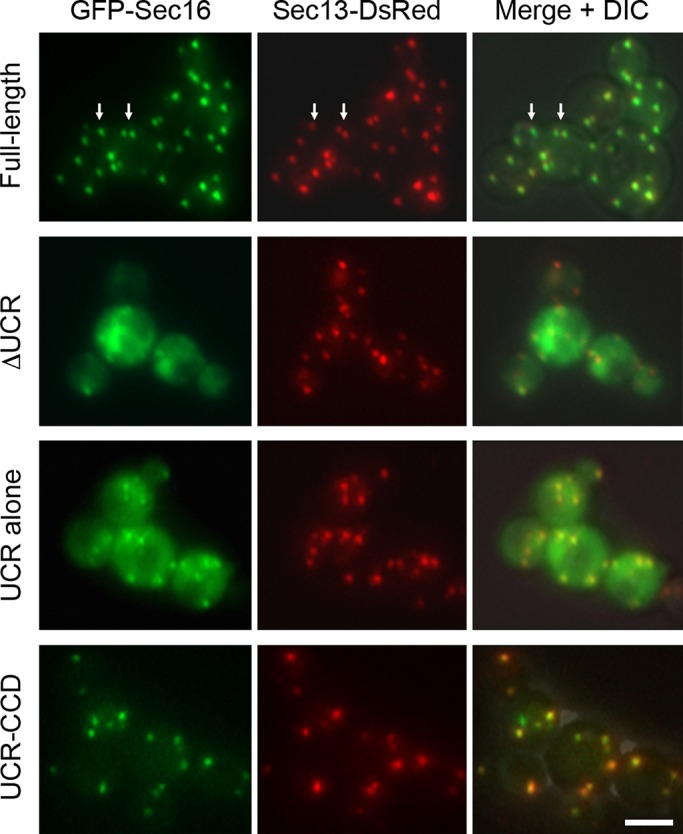
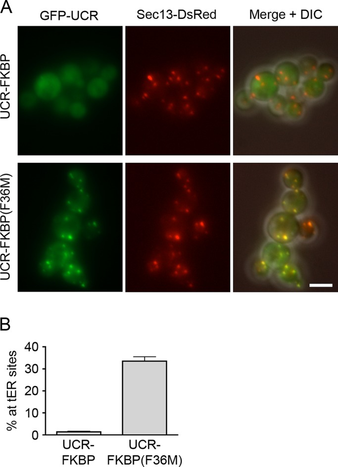
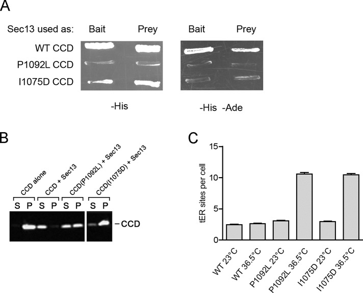
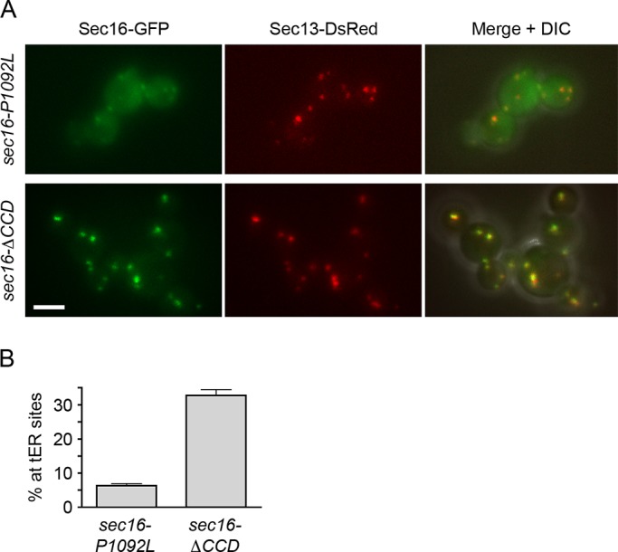
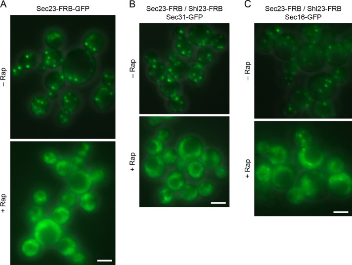
During the budding of coat protein complex II (COPII) vesicles from transitional endoplasmic reticulum (tER) sites, Sec16 has been proposed to play two distinct roles: negatively regulating COPII turnover and organizing COPII assembly at tER sites. We tested these ideas using the yeast Pichia pastoris. Redistribution of Sec16 to the cytosol accelerates tER dynamics, supporting a negative regulatory role for Sec16. To evaluate a possible COPII organization role, we dissected the functional regions of Sec16. The central conserved domain, which had been implicated in coordinating COPII assembly, is actually dispensable for normal tER structure. An upstream conserved region (UCR) localizes Sec16 to tER sites. The UCR binds COPII components, and removal of COPII from tER sites also removes Sec16, indicating that COPII recruits Sec16 rather than the other way around. We propose that Sec16 does not in fact organize COPII. Instead, regulation of COPII turnover can account for the influence of Sec16 on tER sites.


Figures

FIGURE 1:
FIGURE 1:
Domain analysis of P. pastoris Sec16. (A) Summary of the effects of Sec16 partial deletions on cell viability. Top, the UCR (residues 500–868), CCD (residues 1030–1459), a nonconserved glutamine-rich region (Q; residues 1829–1958), and the conserved CTR (residues 2392–2550). The endogenous SEC16 gene was replaced with alleles containing the indicated deletions. Right, the ability of each mutant allele to support growth. Only the deletions marked in red caused a loss of viability. (B) Differential effects of a CCD point mutation and a CCD deletion. The indicated mutations were introduced at the SEC16 locus by gene replacement in a strain expressing Sec13-GFP. Cultures were grown at 23°C, and then half of each culture was shifted to 36.5°C for 1 h. Cells were imaged by differential interference contrast and fluorescence microscopy. Scale bar, 5 μm. (C) Yeast two-hybrid analysis of Sec16-COPII interactions. The “prey” vector encoded the indicated fragments of P. pastoris Sec16, and the “bait” vector encoded the indicated full-length P. pastoris COPII coat proteins. Growth on plates lacking histidine reflects an interaction. With this system, the UCR can self-activate when used as bait, so constructs containing the UCR were tested only as prey. The other constructs gave the same results when the Sec16 fragments were used as bait and the COPII coat proteins were used as prey (unpublished data).
FIGURE 2:
FIGURE 2:
Localization of GFP-tagged fragments of P. pastoris Sec16. These fusions were expressed from the AOX1 promoter as second copies in a P. pastoris strain expressing both wild-type Sec16 and Sec13-DsRed. GFP was fused to the N‑terminus of full-length Sec16, Sec16 lacking residues 500–868 (ΔUCR), residues 500–868 only (UCR alone), or residues 500–1459 only (UCR‑CCD). Expression of the GFP fusions was induced by shifting to methanol medium for 3 h, and then the cells were imaged by fluorescence microscopy. Right, merged fluorescence and differential interference contrast images. Arrows show examples of full-length GFP-Sec16 puncta that do not entirely colocalize with Sec13-DsRed puncta. Scale bar, 5 μm.
FIGURE 3:
FIGURE 3:
tER localization of a dimerized UCR. (A) Replacement of the CCD with monomeric or dimeric FKBP. A GFP-tagged UCR-CCD construct was modified to replace the CCD with either wild-type monomeric FKBP or the dimeric FKBP(F36M) mutant. Localization to Sec13-DsRed-labeled tER sites was then evaluated as in Figure 2. Scale bar, 5 μm. (B) Quantitation of the data from A. The percentage of the total GFP signal at punctate tER sites was measured. Plotted are mean and SEM.
FIGURE 4:
FIGURE 4:
Destabilization of P. pastoris Sec16 by mutations that weaken Sec13 binding. (A) Two-hybrid analysis of CCD-Sec13 interactions. The wild-type (WT) CCD of P. pastoris Sec16, or a mutant CCD carrying the P1092L or I1075D mutation was tested against P. pastoris Sec13 in a yeast two-hybrid screen (James, 2001). Where indicated, Sec13 was used as the “bait” and the CCD as the “prey” or vice versa. Interactions were detected by growth on a plate lacking histidine (–His) or, in a more stringent test, a plate lacking histidine and adenine (–His –Ade). (B) Solubility of wild-type and mutant CCD variants. In E. coli, a C-terminally FLAG-tagged wild-type or mutant CCD was either expressed alone or coexpressed with C‑terminally S peptide–tagged Sec13. The cells were lysed in detergent and centrifuged, and equivalent amounts of the pellet (P) and supernatant (S) fractions were analyzed by SDS–PAGE, followed by immunoblotting with anti-FLAG antibody. (C) Effects of CCD mutations on tER site number. For strains expressing Sec13‑­GFP plus either WT or a mutant Sec16, cultures were grown at 23°C, and then a portion of each culture was shifted for 2 h to 36.5°C. The tER sites were counted in ∼100 cells from each sample. Plotted are mean and SEM.
FIGURE 5:
FIGURE 5:
Redistribution of Sec16 to the cytosol by the P1092L mutation but not by deletion of the entire CCD. (A) Localization of Sec16 mutants. GFP-tagged Sec16‑P1092L or Sec16-ΔCCD was expressed from the SEC16 promoter in cells that also expressed wild-type Sec16 as well as Sec13-DsRed. Cells were grown at 30°C, then imaged by differential interference contrast (DIC) and fluorescence microscopy. Right, merged fluorescence and DIC images. Scale bar, 5 μm. (B) Quantitation of the data from A. The percentage of the total GFP signal at punctate tER sites was measured. Plotted are mean and SEM.
FIGURE 6:
FIGURE 6:
Altered dynamics of tER sites in sec16‑P1092L mutant cells. (A) Shrinkage of tER sites after fusion events in sec16‑P1092L cells. A sec16‑P1092L strain expressing Sec13‑GFP was grown at room temperature, then warmed to 36.5°C for ∼45 min before imaging by 4D microscopy at 36.5°C. Shown are merged fluorescence and differential interference contrast (DIC) images of representative cells. The arrowhead marks a pair of tER sites that underwent fusion followed by shrinkage. Frames are taken from Supplemental Movie S2, and the time from the beginning of the movie is shown in minutes:seconds format. Scale bar, 5 μm. (B) Quantitation of tER site shrinkage in wild-type and sec16‑P1092L cells. From 4D movies of the type shown in A, ∼20 newly fused tER sites were chosen at random for WT or sec16‑P1092L cells. The half-times for shrinkage were determined from plots of the type shown in Supplemental Figure S4A. Each dot represents an individual fused tER site, and the horizontal lines represent the average half-times. (C) Quantitation of de novo tER site formation in wild-type and sec16‑P1092L cells. The cells expressed Sec13-GFP, and, where indicated, they also expressed Sar1(T34N) from the inducible AOX1 promoter. Cultures were shifted to inducing methanol medium for 3 h at room temperature, grown at 36.5°C for an additional 50 min, and then imaged by 4D microscopy at 36.5°C for either 40 min for wild-type cells or 10 min for sec16‑P1092L cells. For each culture, the number of de novo tER site formation events was recorded for ∼20 cells. Plotted are the hourly mean and SEM values. (D) Prevention of tER dispersal by expression of Sar1(T34N). Wild-type or sec16‑P1092L cells expressing Sec13-GFP were transformed with a control vector or a vector encoding Sar1(T34N) expressed from an inducible promoter. Cultures were shifted to inducing methanol medium for 2.5 h at room temperature and then grown at 36.5°C for an additional 1 h before imaging. Shown are merged fluorescence and DIC images of representative cells. Scale bar, 5 μm. (E) Quantitation of the results from D. For each of the four conditions, the tER sites were counted in ∼50 cells. Plotted are mean and SEM. (F) Cytosolic localization of Sec16-P1092L in the presence of Sar1(T34N). In a sec16-P1092L strain, the mutant sec16 gene was tagged with GFP by gene replacement. The resulting strain was transformed with a control vector or a vector encoding Sar1(T34N) expressed from an inducible promoter. Induction and imaging were performed as in D. The expression of Sar1(T34N) was confirmed by measuring growth inhibition (Connerly et al., 2005; unpublished data). Scale bar, 5 μm.
FIGURE 7:
FIGURE 7:
Removal of Sec16 from tER sites by anchoring on cytosolic ribosomes. (A) Drug-induced displacement of Sec16 from tER sites to ribosomes. Endogenous Sec16 was modified with an FRB-GFP dual tag to visualize Sec16 at tER sites in the absence of rapamycin (–Rap). Rapamycin was then added to 1 μg/ml, and cells were imaged after 10 min of incubation with the drug (+Rap). Fluorescence and differential interference contrast images (DIC) were combined, with the same exposure times for both panels. (B) tER dispersal induced by anchoring away Sec16. Gene replacement was used to tag Sec16 with two tandem copies of FRB and to tag Sec31 with GFP. tER sites were then visualized in the absence of rapamycin or after incubation for 10 min in the presence of 1 μg/ml rapamycin. Fluorescence and DIC images were combined, with the same exposure times for both panels. The tER sites per cell were counted in ∼50 cells from each sample. Plotted are mean and SEM. Scale bars, 5 μm.
FIGURE 8:
FIGURE 8:
Displacement of Sec16 by removal of COPII coat proteins from tER sites. (A) Drug-induced displacement of Sec23 from tER sites to ribosomes. The procedure was the same as in Figure 7A, except that Sec23 was tagged with FRB-GFP. (B) Drug-induced displacement of Sec31 from tER sites. Gene replacement was used to tag Sec23 and Shl23 with FRB and to tag Sec31 with GFP. The Sec31-GFP pattern was then visualized in the absence of rapamycin (–Rap) or after incubation for 10 min in the presence of 1 μg/ml rapamycin (+Rap). Fluorescence and differential interference contrast images were combined, with the same exposure times for both panels. (C) Drug-induced displacement of Sec16 from tER sites. The procedure was the same as in B, except that Sec16 was tagged with GFP. Scale bars, 5 μm.
FIGURE 9:
FIGURE 9:
Models for Sec16 function. (A) Diagram summarizing the conserved regions of P. pastoris Sec16 and their inferred functions. See the text for details. (B) Speculative diagram of a cross section through a nascent COPII vesicle. Sar1 has largely dissociated from the interior of the coat lattice due to Sec23- and Sec31-catalyzed GTP hydrolysis. However, a ring of Sar1-GTP is maintained at the edge of the lattice because Sec16 binds to the newly assembled COPII coat subunits and slows Sar1 GTPase activity. Sec16 also recruits Sec12 to the vicinity of the budding vesicle, leading to enhanced local production of Sar1-GTP. See the text for further details.

Similar articles

Cited by

References

    1. Adams PD, et al. PHENIX: a comprehensive Python-based system for macromolecular structure solution. Acta Crystallogr D Biol Crystallogr. 2010;66:213–221. - PMC - PubMed
    1. Antonny B, Madden D, Hamamoto S, Orci L, Schekman R. Dynamics of the COPII coat with GTO and stable analogues. Nat Cell Biol. 2001;3:531–537. - PubMed
    1. Bannykh SI, Balch WE. Membrane dynamics at the endoplasmic reticulum-Golgi interface. J Cell Biol. 1997;138:1–4. - PMC - PubMed
    1. Barlowe C. COPII: a membrane coat formed by Sec proteins that drive vesicle budding from the endoplasmic reticulum. Cell. 1994;77:895–907. - PubMed
    1. Barlowe C, Schekman R. SEC12 encodes a guanine-nucleotide-exchange factor essential for transport vesicle budding from the ER. Nature. 1993;365:347–349. - PubMed

Publication types

Associated data

LinkOut - more resources