

Myeloid cells in atherosclerosis: a delicate balance of anti-inflammatory and proinflammatory mechanisms
- PMID: 24005215
- PMCID: PMC4939820
- DOI: 10.1097/MOL.0b013e328363d298
Myeloid cells in atherosclerosis: a delicate balance of anti-inflammatory and proinflammatory mechanisms
Abstract
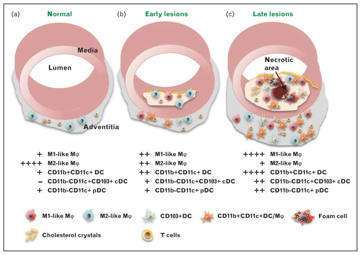
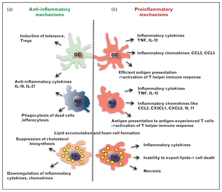
Purpose of review: Atherosclerosis is chronic disease, whose progression is orchestrated by the balance between proinflammatory and anti-inflammatory mechanisms. Various myeloid cells, including monocytes, macrophages, dendritic cells and neutrophils can be found in normal and atherosclerotic aortas, in which they regulate inflammation and progression of atherosclerosis. The lineage relationship between blood monocyte subsets and the various phenotypes and functions of myeloid cells in diseased aortas is under active investigation.
Recent findings: Various subsets of myeloid cells play diverse roles in atherosclerosis. This review discusses new findings in phenotypic and functional characterization of different subsets of macrophages, in part determined by the transcription factors IRF5 and Trib1, and dendritic cells, characterized by the transcription factor Zbtb46, in atherosclerosis.
Summary: Improved understanding proinflammatory and anti-inflammatory mechanisms of macrophages and dendritic cell functions is needed for better preventive and therapeutic measures in atherosclerosis.
Conflict of interest statement
There are no conflicts of interest.
Figures
Similar articles
-
Myeloid dendritic cells: Development, functions, and role in atherosclerotic inflammation.Immunobiology. 2015 Jun;220(6):833-44. doi: 10.1016/j.imbio.2014.12.010. Epub 2015 Jan 5. Immunobiology. 2015. PMID: 25595536 Review.
-
Macrophages and dendritic cells: the usual suspects in atherogenesis.Curr Drug Targets. 2015;16(4):373-82. doi: 10.2174/1389450116666150330115809. Curr Drug Targets. 2015. PMID: 25808566 Review.
-
Single-Cell RNA-Seq Reveals the Transcriptional Landscape and Heterogeneity of Aortic Macrophages in Murine Atherosclerosis.Circ Res. 2018 Jun 8;122(12):1661-1674. doi: 10.1161/CIRCRESAHA.117.312509. Epub 2018 Mar 15. Circ Res. 2018. PMID: 29545365
-
FcγR-TLR Cross-Talk Enhances TNF Production by Human Monocyte-Derived DCs via IRF5-Dependent Gene Transcription and Glycolytic Reprogramming.Front Immunol. 2019 Apr 8;10:739. doi: 10.3389/fimmu.2019.00739. eCollection 2019. Front Immunol. 2019. PMID: 31024565 Free PMC article.
-
The dynamic lives of macrophage and dendritic cell subsets in atherosclerosis.Ann N Y Acad Sci. 2014 Jun;1319(1):19-37. doi: 10.1111/nyas.12392. Epub 2014 Mar 14. Ann N Y Acad Sci. 2014. PMID: 24628328 Free PMC article. Review.
Cited by
-
Atherosclerosis is a major human killer and non-resolving inflammation is a prime suspect.Cardiovasc Res. 2021 Nov 22;117(13):2563-2574. doi: 10.1093/cvr/cvab309. Cardiovasc Res. 2021. PMID: 34609505 Free PMC article. Review.
-
Beyond vascular inflammation--recent advances in understanding atherosclerosis.Cell Mol Life Sci. 2015 Oct;72(20):3853-69. doi: 10.1007/s00018-015-1971-6. Epub 2015 Jun 23. Cell Mol Life Sci. 2015. PMID: 26100516 Free PMC article. Review.
-
Macrophage maturation from blood monocytes is altered in people with HIV, and is linked to serum lipid profiles and activation indices: A model for studying atherogenic mechanisms.PLoS Pathog. 2020 Oct 1;16(10):e1008869. doi: 10.1371/journal.ppat.1008869. eCollection 2020 Oct. PLoS Pathog. 2020. PMID: 33002093 Free PMC article.
-
Vaccination to modulate atherosclerosis.Autoimmunity. 2015 May;48(3):152-60. doi: 10.3109/08916934.2014.1003641. Epub 2015 Feb 16. Autoimmunity. 2015. PMID: 25683179 Free PMC article. Review.
-
Bioinformatics analysis of vascular RNA-seq data revealed hub genes and pathways in a novel Tibetan minipig atherosclerosis model induced by a high fat/cholesterol diet.Lipids Health Dis. 2020 Mar 25;19(1):54. doi: 10.1186/s12944-020-01222-w. Lipids Health Dis. 2020. PMID: 32213192 Free PMC article.
References
-
- Banchereau J, Steinman RM. Dendritic cells and the control of immunity. Nature. 1998;392:245–252. - PubMed
Publication types
MeSH terms
Substances
Grants and funding
LinkOut - more resources
Full Text Sources
Other Literature Sources
Medical
Research Materials
