

Mesenchymal stem cells from human umbilical cord express preferentially secreted factors related to neuroprotection, neurogenesis, and angiogenesis
- PMID: 23991127
- PMCID: PMC3749979
- DOI: 10.1371/journal.pone.0072604
Mesenchymal stem cells from human umbilical cord express preferentially secreted factors related to neuroprotection, neurogenesis, and angiogenesis
Abstract
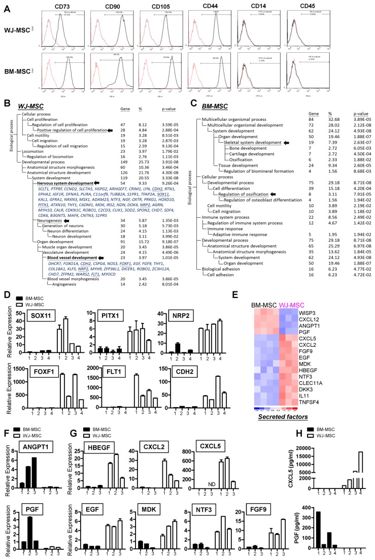
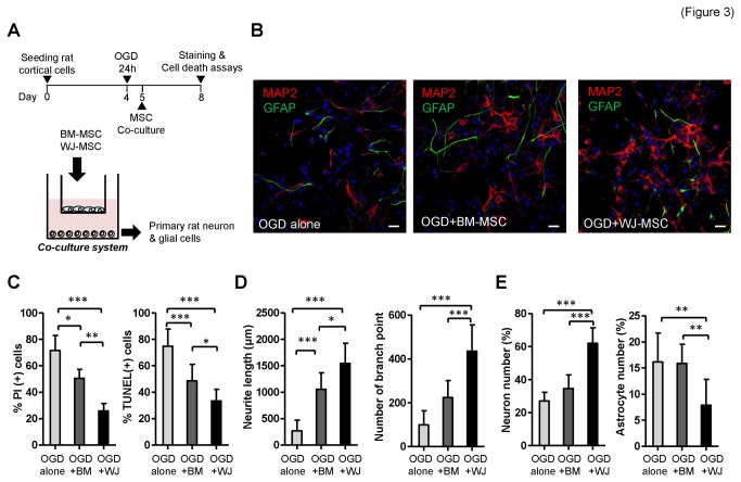
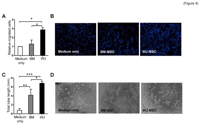
Mesenchymal stem cells (MSCs) are promising tools for the treatment of diseases such as infarcted myocardia and strokes because of their ability to promote endogenous angiogenesis and neurogenesis via a variety of secreted factors. MSCs found in the Wharton's jelly of the human umbilical cord are easily obtained and are capable of transplantation without rejection. We isolated MSCs from Wharton's jelly and bone marrow (WJ-MSCs and BM-MSCs, respectively) and compared their secretomes. It was found that WJ-MSCs expressed more genes, especially secreted factors, involved in angiogenesis and neurogenesis. Functional validation showed that WJ-MSCs induced better neural differentiation and neural cell migration via a paracrine mechanism. Moreover, WJ-MSCs afforded better neuroprotection efficacy because they preferentially enhanced neuronal growth and reduced cell apoptotic death of primary cortical cells in an oxygen-glucose deprivation (OGD) culture model that mimics the acute ischemic stroke situation in humans. In terms of angiogenesis, WJ-MSCs induced better microvasculature formation and cell migration on co-cultured endothelial cells. Our results suggest that WJ-MSC, because of a unique secretome, is a better MSC source to promote in vivo neurorestoration and endothelium repair. This study provides a basis for the development of cell-based therapy and carrying out of follow-up mechanistic studies related to MSC biology.
Conflict of interest statement
Figures

Similar articles
-
Differential expression of cell cycle and WNT pathway-related genes accounts for differences in the growth and differentiation potential of Wharton's jelly and bone marrow-derived mesenchymal stem cells.Stem Cell Res Ther. 2017 Apr 26;8(1):102. doi: 10.1186/s13287-017-0555-9. Stem Cell Res Ther. 2017. PMID: 28446235 Free PMC article.
-
Functional module analysis reveals differential osteogenic and stemness potentials in human mesenchymal stem cells from bone marrow and Wharton's jelly of umbilical cord.Stem Cells Dev. 2010 Dec;19(12):1895-910. doi: 10.1089/scd.2009.0485. Epub 2010 Oct 12. Stem Cells Dev. 2010. PMID: 20367285
-
Netrin-1 acts as a non-canonical angiogenic factor produced by human Wharton's jelly mesenchymal stem cells (WJ-MSC).Stem Cell Res Ther. 2017 Feb 28;8(1):43. doi: 10.1186/s13287-017-0494-5. Stem Cell Res Ther. 2017. PMID: 28241866 Free PMC article.
-
Characteristics and clinical applications of Wharton's jelly-derived mesenchymal stromal cells.Curr Res Transl Med. 2020 Jan;68(1):5-16. doi: 10.1016/j.retram.2019.09.001. Epub 2019 Sep 19. Curr Res Transl Med. 2020. PMID: 31543433 Review.
-
Wharton's Jelly Mesenchymal Stromal Cells as a Feeder Layer for the Ex Vivo Expansion of Hematopoietic Stem and Progenitor Cells: a Review.Stem Cell Rev Rep. 2017 Feb;13(1):35-49. doi: 10.1007/s12015-016-9702-4. Stem Cell Rev Rep. 2017. PMID: 27853939 Review.
Cited by
-
3D culture of alginate-hyaluronic acid hydrogel supports the stemness of human mesenchymal stem cells.Sci Rep. 2024 Feb 23;14(1):4436. doi: 10.1038/s41598-024-54912-1. Sci Rep. 2024. PMID: 38396088 Free PMC article.
-
Longitudinal neuroimaging evaluation of the corticospinal tract in patients with stroke treated with autologous bone marrow cells.Stem Cells Transl Med. 2021 Jul;10(7):943-955. doi: 10.1002/sctm.20-0369. Epub 2021 Mar 10. Stem Cells Transl Med. 2021. PMID: 33689219 Free PMC article.
-
Unraveling the Specific Ischemic Core and Penumbra Transcriptome in the Permanent Middle Cerebral Artery Occlusion Mouse Model Brain Treated with the Neuropeptide PACAP38.Microarrays (Basel). 2015 Jan 21;4(1):2-24. doi: 10.3390/microarrays4010002. Microarrays (Basel). 2015. PMID: 27600210 Free PMC article.
-
Successful Treatment of Noise-Induced Hearing Loss by Mesenchymal Stromal Cells: An RNAseq Analysis of Protective/Repair Pathways.Front Cell Neurosci. 2021 Nov 23;15:656930. doi: 10.3389/fncel.2021.656930. eCollection 2021. Front Cell Neurosci. 2021. PMID: 34887728 Free PMC article.
-
Microvesicles as Emerging Biomarkers and Therapeutic Targets in Cardiometabolic Diseases.Genomics Proteomics Bioinformatics. 2018 Feb;16(1):50-62. doi: 10.1016/j.gpb.2017.03.006. Epub 2018 Feb 17. Genomics Proteomics Bioinformatics. 2018. PMID: 29462670 Free PMC article. Review.
References
-
- Horwitz EM, Prockop DJ, Fitzpatrick LA, Koo WW, Gordon PL et al. (1999) Transplantability and therapeutic effects of bone marrow-derived mesenchymal cells in children with osteogenesis imperfecta. Nat Med 5: 309-313. doi:10.1038/6529. PubMed: 10086387. - DOI - PubMed
-
- Kawada H, Fujita J, Kinjo K, Matsuzaki Y, Tsuma M et al. (2004) Nonhematopoietic mesenchymal stem cells can be mobilized and differentiate into cardiomyocytes after myocardial infarction. Blood 104: 3581-3587. doi:10.1182/blood-2004-04-1488. PubMed: 15297308. - DOI - PubMed
-
- Ripa RS, Haack-Sørensen M, Wang Y, Jørgensen E, Mortensen S et al. (2007) Bone marrow derived mesenchymal cell mobilization by granulocyte-colony stimulating factor after acute myocardial infarction: results from the Stem Cells in Myocardial Infarction (STEMMI) trial. Circulation 116: I24-I30. PubMed: 17846310. - PubMed
-
- Bang OY, Lee JS, Lee PH, Lee G (2005) Autologous mesenchymal stem cell transplantation in stroke patients. Ann Neurol 57: 874-882. doi:10.1002/ana.20501. PubMed: 15929052. - PubMed
-
- Chen J, Li Y, Wang L, Zhang Z, Lu D et al. (2001) Therapeutic benefit of intravenous administration of bone marrow stromal cells after cerebral ischemia in rats. Stroke 32: 1005-1011. doi:10.1161/01.STR.32.4.1005. PubMed: 11283404. - DOI - PubMed
Publication types
MeSH terms
Substances
Grants and funding
LinkOut - more resources
Full Text Sources
Other Literature Sources
Molecular Biology Databases
