. 2013 Aug 26:4:222.
doi: 10.3389/fphys.2013.00222. eCollection 2013.

The effect of heart rate on the heart rate variability response to autonomic interventions

Affiliations

The effect of heart rate on the heart rate variability response to autonomic interventions

George E Billman. Front Physiol. .

Abstract

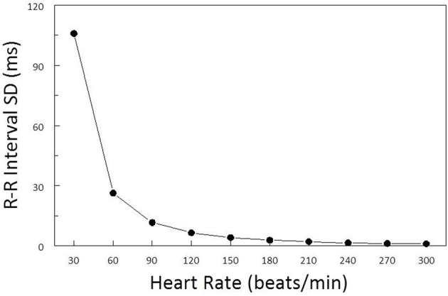
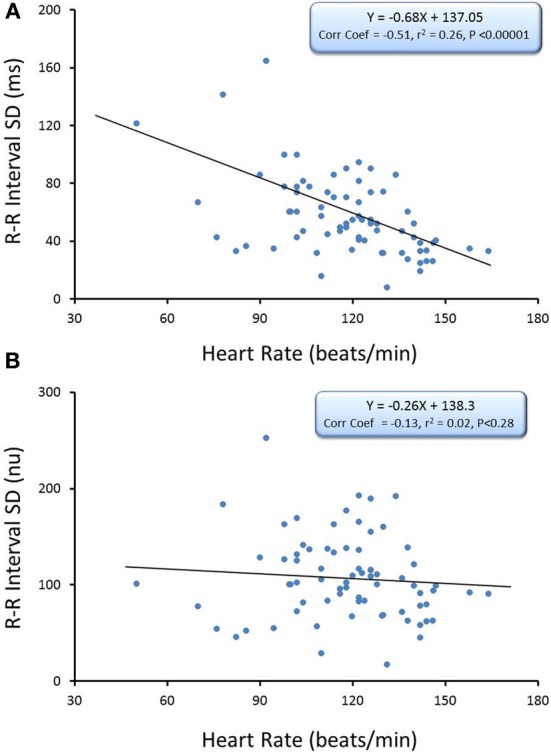
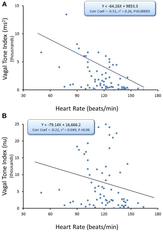
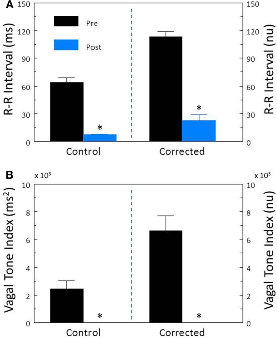
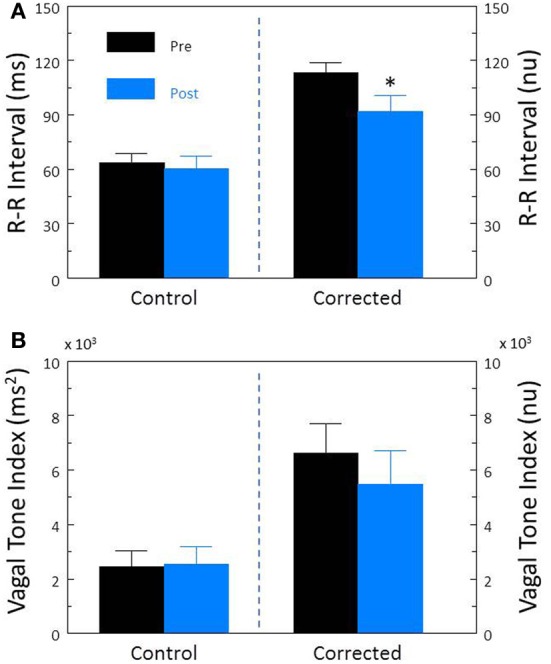
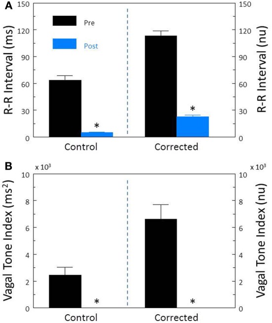
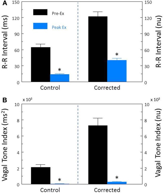
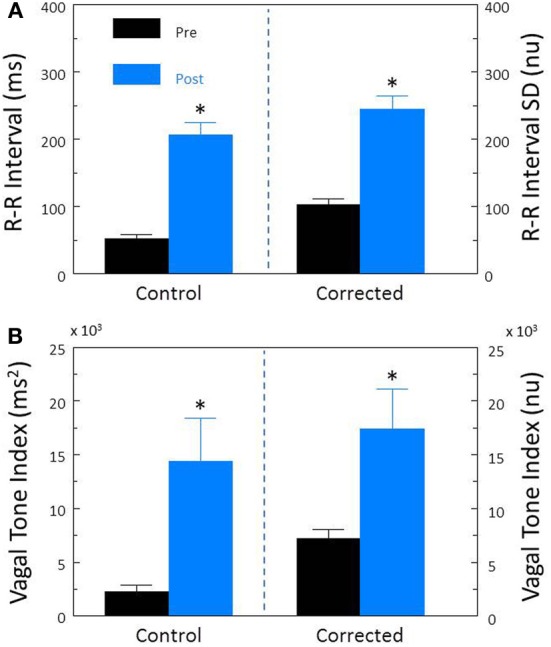
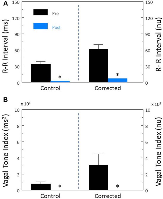
Heart rate variability (HRV), the beat-to-beat variation in either heart rate (HR) or heart period (R-R interval), has become a popular clinical and investigational tool to quantify cardiac autonomic regulation. However, it is not widely appreciated that, due to the inverse curvilinear relationship between HR and R-R interval, HR per se can profoundly influence HRV. It is, therefore, critical to correct HRV for the prevailing HR particularly, as HR changes in response to autonomic neural activation or inhibition. The present study evaluated the effects of HR on the HRV response to autonomic interventions that either increased (submaximal exercise, n = 25 or baroreceptor reflex activation, n = 20) or reduced (pharmacological blockade: β-adrenergic receptor, muscarinic receptor antagonists alone and in combination, n = 25, or bilateral cervical vagotomy, n = 9) autonomic neural activity in a canine model. Both total (RR interval standard deviation, RRSD) and the high frequency (HF) variability (HF, 0.24-1.04 Hz) were determined before and in response to an autonomic intervention. All interventions that reduced or abolished cardiac parasympathetic regulation provoked large reductions in HRV even after HR correction [division by mean RRsec or (mean RRsec)(2) for RRSD and HF, respectively] while interventions that reduced HR yielded mixed results. β-adrenergic receptor blockade reduced HRV (RRSD but not HF) while both RRSD and HF increased in response to increases in arterial blood (baroreceptor reflex activation) even after HR correction. These data suggest that the physiological basis for HRV is revealed after correction for prevailing HR and, further, that cardiac parasympathetic activity is responsible for a major portion of the HRV in the dog.

Keywords: autonomic nervous system; baroreceptor reflex; cholinergic receptor antagonists; exercise; heart rate; heart rate variability; β-adrenergic receptors.


Figures

Figure 1
Figure 1
Effect of baseline heart rate on heart rate variability. The standard deviation of R-R interval (RRSD) was calculated for a set of 5 simulated heart beats (X − 2, X − 1, X, X + 1, X + 2) over a range of mean heart rates (HR, from 30 to 300 beats/min) (solid black line). The standard deviation for HR was ±1.6 beats/min at each HR level. Note that RRSD was inversely related to HR, identical changes in HR were accompanied by much larger R-R interval variability at low as compared to high prevailing HRs.
Figure 2
Figure 2
The relationship between baseline heart rate and heart rate variability. The total heart rate (HR) variability (standard deviation of R-R interval, RRSD) was calculated for over the last 30 s before an autonomic intervention was administered and plotted against the average HR for that interval. One data point is displayed for each animal (n = 74). The data without and with HR correction (RRSD/mean RR) are displayed in (A,B), respectively. Note that HR only accounted for less than 30% (r2 = 0.26) of the variability before correction for HR. nu, normalized units following HR correction.
Figure 3
Figure 3
The relationship between baseline heart rate and heart rate variability. The high frequency (HF) component of the R-R interval variability (cardiac vagal tone index, 0.24–1.04 Hz) was calculated for over the last 30 s before an autonomic intervention was administered and plotted against the average HR for that interval. One data point is displayed for each animal (n = 74). The data without and with HR correction [cardiac vagal tone index/(mean RRsec)2] are displayed in (A,B), respectively. Note that HR only accounted for less than 30% (r2 = 0.26) of the variability before correction for HR. nu, normalized units following HR correction.
Figure 4
Figure 4
The effect of the cholinergic receptor antagonist atropine on heart rate variability. The effect of atropine sulfate (50 μ g/kg i.v.; n = 25) on total heart rate variability (standard deviation of R-R interval, RRSD) without and with correction (RRSD/mean RRsec) are displayed in (A). The effects of this drug on the high frequency variability (cardiac vagal tone index, 0.24–1.04 Hz) without and with correction [cardiac vagal tone/(mean RRsec)2] are shown in (B). Note that despite correction for large increases in heart rate, atropine still provoked large reductions in both RRSD and the cardiac vagal tone index. Thus, cardiac parasympathetic activity is responsible for a large portion of the heart rate variability independent of changes in HR. *P < 0.01 pre (black bars) vs. post (blue bars); pre = last 30 s before atropine administration, post = 30 s interval recorded 5 min after atropine treatment. nu, normalized units following HR correction.
Figure 5
Figure 5
The effect of the β-adrenergic receptor antagonist propranolol on heart rate variability. The effect of propranolol HCl (1.0 mg/kg i.v.; n = 25) on total heart rate variability (standard deviation of R-R interval, RRSD) without and with correction (RRSD/mean RRsec) are displayed in (A). The effects of this drug on the high frequency variability (cardiac vagal tone index, 0.24–1.04 Hz) without and with correction [cardiac vagal tone/(mean RRsec)2] are shown in (B). Note that after correction for propranolol-induced reductions in baseline HR, total (RRSD) heart rate variability significantly decreased following this treatment. *P < 0.01 pre (black bars) vs. post (blue bars); pre = last 30 s before propranolol administration, post = 30 s interval recorded 5 min after this drug treatment. nu, normalized units following HR correction.
Figure 6
Figure 6
The effect of total autonomic neural inhibition on heart rate variability. Cardiac autonomic blockade (n = 25) was achieved using the combination of a cholinergic receptor antagonist (atropine sulfate, 50 μ g/kg i.v.) and a non-selective β-adrenergic receptor (propranolol HCl, 1.0 mg/kg i.v.). The effects of autonomic blockade on total heart rate variability (standard deviation of R-R interval, RRSD) without and with correction (RRSD/mean RRsec) are displayed in (A). The effects of this treatment on the high frequency variability (cardiac vagal tone index, 0.24–1.04 Hz) without and with correction [cardiac vagal tone/(mean RRsec)2] are shown in (B). Note that despite correction for large increases in heart rate, this treatment still provoked large reductions in both RRSD and the cardiac vagal tone index. As baseline HR increased following autonomic blockade, these data indicate the presence of a tonic parasympathetic restraint of intrinsic HR under basal conditions in the dog. *P < 0.01 pre (black bars) vs. post (blue bars); pre = last 30 s before atropine + propranolol administration, post = 30 s interval recorded 5 min after this drug treatment. nu, normalized units following HR correction.
Figure 7
Figure 7
Effect of submaximal exercise on heart rate variability. The effect of exercise (n = 25) on total heart rate variability (standard deviation of R-R interval, RRSD) without and with correction (RRSD/mean RRsec) is displayed in (A). The effect of exercise on the high frequency variability (cardiac vagal tone index, 0.24–1.04 Hz) without and with correction [cardiac vagal tone/(mean RRsec)2] are shown in (B). Note that despite correction for large increases in heart rate, exercise provoked even large reductions in both RRSD and the cardiac vagal tone index than were noted before correction. The data were averaged over the last 30 s before exercise onset (Pre-Ex, black bars) and during the last 30 s of exercise (Peak Ex, blue bars) level. Peak exercise = 6.4 kph and 16% grade. *P < 0.01 Pre-Ex vs. Peak Ex. nu, normalized units following HR correction.
Figure 8
Figure 8
The effect of activation of the baroreceptor reflex on heart rate variability. The α-adrenergic agonist phenylephrine HCl (10 μ g/kg, i.v; n = 20) was used to increase arterial blood 30–50 mm Hg and thereby reflexively induce reductions in heart rate. The effects of this intervention on total heart rate variability (standard deviation of R-R interval, RRSD) without and with correction (RRSD/mean RRsec) are displayed in (A). The effects of this treatment on the high frequency variability (cardiac vagal tone index, 0.24–1.04 Hz) without and with correction [cardiac vagal tone/(mean RRsec)2] are shown in (B). Note that both before and after correction for the phenylephrine-induced decreases in HR, baroreceptor reflex activation provoked significant increases in both RRSD and the cardiac vagal tone index. These data suggest that a reflexively mediated increase in cardiac parasympathetic activity is responsible for a large portion of the heart rate variability response to increases in arterial pressure independent of changes in HR. *P < 0.01 pre (black bars) vs. post (blue bars); pre = last 30 s before phenylephrine administration, post = 30 s interval recorded 5 min after this physiological intervention. nu, normalized units following HR correction.
Figure 9
Figure 9
The effect of bilateral cervical vagotomy on heart rate variability. The effect of surgical disruption of the vagus nerves (n = 9) on total heart rate variability (standard deviation of R-R interval, RRSD) without and with correction (RRSD/mean RRsec) are displayed in (A). The effects of this interval on the high frequency variability (cardiac vagal tone index, 0.24–1.04 Hz) without and with correction [cardiac vagal tone/(mean RRsec)2] are shown in (B). Note that despite correction for large increases in heart rate, the vagotomy still provoked large reductions in both RRSD and the cardiac vagal tone index. These changes are very similar to those noted after treatment with the cholinergic receptor antagonist atropine. Thus, the vagotomy data further indicate that cardiac parasympathetic activity is responsible for a large portion of the heart rate variability independent of changes in HR *P < 0.01 pre (black bars) vs. post (blue bars); pre = last 30 s before vagotomy, post = 30 s interval recorded 5 min after this treatment. nu, normalized units following HR correction.

Similar articles

Cited by

References

    1. Appel M. L., Berger R. D., Saul J. P., Smith J. M., Cohen R. J. (1989). Beat to beat variability in cardiovascular variables: noise or music? J. Am. Coll. Cardiol. 14, 139–1148 10.1016/0735-1097(89)90408-7 - DOI - PubMed
    1. Berntson G. G., Bigger J. T., Eckberg D. L., Grossman P., Kaufmann P. G., Malik M., et al. (1997). Heart rate variability: origins, methods, and interpretive caveats. Pyschophysiology 34, 623–648 10.1111/j.1469-8986.1997.tb02140.x - DOI - PubMed
    1. Billman G. E. (2006). A comprehensive review and analysis of 25 years of data from an in vivo canine model of sudden cardiac death: implications for future anti-arrhythmic drug development. Pharmacol. Therap. 111, 808–835 10.1016/j.pharmthera.2006.01.002 - DOI - PubMed
    1. Billman G. E. (2009). Cardiac autonomic neural “remodeling” and susceptibility to sudden cardiac death: effect of endurance exercise training. Am. J. Physiol. Heart Circ. Physiol. 297, H1171–H1193 10.1152/ajpheart.00534.2009 - DOI - PubMed
    1. Billman G. E. (2011). Heart variability – a historical perspective. Front. Physiol. 2:86 10.3389/fphys.2011.00086 - DOI - PMC - PubMed