. 2013 Oct;24(20):3227-37.
doi: 10.1091/mbc.E13-05-0228. Epub 2013 Aug 28.

Wounded cells drive rapid epidermal repair in the early Drosophila embryo

Affiliations

Wounded cells drive rapid epidermal repair in the early Drosophila embryo

Rodrigo Fernandez-Gonzalez et al. Mol Biol Cell. 2013 Oct.

Abstract

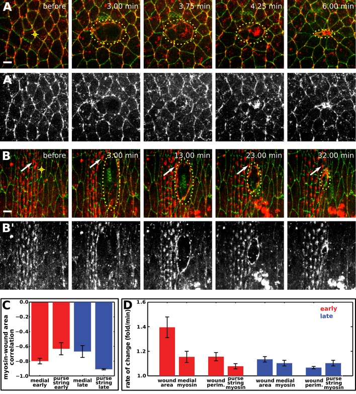
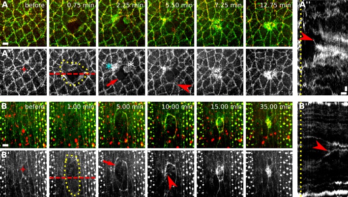
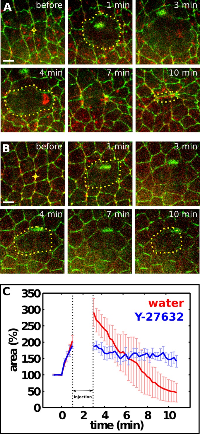
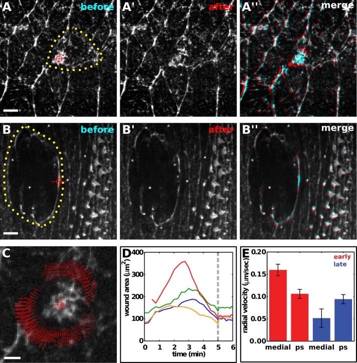
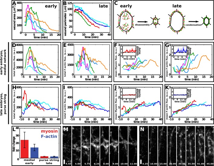
Epithelial tissues are protective barriers that display a remarkable ability to repair wounds. Wound repair is often associated with an accumulation of actin and nonmuscle myosin II around the wound, forming a purse string. The role of actomyosin networks in generating mechanical force during wound repair is not well understood. Here we investigate the mechanisms of force generation during wound repair in the epidermis of early and late Drosophila embryos. We find that wound closure is faster in early embryos, where, in addition to a purse string around the wound, actomyosin networks at the medial cortex of the wounded cells contribute to rapid wound repair. Laser ablation demonstrates that both medial and purse-string actomyosin networks generate contractile force. Quantitative analysis of protein localization dynamics during wound closure indicates that the rapid contraction of medial actomyosin structures during wound repair in early embryos involves disassembly of the actomyosin network. By contrast, actomyosin purse strings in late embryos contract more slowly in a mechanism that involves network condensation. We propose that the combined action of two force-generating structures--a medial actomyosin network and an actomyosin purse string--contributes to the increased efficiency of wound repair in the early embryo.


Figures

FIGURE 1:
FIGURE 1:
Wound closure is faster in early Drosophila embryos. (A, B) Epidermal cells expressing E-cadherin:GFP in a stage 7 (A, early) and a stage 14 (B, late) embryo. Red targets indicate the interface ablated for wounding. Yellow dots delineate the wound. The time after wounding is indicated. Red brackets at t = 4 min indicate laser-induced holes in the vitelline membrane. Wounded cells move away from the wound site (A) due to cell rearrangement. Anterior, left; dorsal, up. Bars, 5 μm. (C) Wound healing in early (top) and late (bottom) embryos, corresponding to image sequences in A and B, respectively. Wound perimeter was measured at 1-min intervals using the LiveWire algorithm. Bar, 5 μm. (D) Wound area normalized to the total area of the prewounded cells in early (n = 9, red) and late (n = 10, blue) embryos. Error bars, SEM. (E, F) Wound area over time in early (E) and late (F) embryos. Different colors indicate different wounds. t = 0 min is the time of wounding. (G) Linear fits (black, dashed lines) to the fast and slow phases of wound closure (green). The rates of wound closure during the fast (yellow shade) and slow (gray shade) phases were calculated as the slopes of these lines. (H) Duration of the fast phase in early (n = 9, red) and late (n = 10, blue) embryos. Error bars, SEM. (I) Wound closure rate in the fast (left) and slow (right) phases of wound closure in early (n = 9, red) and late (n = 10, blue) embryos. Error bars, SEM.
FIGURE 2:
FIGURE 2:
Distinct myosin structures form during epidermal wound closure in early and late Drosophila embryos. (A, B) Epidermal cells expressing E-cadherin:GFP and myosin:mCherry in a stage 7 (A) and a stage 14 (B) embryo. The time after wounding is indicated. Yellow target indicates the site of wounding, dots delineate the wound margin. White arrows track the position of a denticle precursor. Anterior, left; dorsal, up. Bars, 5 μm. (A′, B′) The myosin:mCherry signal in A and B. (C) Correlation coefficient between wound area and mean myosin fluorescence in the wounded cells or the purse string in early (n = 10, red bars) and late (n = 8, blue bars) embryos. Error bars, SEM. (D) Rate of change of wound area, perimeter, or mean myosin fluorescence in the medial region of wounded cells or at the purse string in early (n = 10, red bars) and late (n = 8, blue bars) embryos. The rate of wound area contraction is the average ratio of area(t − 1 min)/area(t). Perimeter reduction was similarly quantified. The rate of increase in mean myosin fluorescence was quantified with the ratio myosin(t)/myosin(t − 1 min) to account for the fact that area and mean fluorescence are inversely correlated. Error bars, SEM.
FIGURE 3:
FIGURE 3:
Complex actin dynamics during embryonic wound closure in Drosophila. (A, B) Epidermal cells expressing GFP:moesin (green) to visualize filamentous actin and myosin:mCherry (red) in a stage 7 (A) and a stage 14 (B) embryo. The time after wounding is indicated. Anterior, left; dorsal, up. Bars, 5 μm. (A′, B′) GFP:moesin signal in A and B. Red targets indicate the interface ablated for wounding. Yellow dots delineate the wound outline. Actin accumulates around the wound (arrows), at the medial cortex of wounded and adjacent cells (cyan and white asterisk, respectively), and in protrusions (arrowheads). (A′′, B′′) Kymographs showing the pattern of GFP:moesin along the red dashed lines in A′ and B′. Yellow line indicates time of wounding. Arrowheads indicate protrusions formed by wounded cells (A′′) or cells adjacent to the wound (B′′). Anterior, down. Bars, horizontal, 3 min; vertical, 2 μm.
FIGURE 4:
FIGURE 4:
Myosin activity is necessary for rapid wound repair in early embryos. (A, B) Epidermal cells expressing E-cadherin:GFP and myosin:mCherry in stage 7 embryos injected with water (A) or 100 mM of the Rho-kinase inhibitor Y-27632 (B). Red targets indicate the interface ablated for wounding. Yellow dots delineate the wound in alternating time points. The time after wounding is indicated. Injection is between 1 and 3 min. Anterior, left; dorsal, up. Bars, 5 μm. (C) Wound area normalized to the total area of the prewounded cells in embryos injected with water (n = 5, red) or 100 mM Y-27632 (n = 4, blue). The dotted lines delimit the time of injection. Error bars, SEM.
FIGURE 5:
FIGURE 5:
Contractile actomyosin networks in wounded cells drive epidermal repair in early Drosophila embryos. (A–A′′, B–B′′) Epidermal cells expressing myosin:GFP in a wounded stage 7 (A) or stage 14 (B) embryo. Yellow dots delineate the wound margin, red targets indicate the site of ablation. Myosin networks are shown immediately before ablation (A, B) and 3.3 s after ablation (A′, B′). (A′′, B′′) Overlay of images before (cyan) and after (red) ablation of myosin networks. Anterior, left; dorsal, up. Bars, 5 μm. (C) Particle image velocimetry analysis of the response to ablation in A. Red target indicates the site of ablation, red arrows indicate the direction and magnitude of recoil. Bar, 2 μm. (D) Wound area over time in early embryos. Medial actomyosin networks that form after wounding were ablated at the time indicated by the gray line. (E) Radial recoil velocity after spot ablation of distinct myosin networks during wound repair. Red bars correspond to medial networks in wounded cells (medial, n = 8) and purse strings around wounds in early embryos (ps, n = 6); blue bars correspond to the medial surface of the wounded cells (medial, n = 6) and purse strings around wounds in late embryos (ps, n = 8). Error bars, SEM.
FIGURE 6:
FIGURE 6:
Distinct F-actin and myosin dynamics during embryonic wound closure in early and late embryos. (A, B) Wound area over time in early embryos (A) and wound perimeter over time in late embryos (B). t = 0 min is the time of wounding. (C) Model of wound closure in early and late embryos. (D, E) Total myosin (D) and F-actin (E) fluorescence in the medial cortex of wounded cells in early embryos decreased rapidly during wound closure. (F, G) Mean myosin (F) and F-actin (G) fluorescence in the medial cortex of wounded cells in early embryos increased during wound closure. Insets, ratio of the rates of change of wound area to the rate of change of mean myosin (F) or F-actin (G) fluorescence. The black dashed line indicates the point where wound area and mean fluorescence change at the same rate. Error bars, SEM. (H, I) Total myosin (H) and F-actin (I) fluorescence in the purse string in late embryos decreased only slightly during wound closure. Note the different scales in the x- and y-axes with respect to D and E. (J, K) Mean myosin (J) and F-actin (K) fluorescence in the purse string in late embryos increased during wound closure. Insets, ratio of the rates of change of wound perimeter to the rate of change of mean myosin (J) or F-actin (K) fluorescence. The black dashed line indicates the point where wound perimeter and mean fluorescence change at the same rate. Error bars, SEM. (A, B, D–K) Embryos expressed both GFP:moesin and myosin:mCherry. Different colors indicate different wounds. Circles indicate the onset of wound closure. (D–K) Data were smoothed using a Gaussian filter with σ = 15 s. Here t = 0 min indicates the onset of wound closure. (L) Fold change in total myosin (red) and F-actin (blue) levels in medial networks in early embryos or purse strings in late embryos during wound repair. Error bars, SEM. (M, N) myosin:mCherry in a medial network in an early embryo (M) and a purse string in a late embryo (N). Times shown in minutes after wounding. Anterior, left; ventral, down. Bar, 2 μm.

Similar articles

Cited by

References

    1. Abreu-Blanco MT, Verboon JM, Liu R, Watts JJ, Parkhurst SM. Drosophila embryos close epithelial wounds using a combination of cellular protrusions and an actomyosin purse string. J Cell Sci. 2012;125:5984–5997. - PMC - PubMed
    1. Abreu-Blanco MT, Verboon JM, Parkhurst SM. Cell wound repair in Drosophila occurs through three distinct phases of membrane and cytoskeletal remodeling. J Cell Biol. 2011;193:455–464. - PMC - PubMed
    1. Amano M, Ito M, Kimura K, Fukata Y, Chihara K, Nakano T, Matsuura Y, Kaibuchi K. Phosphorylation and activation of myosin by Rho-associated kinase (Rho-kinase) J Biol Chem. 1996;271:20246–20249. - PubMed
    1. Bement WM, Forscher P, Mooseker MS. A novel cytoskeletal structure involved in purse string wound closure and cell polarity maintenance. J Cell Biol. 1993;121:565–578. - PMC - PubMed
    1. Bement WM, Mandato CA, Kirsch MN. Wound-induced assembly and closure of an actomyosin purse string in Xenopus oocytes. Curr Biol. 1999;9:579–587. - PubMed

Publication types

MeSH terms

LinkOut - more resources