. 2013 Nov;33(21):4152-65.
doi: 10.1128/MCB.01691-12. Epub 2013 Aug 26.

Cross talk between the Akt and p38α pathways in macrophages downstream of Toll-like receptor signaling

Affiliations

Cross talk between the Akt and p38α pathways in macrophages downstream of Toll-like receptor signaling

Victoria A McGuire et al. Mol Cell Biol. 2013 Nov.

Abstract

The stimulation of Toll-like receptors (TLRs) on macrophages by pathogen-associated molecular patterns (PAMPs) results in the activation of intracellular signaling pathways that are required for initiating a host immune response. Both phosphatidylinositol 3-kinase (PI3K)-Akt and p38 mitogen-activated protein kinase (MAPK) signaling pathways are activated rapidly in response to TLR activation and are required to coordinate effective host responses to pathogen invasion. In this study, we analyzed the role of the p38-dependent kinases MK2/3 in the activation of Akt and show that lipopolysaccharide (LPS)-induced phosphorylation of Akt on Thr308 and Ser473 requires p38α and MK2/3. In cells treated with p38 inhibitors or an MK2/3 inhibitor, phosphorylation of Akt on Ser473 and Thr308 is reduced and Akt activity is inhibited. Furthermore, BMDMs deficient in MK2/3 display greatly reduced phosphorylation of Ser473 and Thr308 following TLR stimulation. However, MK2/3 do not directly phosphorylate Akt in macrophages but act upstream of PDK1 and mTORC2 to regulate Akt phosphorylation. Akt is recruited to phosphatidylinositol 3,4,5-trisphosphate (PIP3) in the membrane, where it is activated by PDK1 and mTORC2. Analysis of lipid levels in MK2/3-deficient bone marrow-derived macrophages (BMDMs) revealed a role for MK2/3 in regulating Akt activity by affecting availability of PIP3 at the membrane. These data describe a novel role for p38α-MK2/3 in regulating TLR-induced Akt activation in macrophages.


Figures
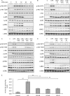
Fig 1
Fig 1
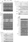
LPS-induced phosphorylation of Akt on Thr308 and Ser473 requires p38. (A) RAW264.7 cells were stimulated for the indicated times with 100 ng/ml of LPS. Cells were lysed and the levels of phospho-Thr308 Akt, phospho-Ser473 Akt, total Akt, phospho-p38 and total p38, phospho-MK2 and total MK2, and total ERK determined by immunoblotting. (B) Same as panel A except that cells were preincubated for 1 h with 1 μM PI-103 (PI), 1 μM GDC-0941 (GDC), or 1 μM MK-2206 (MK) where indicated. (C) Same as panel A except that cells were preincubated for 1 h with 1 μM GDC-0941 (GDC), 0.5 μM PIK75 (PIK), 1 μM TGX221 (TGX), or 1 μM IC87114 (IC8) where indicated. (D) Same as panel A except that cells were preincubated for 1 h with 5 μM SB203580 (SB), 1 μM VX-745 (VX), or 0.1 μM BIRB-0796 (BI) where indicated. (E) RAW264.7 cells were preincubated for 1 h with 5 μM SB203580 (SB), 0.1 μM BIRB0796 (BI), or 1 μM PI-103 (PI) where indicated. Cells were then stimulated with 100 ng/ml of LPS for 30 min and lysed, and Akt activity was determined as described in Materials and Methods. Error bars represent the standard deviations for 5 independent stimulations. A P value of less than 0.01 relative to the LPS-stimulated sample is indicated by double asterisks. Each panel is representative of at least 2 or 3 independent experiments.
Fig 2
Fig 2
LPS-induced Akt phosphorylation requires p38α and MK2/3. (A) BMDMs were cultured from wild-type, T106M p38α knock-in (p38α KI), or T106M p38α/T106M p38β double-knock-in (p38αβ KI) mice. Where indicated, cells were preincubated for 1 h with 5 μM SB203580 (SB) and then stimulated with 100 ng/ml of LPS for 30 min. Cells were lysed and levels of the indicated phosphorylated and total proteins determined by immunoblotting. (B) BMDMs were cultured from wild-type, MK2, MK3, or MK2/MK3 knockouts. Cells were stimulated for the indicated times with 100 ng/ml of LPS and lysed, and the levels of the indicated phosphorylated and total proteins were determined by immunoblotting. Gray arrowheads indicate MK2 bands, while the open arrowhead indicates a nonspecific band. (C) HeLa cells were preincubated for 1 h with PF3644022 (PF) and then stimulated for 30 min with 10 μg/ml of anisomycin. Cells were lysed and the levels of phospho-Hsp27 and total Hsp27, phospho-MK2 and total MK2, phospho-p38 and total p38, and total ERK measured by immunoblotting. (D) RAW264.7 cells were preincubated for 1 h with PF3644022 (PF) and then stimulated for 30 min with 100 ng/ml of LPS. Cells were lysed and the levels of the indicated phosphorylated and total proteins determined by immunoblotting. (E) BMDMs were cultured from wild-type mice and stimulated with 100 ng/ml of LPS for 15 min. Where indicated, cells were pretreated with 1 μM MK-2206, 2 μM PD184352, or 3 μM BI-D1870 for 1 h. Following stimulation, cells were lysed and the levels of the indicated phosphorylated and total proteins determined by immunoblotting. Blots are representative of a minimum of two independent experiments.
Fig 3
Fig 3
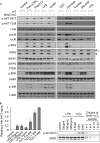
MK2 and -3 regulate Akt phosphorylation in response to multiple TLR agonists. (A) BMDMs were cultured from wild-type or MK2/3 knockouts and stimulated with 1 μg/ml of Pam3CSK4, 10 μg/ml of poly(I·C), 100 ng/ml of LPS, 1 μg/ml of CL097, 2 μM CpG-ODN1826, 200 μg/ml of zymosan, 10 μg/ml of curdlan, 50 ng/ml of GM-CSF, or 10 nM C5a for 30 min. Cells were then lysed and the levels of the indicated phosphorylated and total proteins determined by immunoblotting. Gray arrowheads indicate MK2 bands, while the open arrowhead indicates a nonspecific band. Blots are representative of a minimum of two independent experiments. (B) BMDMs were cultured from wild-type mice and stimulated for 15 min with LPS or H2O2 as indicated. Cells were then lysed and the levels of phospho-Ser473 Akt and total ERK1/2 measured by immunoblotting on the same membrane. Akt Ser473 phosphorylation was quantified as described in Materials and Methods. Results of the quantification are shown on the left, and a representative membrane is shown on the right. Error bars represent the standard deviations of 3 independent experiments.
Fig 4
Fig 4
Hsp25 (HspB1) is dispensable for regulating Akt activity in BMDMs. BMDMs were cultured from wild-type or HspB1 knockouts. Cells were preincubated for 1 h with 5 mM SB203580 and then stimulated for the indicated times with 100 ng/ml of LPS. Cells were lysed and the levels of phospho-Thr308 Akt, phospho-Ser473 Akt, total Akt, phospho-MK2 and total MK2, and total ERK measured by immunoblotting. Data are shown from two mice per genotype.
Fig 5
Fig 5
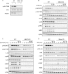
Phosphorylation of Akt downstream of MK2/3 is indirect. (A) Splenic macrophages were cultured from wild-type or conditional PDK1 knockouts. Cells were stimulated for 30 min with 100 ng/ml of LPS and lysed, and the levels of phospho-Thr308 Akt, total Akt, and total PDK1 were measured by immunoblotting. (B) BMDMs were cultured from wild-type or MK2/3 knockouts. Cells were pretreated with 5 μM SB203580 (SB) or 1 μM Ku-0063794 for 1 h and then stimulated for the indicated times with 100 ng/ml of LPS. Cells were lysed and the levels of phospho-Thr308 Akt, phospho-Ser473 Akt, total Akt, phospho- and total p38, phospho- and total MK2, and total ERK measured by immunoblotting. For total Akt and total ERK, membranes were stripped and reprobed from the phospho-Thr308 Akt and total p38, respectively. (C) BMDMs were cultured from wild-type or MK2/3 knockouts. Cells were pretreated with 5 mM SB203580 (SB) or 1 mM KU-0063794 for 1 h and then stimulated for the indicated times with 200 mg/ml of zymosan. Cells were lysed and the levels of phospho-Thr308 Akt, phospho-Ser473 Akt, total Akt, phospho-p38 and total p38, phospho-MK2 and total MK2, and total ERK measured by immunoblotting. For total Akt and total ERK, membranes were stripped and reprobed from the phospho-Thr308 Akt and total p38, respectively. (D) BMDMs were cultured from wild-type or conditional Rictor knockouts. Cells were preincubated for 1 h with 5 μM SB203580 (SB) or 1 μM Ku-0063794 (KU) and then stimulated for the indicated times with 100 ng/ml of LPS. Cells were lysed and the levels of phospho-Thr308 Akt, phospho-Ser473 Akt, total Akt, phospho-p38 and total p38, phospho-MK2 and total MK2, and total ERK measured by immunoblotting. For total Akt and total ERK, membranes were stripped and reprobed from the phospho-Thr308 Akt and total p38, respectively.
Fig 6
Fig 6
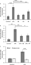
Accumulation of PIP3 following LPS stimulation requires MK2/3. (A) RAW264.7 cells were preincubated for 1 h with 5 μM SB203580, 1 μM VX-745, or 0.1 μM BIRB0796 and then stimulated for 30 min with 100 ng/ml of LPS. Lipids were isolated and relative levels of PIP3 were measured using a TRFRET displacement assay as described in Materials and Methods. Error bars represent the SEMs of 12 replicates per condition. (B) RAW264.7 cells were preincubated for 1 h with 5 μM SB203580 or 10 μM PF-3644022 and then stimulated for 30 min with 100 ng/ml of LPS. PIP3 levels were determined as for panel A. Error bars represent the SEMs of 5 to 8 replicates per condition. (C) BMDMs were cultured from wild-type or MK2/3 knockouts and expanded as described in Materials and Methods. Cells were stimulated for 30 min with 100 ng/ml of LPS and PIP3 levels determined as for panel A. Error bars represent the SEMs of 4 replicates from 2 mice per genotype. In all panels, a P value (Student's t test) of less than 0.05 is indicated by a single asterisk, a P value of less than 0.01 is indicated by double asterisks, and a P value of less than 0.001 is indicated by triple asterisks.

Similar articles

Cited by

References

    1. Manning BD, Cantley LC. 2007. AKT/PKB signaling: navigating downstream. Cell 129:1261–1274 - PMC - PubMed
    1. Leevers SJ, Vanhaesebroeck B, Waterfield MD. 1999. Signalling through phosphoinositide 3-kinases: the lipids take centre stage. Curr. Opin. Cell Biol. 11:219–225 - PubMed
    1. Franke TF. 2008. PI3K/Akt: getting it right matters. Oncogene 27:6473–6488 - PubMed
    1. So L, Fruman DA. 2012. PI3K signalling in B- and T-lymphocytes: new developments and therapeutic advances. Biochem. J. 442:465–481 - PMC - PubMed
    1. Hazeki K, Nigorikawa K, Hazeki O. 2007. Role of phosphoinositide 3-kinase in innate immunity. Biol. Pharm. Bull. 30:1617–1623 - PubMed

Publication types

MeSH terms

Substances