. 2013 Sep;123(9):4050-62.
doi: 10.1172/JCI70026. Epub 2013 Aug 27.

A feed-forward spinal cord glycinergic neural circuit gates mechanical allodynia

Affiliations

A feed-forward spinal cord glycinergic neural circuit gates mechanical allodynia

Yan Lu et al. J Clin Invest. 2013 Sep.

Abstract

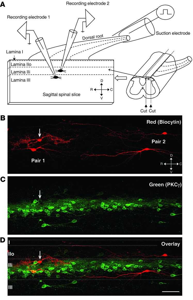
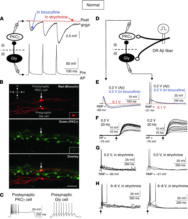
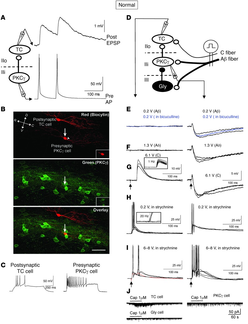
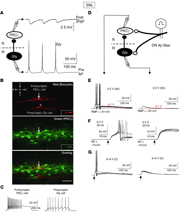
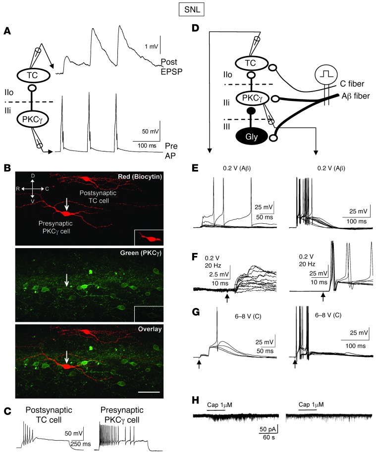
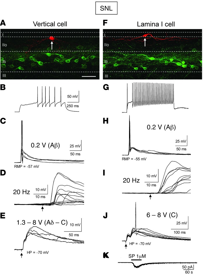
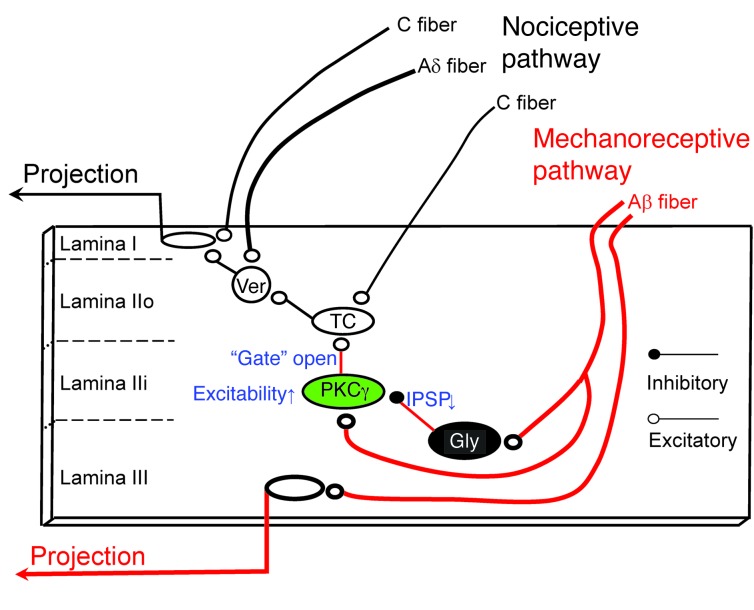
Neuropathic pain is characterized by mechanical allodynia induced by low-threshold myelinated Aβ-fiber activation. The original gate theory of pain proposes that inhibitory interneurons in the lamina II of the spinal dorsal horn (DH) act as "gate control" units for preventing the interaction between innocuous and nociceptive signals. However, our understanding of the neuronal circuits underlying pain signaling and modulation in the spinal DH is incomplete. Using a rat model, we have shown that the convergence of glycinergic inhibitory and excitatory Aβ-fiber inputs onto PKCγ+ neurons in the superficial DH forms a feed-forward inhibitory circuit that prevents Aβ input from activating the nociceptive pathway. This feed-forward inhibition was suppressed following peripheral nerve injury or glycine blockage, leading to inappropriate induction of action potential outputs in the nociceptive pathway by Aβ-fiber stimulation. Furthermore, spinal blockage of glycinergic synaptic transmission in vivo induced marked mechanical allodynia. Our findings identify a glycinergic feed-forward inhibitory circuit that functions as a gate control to separate the innocuous mechanoreceptive pathway and the nociceptive pathway in the spinal DH. Disruption of this glycinergic inhibitory circuit after peripheral nerve injury has the potential to elicit mechanical allodynia, a cardinal symptom of neuropathic pain.


Figures

Figure 1
Figure 1. Illustration of paired patch-clamp recordings in sagittal spinal cord slices.
(A) Diagram of the typical experimental paradigm. Sagittal spinal slices (400- to 600-μm thick) with an attached DR (8- to 10-mm long) were cut from the lumber spinal cord. Separate recording electrodes were used to make simultaneous whole-cell recordings from 2 neurons located in laminae IIo and IIi or laminae IIi and III. A suction electrode was used to stimulate DR to evoke synaptic responses in recorded neurons. C, caudal; D, dorsal; R, rostral; V, ventral. (B) Confocal image of 2 biocytin-labeled pairs of the recorded neurons. (C) Immunostaining shows that PKCγ-positive neurons are mainly distributed in lamina IIi. The boundaries of PKCγ neuron plexus were used to define lamina IIo, IIi, and III. (D) The overlay confocal image shows that 1 cell in pair 1 is PKCγ positive (arrow). Scale bar: 100 μm. I, lamina I; IIo, lamina IIo; IIi, lamina IIi; III, lamina III.
Figure 2
Figure 2. A feed-forward inhibitory circuit gates the output of PKCγ+ neurons following low-threshold Aβ-fiber stimulation.
(A) Gly unitary inhibitory connection between Gly and PKCγ+ neurons. The bottom trace shows the APs initiated by 3 successive depolarizing pulses in Gly neuron. Top traces show the evoked unitary IPSPs in PKCγ+ neuron. (B) Confocal images show 30-μm-thick optical stacks of the recorded neuronal pair. Arrows indicate the PKCγ+ cell; an arrowhead indicates putative axon. Insets show 1-μm-thick optical stacks of the PKCγ+ cell. Scale bar: 100 μm. (C) AP patterns of the recorded neurons. (D) Schematic diagram of the feed-forward inhibitory circuit. (E) DR stimulation at Aβ-fiber strength evokes a biphasic response in the PKCγ+ cells (monosynaptic EPSPs and polysynaptic IPSPs) and EPSPs with APs in the Gly cells. (F) Repetitive DR stimulation (20 Hz) indicates the evoked EPSPs are monosynaptic. Arrows indicate the stimulus artifacts. (G) Strychnine blocks the Aβ-fiber strength–evoked polysynaptic IPSPs and generates long-lasting EPSPs with APs in PKCγ+ neurons. (H) The C-fiber strength stimulation does not recruit C-fiber inputs in both PKCγ+ and Gly neurons in the presence of strychnine.
Figure 3
Figure 3. Feed-forward excitatory connections from PKCγ+ neurons to nociceptive TC neurons are normally silent after Aβ-fiber stimulation.
(A) Unitary excitatory connection between PKCγ+ and TC neurons. post, postsynaptic; pre, presynaptic. (B) Confocal images show 30-μm-thick optical stacks of the recorded neuronal pair. Arrows indicate the PKCγ+ cell, and the arrowhead indicates putative axon. Insets indicate 1-μm-thick optical stacks of the PKCγ+ cell. Scale bar: 100 μm. (C) AP patterns of the recorded neurons. (D) Schematic diagram of the excitatory connection and the feed-forward inhibitory circuit. (E and F) DR stimulation at Aβ- or Aδ-fiber strength evokes a biphasic synaptic response in PKCγ+ neurons but fails to evoke a synaptic response in TC cell. (G) DR stimulation at C-fiber strength evokes monosynaptic C-fiber EPSPs in TC cell. The inset shows the consistent latency and lack of synaptic failure during 1-Hz trials. (H) Strychnine blocks the DR-evoked polysynaptic IPSPs and generates long-lasting EPSPs with APs in PKCγ+ neurons and recruits polysynaptic Aβ-fiber EPSPs in TC cells. The inset shows the variation of the latency and synaptic failure during 20-Hz trials. (I) Application of strychnine does not recruit additional C-fiber inputs, aside from the original C-fiber inputs in TC neurons. (J) Capsaicin evokes marked increases in mEPSC frequency in TC neurons but has no effect on PKCγ+ and Gly neurons.
Figure 4
Figure 4. The feed-forward inhibition is impaired after spinal nerve injury.
(A) Gly unitary inhibitory connection between Gly and PKCγ+ neurons recorded from a SNL rat. (B) Confocal images show 30-μm-thick optical stacks of the recorded neuronal pair. Arrows indicate the PKCγ+ cell, and an arrowhead indicates putative axon. Insets show 1-μm-thick optical stacks of the PKCγ+ cell. Scale bar: 100 μm. (C) AP patterns of the recorded neurons. (D) Schematic diagram of the feed-forward inhibitory circuit. (E) DR stimulation evokes Aβ-fiber EPSPs in both Gly and PKCγ+ neurons. The amplitudes of the evoked EPSPs in Gly neurons were significantly smaller than those recorded in naive rats. The polysynaptic inhibitory components revealed in the naive slices almost completely disappeared after SNL. (F) Repetitive DR stimulation (20 Hz) indicates the evoked EPSPs are monosynaptic. Arrows indicate the stimulus artifacts. (G) DR stimulation at C-fiber strength fails to recruit additional Aδ- or C-fiber inputs to PKCγ and Gly neurons.
Figure 5
Figure 5. The excitatory connections from PKCγ+ neurons to TC neurons are enhanced after nerve injury.
(A) Unitary excitatory connection between PKCγ+ and TC neurons recorded from a SNL rat. (B) Confocal images show 30-μm-thick optical stacks of the recorded neuronal pair. Arrows indicate the PKCγ+ cell. Insets show 1-μm-thick optical stacks of the PKCγ+ cell. The axon of the PKCγ+ cell was not well labeled. Scale bar: 100 μm. (C) AP patterns of the recorded neurons. (D) Schematic diagram of the excitatory connection and the feed-forward inhibitory circuit. (E) DR stimulation evokes Aβ-fiber EPSPs in both PKCγ+ and TC neurons after SNL. (F) Repetitive DR stimulation (20 Hz) indicates the evoked EPSPs are monosynaptic in PKCγ neurons and polysynaptic in TC cell. (G) DR stimulation at C-fiber strength fails to recruit additional Aδ- or C-fiber inputs to TC neurons, aside from the original C-fiber inputs. (H) Capsaicin increases mEPSC frequency in TC neurons but not in PKCγ+ neurons.
Figure 6
Figure 6. Activation of low-threshold Aβ fibers evokes AP output of the nociceptive pathway after spinal nerve injury.
(A and F) Confocal images of morphology and location of the recorded (A) vertical cell and (F) lamina I cell. Arrows point out the recorded cells. Scale bar: 100 μm. (B and G) AP firing patterns of vertical and lamina I neurons. (C and H) DR stimulation at Aβ strength recruits polysynaptic EPSPs with APs in (C) vertical and (H) lamina I neurons. (D and I) The EPSPs are polysynaptic, as demonstrated by the varied latency and synaptic failure during 20-Hz stimulation. (E and J) DR stimulation at C-fiber strength fails to recruit additional Aδ- or C-fiber inputs to vertical and lamina I neurons, aside from their original Aδ- and C-fiber inputs. (K) Substance P (SP) (1 μM) evokes inward current in lamina I neurons.
Figure 7
Figure 7. Blockage or activation of spinal Gly synaptic transmission differentially regulates mechanical allodynia.
(A and B) SNL induced (A and C) mechanical allodynia and (B and D) thermal hyperalgesia manifested as a lowered threshold of mechanical or thermal withdrawal in rats. Ten rats were included in each group. (C and D) The SNL-induced mechanical allodynia was attenuated by intrathecal glycine, and this effect was blocked by strychnine. The same dose of glycine had no effect on the SNL-induced thermal hyperalgesia. Six rats were included in each group. (E and F) Intrathecal injection of strychnine induced robust mechanical allodynia but mild thermal hyperalgesia in naive rats. Six rats were included in each group. (*P < 0.05, **P < 0.01, compared with vehicle or sham controls, 1-way ANOVA with Bonferroni post-hoc test).
Figure 8
Figure 8. Working hypothesis of the “gate control” circuit for the generation of mechanical allodynia after nerve injury.
There is a preexisting but normally silent excitatory linkage from PKCγ+ cell to TC cell. This connection is normally under strong Gly control of a feed-forward inhibitory circuit. Nerve injury results in disinhibition of PKCγ+ neurons and allows the low-threshold mechanoreceptive signals to activate the nociceptive pathway.

Similar articles

Cited by

References

    1. Melzack R, Wall PD. Pain mechanisms: a new theory. Science. 1965;150(3699):971–979. doi: 10.1126/science.150.3699.971. - DOI - PubMed
    1. Sandkuhler J. Models and mechanisms of hyperalgesia and allodynia. Physiol Rev. 2009;89(2):707–758. doi: 10.1152/physrev.00025.2008. - DOI - PubMed
    1. Todd AJ. Neuronal circuitry for pain processing in the dorsal horn. Nat Rev Neurosci. 2010;11(12):823–836. - PMC - PubMed
    1. Zeilhofer HU, Wildner H, Yevenes GE. Fast synaptic inhibition in spinal sensory processing and pain control. Physiol Rev. 2012;92(1):193–235. doi: 10.1152/physrev.00043.2010. - DOI - PMC - PubMed
    1. Takazawa T, MacDermott AB. Synaptic pathways and inhibitory gates in the spinal cord dorsal horn. Ann N Y Acad Sci. 2010;1198:153–158. doi: 10.1111/j.1749-6632.2010.05501.x. - DOI - PMC - PubMed

Publication types