. 2013 Aug 22;39(2):311-323.
doi: 10.1016/j.immuni.2013.08.001. Epub 2013 Aug 15.

Mitochondrial cardiolipin is required for Nlrp3 inflammasome activation

Affiliations

Mitochondrial cardiolipin is required for Nlrp3 inflammasome activation

Shankar S Iyer et al. Immunity. .

Abstract

Nlrp3 inflammasome activation occurs in response to numerous agonists but the specific mechanism by which this takes place remains unclear. All previously evaluated activators of the Nlrp3 inflammasome induce the generation of mitochondrial reactive oxygen species (ROS), suggesting a model in which ROS is a required upstream mediator of Nlrp3 inflammasome activation. Here we have identified the oxazolidinone antibiotic linezolid as a Nlrp3 agonist that activates the Nlrp3 inflammasome independently of ROS. The pathways for ROS-dependent and ROS-independent Nlrp3 activation converged upon mitochondrial dysfunction and specifically the mitochondrial lipid cardiolipin. Cardiolipin bound to Nlrp3 directly and interference with cardiolipin synthesis specifically inhibited Nlrp3 inflammasome activation. Together these data suggest that mitochondria play a critical role in the activation of the Nlrp3 inflammasome through the direct binding of Nlrp3 to cardiolipin.


Figures

Figure 1
Figure 1. Linezolid induces caspase-1-dependent IL-1β secretion
(A) J774A.1 mouse macrophages were stimulated with either linezolid or LPS; culture supernatants were collected 18 hrs later and TNFα and IL-6 release measured by ELISA. (B) LPS-primed J774A.1 macrophages were stimulated with either silica or linezolid; culture supernatants were collected 18 hrs later and IL-1β release measured by ELISA. (C) LPS-primed J774A.1 macrophages were stimulated with linezolid or silica; culture supernatants were collected at the indicated times and cytotoxicity measured by LDH release and expressed as a percentage of LDH release by Triton X-100 detergent. (D) LPS-primed human PBMC were incubated with 20 μM z-YVAD-fmk for 1 hr prior to the addition of silica or linezolid. 18 hrs after stimulation culture supernatants were collected and IL-1β release measured by ELISA. (E) LPS-primed J774A.1 macrophages were challenged with silica or 1, 10 or 100 μg/ml of the indicated antibiotic for 18 hrs; IL-1β release into the culture supernatant was measured by ELISA. (A-E) Determinations were performed in triplicate and expressed as the mean ± SD. Results are representative of two separate experiments. See also Figure S1.
Figure 2
Figure 2. Linezolid induces Nlrp3-dependent inflammation in vitro and in vivo
(A) LPS-primed BMM from WT, Nlrp3-, ASC-, caspase-1-, or Nlrc4-deficient mice were stimulated with either linezolid or silica. Culture supernatants were collected at 18 hrs after stimulation and IL-1β release measured by ELISA. (B) Lysates from LPS-primed WT, Nlrp3-, ASC-, or Nlrc4-deficient BMM stimulated with linezolid for 18 hrs were immunoblotted with antibodies against the p10 subunit of caspase-1. Determinations were performed in triplicate and expressed as the mean ± SD. Results are representative of four (A) and three (B) separate experiments. (C) WT, Nlrp3-, ASC- and caspase-1-deficient mice (n=5) were challenged with 200 mg/kg of linezolid intraperitoneally; 16 hrs later neutrophil influx into the peritoneum was determined. Control WT mice (n=3) were challenged intraperitoneally with D5W. Results are representative of at least three independent experiments. * p < 0.01 by two-tailed Mann-Whitney test. (D, E) WT and Nlrp3-deficient mice received daily intraperitoneal injections of linezolid (200 mg/kg) (WT, n=9; Nlrp3−/−, n=9) or an equal volume of control D5W (WT, n=10; Nlrp3−/−, n=6). 12 days later femurs were sectioned and stained with H&E. (D) Representative 600X images are shown. Open arrows indicate myeloid cells and black arrows represent erythroid cells. (E) The mature stages of myeloid and erythroid cells were counted in each section with two sections counted per femur. Results are pooled from two independent experiments and expressed as the mean ± SEM. * p < 0.01 by two-tailed Mann-Whitney test.
Figure 3
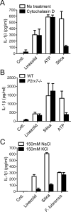
Figure 3. Linezolid-induced IL-1β secretion requires a potassium efflux
(A) LPS-primed J774A.1 macrophages were incubated with 20 μM cytochalasin D for 30 min prior to the addition of linezolid, silica or ATP. 18 hrs later culture supernatants were collected and IL-1β release measured by ELISA. (B) LPS-primed WT and P2rx7−/− BMM were stimulated with linezolid, silica or ATP. 18 hrs after stimulation culture supernatants were collected and IL-1β release measured by ELISA. (C) LPS-primed J774A.1 macrophages were incubated in either high Na+ or high K+ containing media and then stimulated with either linezolid, silica or F. tularensis LVS (MOI 50:1). 18 hrs later culture supernatants were collected and IL-1β release measured by ELISA. (A-C) Determinations were performed in triplicate and expressed as the mean ± SD. Results are representative of three (A) and (B-C) two separate experiments.
Figure 4
Figure 4. Linezolid-induced Nlrp3 inflammasome activation is independent of ROS generation
(A) LPS-primed J774A.1 macrophages were pretreated with the ROS inhibitors DPI (20 μM), APDC (20 μM) or NAC (1 mM) for 30 min prior to stimulation with linezolid, silica, nigericin, ATP or alum. 18 hrs later supernatants were collected and IL-1β secretion measured by ELISA. (B) Lysates from LPS-primed J774A.1 macrophages challenged with linezolid or silica in the absence or presence of DPI were immunoblotted with antibodies against the p10 subunit of caspase-1. (C) LPS-primed J774A.1 macrophages were pretreated with the antioxidant Mito-TEMPO (500 μM) for 1 hr prior to stimulation with linezolid or ATP. 18 hrs later supernatants were collected and IL-1β secretion measured by ELISA. * p = 0.003 by two-tailed unpaired Student's t-test. (D) Lysates from LPS-primed J774A.1 macrophages challenged with linezolid or ATP in the absence or presence of Mito-TEMPO were immunoblotted with antibodies against the p10 subunit of caspase-1. (E) LPS primed J774A.1 macrophages were treated with antimycin A (5 μg/ml) or rotenone (4 μM) for 30 min prior to stimulation with ATP, linezolid or silica. Supernatants were collected after 12 hrs and IL-1β secretion measured by ELISA. (F) LPS-primed J774A.1 macrophages were treated with ATP (5 mM), Linezolid (25 μg/ml) or media for one hour and then stained with MitoSOX red (2.5 μM) for the final 15 min of the stimulation. (G) LPS-primed J774A.1 cells were pretreated with 20 μM CsA for 1 hr and then challenged with linezolid, silica or nigericin for 6 hrs. Supernatants were collected and cytokine secretions analyzed by ELISA. * p = 0.037, ** p = 0.003 by two-tailed unpaired Student's t-test. Determinations were performed in triplicate and expressed as the mean ± SD. Results are representative of four (A (left panel), C), three (G) and two (A (right panel), B, D-F) separate experiments. See also Figure S2.
Figure 5
Figure 5. Cardiolipin binds to Nlrp3
(A, B) J774A.1 cells were radiolabeled for 24 hrs with 14C linoleic acid (10 μCi); cells were then LPS-primed and stimulated for 12 hrs with ATP or silica. The mitochondrial fraction was isolated, solubilized in 1% Triton X-100 containing lysis buffer and subjected to immunoprecipitation with anti-Nlrp3 antibody or isotype control antibody. Lipids associated with the immunoprecipitates were extracted with chloroform: methanol (2:1 v/v). Radioactivity associated with the organic fraction following immunoprecipitation was quantified (A) and subjected to 1-D HPTLC; the location of migration of cardiolipin (CL) was determined by running a cardiolipin standard in parallel (B). (A) Results are expressed as the mean ± SEM of three independent experiments; ** p < 0.01, *** p < 0.001 by two-tailed unpaired Student's t-test. (C, D) LPS-primed BMM were treated with staurosporine, silica or nigericin for 8 hrs. Cell lysates and supernatants were collected and lysates assessed for caspase-1 and caspase-3 via immunoblot (C) and supernatants for IL-1β via ELISA (D); determinations were performed in triplicate and expressed as the mean ± SD. (E) Lipid strips were incubated with lysates of RAW264.7 cells expressing HA-tagged mouse Nlrp3 or control buffer; strips were then washed and developed with a mouse anti-HA antibody followed by anti-mouse IgG-HRP. (F) Lipid strips were incubated with 10 μg/ml of His-tagged human Nlrp3, Nlrc4 or TLR4; strips were then washed and developed with a mouse anti-His antibody followed by anti-mouse IgG-HRP. (G) The indicated phospholipid-coated beads were incubated with 10 μg/ml of His-tagged Nlrp3; following washes, bound and unbound His tagged-Nlrp3 was determined by immunoblotting with an anti-Nlrp3 antibody. (H) Lipid strips were incubated with lysates of HEK293T cells expressing Flag-tagged full length Nlrp3.1-1036, or truncation mutants Nlrp3ΔLRR.1-719, PYD.1-95, NACHT.69-546 or LRR.535-719; strips were then washed and developed with a mouse anti-FLAG antibody followed by anti-mouse IgG-HRP. Results are representative of two (C, E, F, H), three (B, G) or four (D) separate experiments. See also Figure S3.
Figure 6
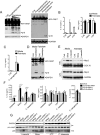
Figure 6. Cardiolipin is required and sufficient for Nlrp3 inflammasome activation
(A) LPS-primed J774A.1 cells were lysed by nitrogen cavitation in the presence of liposomes containing phosphatidylcholine (300 μM), phosphatidylserine (300 μM), phosphatidic acid (300 μM), or cardiolipin (300, 100, or 30 μM left panel; 300 μM right panel), incubated for one hour and analyzed for caspase-1 and GADPH by immunoblot. (B, C) J774A.1 macrophages grown in the presence or absence of 0.5 mM palmitate were LPS-primed followed by stimulation with silica, ATP or P. aeruginosa PAK strain (MOI 1:1). 12 hrs after stimulation culture supernatants were collected and IL-1β and TNFα release measured by ELISA. (D) J774A.1 macrophages grown in the presence or absence of 0.5 mM palmitate were LPS-primed followed by nitrogen cavitation lysis in the presence or absence of 300 μM of cardiolipin liposomes. Lysates were incubated for one hour and analyzed by immunoblot. (E) J774A.1 cells grown in the presence or absence of 0.5 mM palmitate were LPS-primed followed by stimulation with silica or linezolid. 12 hrs post stimulation cells were fractionated into mitochondrial and cytosolic fractions and subjected to SDS-PAGE followed by immunoblotting with antibodies to Nlrp3, COX IV (mitochondrial marker) and GAPDH (cytosolic marker). (F) THP-1 cells treated with the indicated siRNA were challenged with linezolid, silica, ATP, F. tularensis LVS (MOI 50:1) for 18 hrs or P. aeruginosa PAK strain (MOI 10:1) for 6 hrs. Culture supernatants were collected and IL-1β and TNFα release measured by ELISA (E). Lysates and supernatants were immunoblotted with antibodies against pro-caspase-1 and the p20 subunit of caspase-1 (F). Determinations were performed in triplicate and expressed as the mean ± SD. Results are representative of two (A-C, E, G) and three (D, F) separate experiments. * p < 0.05, ** p < 0.01, *** p < 0.005 by one-way ANOVA with Bonferroni's multiple comparison test. See also Figure S4.

Comment in

Similar articles

Cited by

References

    1. Agostini L, Martinon F, Burns K, McDermott MF, Hawkins PN, Tschopp J. NALP3 forms an IL-1beta-processing inflammasome with increased activity in Muckle-Wells autoinflammatory disorder. Immunity. 2004;20:319–325. - PubMed
    1. Allam R, Darisipudi MN, Rupanagudi KV, Lichtnekert J, Tschopp J, Anders HJ. Cutting edge: cyclic polypeptide and aminoglycoside antibiotics trigger IL-1β secretion by activating the NLRP3 inflammasome. J Immunol. 2011;186:2714–2718. - PubMed
    1. Ament PW, Jamshed N, Horne JP. Linezolid: its role in the treatment of gram-positive, drug-resistant bacterial infections. Am Fam Physician. 2002;65:663–670. - PubMed
    1. Attassi K, Hershberger E, Alam R, Zervos MJ. Thrombocytopenia associated with linezolid therapy. Clin Infect Dis. 2002;34:695–698. - PubMed
    1. Bauernfeind F, Bartok E, Rieger A, Franchi L, Nunez G, Hornung V. Cutting edge: reactive oxygen species inhibitors block priming, but not activation, of the NLRP3 inflammasome. J Immunol. 2011;187:613–617. - PMC - PubMed

Publication types

MeSH terms