. 2013 Oct;41(19):9129-40.
doi: 10.1093/nar/gkt667. Epub 2013 Aug 5.

Dissecting the interactions of SERRATE with RNA and DICER-LIKE 1 in Arabidopsis microRNA precursor processing

Affiliations

Dissecting the interactions of SERRATE with RNA and DICER-LIKE 1 in Arabidopsis microRNA precursor processing

Yuji Iwata et al. Nucleic Acids Res. 2013 Oct.

Abstract

Efficient and precise microRNA (miRNA) biogenesis in Arabidopsis is mediated by the RNaseIII-family enzyme DICER-LIKE 1 (DCL1), double-stranded RNA-binding protein HYPONASTIC LEAVES 1 and the zinc-finger (ZnF) domain-containing protein SERRATE (SE). In the present study, we examined primary miRNA precursor (pri-miRNA) processing by highly purified recombinant DCL1 and SE proteins and found that SE is integral to pri-miRNA processing by DCL1. SE stimulates DCL1 cleavage of the pri-miRNA in an ionic strength-dependent manner. SE uses its N-terminal domain to bind to RNA and requires both N-terminal and ZnF domains to bind to DCL1. However, when DCL1 is bound to RNA, the interaction with the ZnF domain of SE becomes indispensible and stimulates the activity of DCL1 without requiring SE binding to RNA. Our results suggest that the interactions among SE, DCL1 and RNA are a potential point for regulating pri-miRNA processing.


Figures
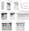
Figure 1.
Figure 1.
Characterization of pri-miRNA processing by DCL1. (A) Purified recombinant DCL1 protein. Coomassie blue-stained SDS–PAGE of purified DCL1 expressed in a baculovirus/insect cell expression system. (B) pri-miR167b processing by DCL1. The reaction was carried out under the same conditions as described previously (29) and at different concentrations of MgCl2, ATP and GTP. Pri-miR167b (150 nM) and DCL1 (50 nM) were incubated in a buffer [20 mM Tris–HCl (pH 7.0), 50 mM NaCl and indicated concentrations of MgCl2, ATP and GTP] at 37°C for 60 min. RNA was fractionated on 12% polyacrylamide/7.5 M urea denaturing gel and visualized by SYBR Gold staining. (C) Time course analysis of pri-miR167b processing. Pri-miR167b (150 nM) and DCL1 (50 nM) were incubated in a buffer [20 mM Tris–HCl (pH 7.0), 50 mM NaCl, 5 mM MgCl2 and 1 mM ATP] at 37°C for the indicated times. RNA was fractionated and detected as in (B). (D) pri-miR167b processing with a varying concentration of DCL1. The reaction was done at 37°C for 7 min with the same buffer components as in (C). The RNA was fractionated and detected as in (B). The amount of small RNA with 21mer and 23mer lengths was determined based on a series of known amounts of 20mer RNA fractionated alongside, and the velocity was plotted as a function of DCL1 concentration. (E) Effect of NaCl concentration. Pri-miR167b (150 nM) and DCL1 (50 nM) were incubated in a reaction buffer [20 mM Tris–HCl (pH 7.0), 5 mM MgCl2, 1 mM ATP and the indicated concentrations of NaCl] at 37°C for 20 min. The RNA was fractionated and detected as in (B). (F) Effect of pH. Pri-miR167b (150 nM) and DCL1 (50 nM) were incubated in a reaction buffer (50 mM NaCl, 5 mM MgCl2, 1 mM ATP and 20 mM Tris–HCl at the indicated pH) at 37°C for 20 min. The RNA was fractionated and detected as in (B). (G) Effect of nucleoside triphosphate. Pri-miR167b (150 nM) and DCL1 (50 nM) were incubated in a reaction buffer [20 mM Tris–HCl (pH 7.0), 50 mM NaCl, 5 mM MgCl2, and the indicated nucleoside triphosphates at 1 mM] at 37°C for 60 min. The RNA was fractionated and detected as in (B). (H) Effect of ATP on four pri-miRNA substrates. The reaction was done with the indicated pri-miRNA (150 nM) and DCL1 (50 nM) with or without 1 mM ATP as in (G). The RNA was fractionated and detected as in (B). (I) Effect of temperature. The reaction was done with the indicated pri-miRNA (150 nM) and DCL1 (50 nM) in a reaction buffer [20 mM Tris–HCl (pH 7.0), 5 mM MgCl2, 1 mM ATP and 50 mM NaCl] at 37°C or 25°C for 20 min. The RNA was fractionated and detected as in (B).
Figure 2.
Figure 2.
Effect of SE on pri-miRNA processing. (A) Purified recombinant SE protein. Coomassie blue stained SDS–PAGE of purified SE expressed in a baculovirus/insect cell expression system. (B) Effect of SE on pri-miR167b processing by DCL1. Pri-miR167b (150 nM) was incubated with DCL1 (50 nM) and SE (240 nM) in a reaction buffer [20 mM Tris–HCl (pH 7.0), 50 mM NaCl, 5 mM MgCl2 and 1 mM ATP] at 37°C for 20 min. The RNA was fractionated on 12% polyacrylamide/7.5 M urea denaturing gel and visualized by SYBR Gold staining. (C) Effect of SE using three different pri-miRNA substrates. The reaction was done as in (B) with the indicated pri-miRNA substrates (150 nM). The RNA was fractionated and visualized as in (B). (D) Effect of SE on DCL1 at different NaCl concentrations. The reaction was done as in (B) with pri-miR167b, and the RNA was fractionated and visualized as in (B). (E) SPR analysis of interaction of DCL1 and SE with RNA. In all, 240 RU of biotin-labeled forked-RNA consisting of a 40-base pairs dsRNA region with a forked structure on one end that consist of a 20-base ssRNA on both strands was immobilized on the SA-coated sensor chip as shown in the schematic diagram, and DCL1 or SE was introduced for 70 s at a concentrations of 42 nM, in a buffer containing 20 mM Tris–HCl (pH 7.0), 2 mM β-ME, and either 50 mM or 150 mM NaCl. The up and down arrows indicate the start and end of protein injections, respectively. (F) SPR analysis of interaction of SE with DCL1. 1750 RU of DCL1 was immobilized via its amine group on the CM-5 sensor chip, and SE was injected for 70 s at a concentration of 20 nM in a buffer containing 20 mM Tris–HCl (pH 7.0), 2 mM β-ME, 0.005% surfactant P20 and 50 mM or 150 mM NaCl. The up and down arrows indicate the start and end of protein injections, respectively.
Figure 3.
Figure 3.
Effect of SE and its mutant variants on pri-miRNA processing by DCL1. (A) Schematic representation of the SE domain structure and SE mutant proteins used in the following experiments. The N-terminal, proline/arginine-rich region (Pro/Arg-rich), ZnF domain and the C-terminal region (C-ter) are indicated. The inset shows the alignment of the C-ter end from plant SE homologs (amino acid residues conserved in four or more SE species are shaded in gray, and those conserved in all SE species are shaded in black. At; Arabidopsis thaliana, Vv; Vitis vinifera, Zm; Zea mays, Sb; Sorghum bicolor, Os; Oryza sativa). The mutant SE proteins, in which ZnF, N-terminal, and C-terminal domains are deleted, are designated as SEΔ497–547, SE143–720 and SE1–701, respectively. (B) Purified recombinant SE and its mutant proteins. Coomassie blue-stained SDS–PAGE of purified SE proteins expressed in a baculovirus/insect cell expression system. (C) Effect of SE and its mutant variants on pri-miR167b processing. Pri-miR167b (150 nM) was incubated with DCL1 (50 nM) and wild-type SE and its deletion mutants (240 nM) in a reaction buffer [20 mM Tris–HCl (pH 7.0), 100 mM NaCl, 5 mM MgCl2 and 1 mM ATP] at 37°C for 20 min. The RNA was fractionated on a 12% polyacrylamide/7.5 M Urea denaturing gel and visualized by SYBR Gold staining. (D) Effect of SE and its mutant variants on DCL1’s processing reaction on two more pri-miRNA substrates. The reaction and detection were done as in (C) with the indicated pri-miRNA substrates (150 nM). The RNA was fractionated and visualized as in (C).
Figure 4.
Figure 4.
Interaction of SE and its mutant variants with DCL1 and/or RNA. (A) Interaction of SE and its mutant variants with DCL1. Approximately 1750 RU of DCL1 was immobilized via its amine group on the CM-5 sensor chip and SE protein was injected for 70 s in a buffer containing 20 mM Tris–HCl (pH 7.0), 50 mM NaCl, 2 mM β-ME and 0.005% surfactant P20. The concentration of SE and its mutants was 20 nM. The up and down arrows indicate the start and end of protein injection, respectively. (B) Interaction of SE and its mutant variants with RNA. Approximately 210 RU of RNA was immobilized as in Figure 2E, and SE and its mutant variants were injected in a buffer containing 20 mM Tris–HCl (pH 7.0), 50 mM NaCl, 2 mM β-ME and 0.005% surfactant P20 at concentration of 20 nM. The up and down arrows indicate the start and end of protein injections, respectively. (C) Interaction of SE and its mutant variants with DCL1 in the presence of RNA. Approximately 1200 RU of DCL1 was bound to 60 RU of biotin-labeled RNA immobilized on the SA-coated sensor chip as in Figure 2E, and then SE and its mutant variants were injected for 70 s as shown in the diagram in a buffer containing 20 mM Tris–HCl (pH 7.0), 50 mM NaCl, 2 mM β-ME and 0.005% surfactant P20 at a concentration of 20 nM. The up and down arrows indicate the start and end, respectively, of injections of either DCL1 or SE.

Similar articles

Cited by

References

    1. Voinnet O. Origin, biogenesis, and activity of plant microRNAs. Cell. 2009;136:669–687. - PubMed
    1. Kim VN, Han J, Siomi MC. Biogenesis of small RNAs in animals. Nat Rev Mol Cell Biol. 2009;10:126–139. - PubMed
    1. Han MH, Goud S, Song L, Fedoroff N. The Arabidopsis double-stranded RNA-binding protein HYL1 plays a role in microRNA-mediated gene regulation. Proc. Natl Acad. Sci. USA. 2004;101:1093–1098. - PMC - PubMed
    1. Lobbes D, Rallapalli G, Schmidt DD, Martin C, Clarke J. SERRATE: a new player on the plant microRNA scene. EMBO Rep. 2006;7:1052–1058. - PMC - PubMed
    1. Yang L, Liu Z, Lu F, Dong A, Huang H. SERRATE is a novel nuclear regulator in primary microRNA processing in Arabidopsis. Plant J. 2006;47:841–850. - PubMed

Publication types

MeSH terms