Review
. 2013 Aug 1;5(8):a017780.
doi: 10.1101/cshperspect.a017780.

Position-effect variegation, heterochromatin formation, and gene silencing in Drosophila

Affiliations
Review

Position-effect variegation, heterochromatin formation, and gene silencing in Drosophila

Sarah C R Elgin et al. Cold Spring Harb Perspect Biol. .

Abstract

Position-effect variegation (PEV) results when a gene normally in euchromatin is juxtaposed with heterochromatin by rearrangement or transposition. When heterochromatin packaging spreads across the heterochromatin/euchromatin border, it causes transcriptional silencing in a stochastic pattern. PEV is intensely studied in Drosophila using the white gene. Screens for dominant mutations that suppress or enhance white variegation have identified many conserved epigenetic factors, including the histone H3 lysine 9 methyltransferase SU(VAR)3-9. Heterochromatin protein HP1a binds H3K9me2/3 and interacts with SU(VAR)3-9, creating a core memory system. Genetic, molecular, and biochemical analysis of PEV in Drosophila has contributed many key findings concerning establishment and maintenance of heterochromatin with concomitant gene silencing.


Figures

Figure 1.
Figure 1.
A schematic illustration of white variegation in the X chromosome inversion In(1)wm4. (A) Rearrangement attributable to an X-ray-induced inversion places the white locus, normally located in the distal euchromatin (white bar) of the X chromosome (see top line), ∼25 kb from a breakpoint in the pericentric heterochromatin (black bar; bottom rearranged line). Spreading of heterochromatin packaging into the euchromatic domain results in silencing (causing a white eye in this case); loss of silencing in some cells during differentiation results in a variegating phenotype (bottom line, right). (B) Given a variegating phenotype, screens for second site mutations can recover suppressors (Su(var)s) and enhancers (E(var)s) as described in the text. (C) Some Su(var) loci (e.g., Su(var)3-9, shown here) show an antipodal dosage-dependent effect, and are consequently thought to be structural proteins of heterochromatin. The presence of only one copy of the modifier gene results in less heterochromatin formation, and more expression from the reporter gene (suppression of PEV, top fly eye); conversely, the presence of three copies of such a modifier gene will drive more extensive heterochromatin formation, resulting in an enhancement of reporter gene silencing (enhancement of PEV, bottom fly eye).
Figure 2.
Figure 2.
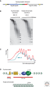
Heterochromatin is packaged into a regular nucleosome array. A TE such as that shown (A), carrying a marked copy of a heat shock gene for study and an hsp70-driven copy of white as a visual marker, can be used to study the same gene in different chromatin domains. (B) Nuclei from Drosophila lines carrying the transgene in a euchromatic domain (39C-X; red eye) and a heterochromatic domain (HS-2; variegating eye) were digested with increasing amounts of micrococcal nuclease (MNase), the DNA was purified and size-separated on an agarose gel, and the resulting Southern blot was hybridized with a probe unique to the transgene. Linker sites cleaved by MNase are marked with arrowheads. (C) Densitometer scans from the last lane of each sample are compared (top to bottom is left to right). An array of nine to 10 nucleosomes can be detected in heterochromatin (red line), compared with five to six in euchromatin (blue line), indicating more uniform spacing in the former case. (D) A diagrammatic representation of the results. DH site, deoxyribonuclease (DNase)-hypersensitive site; HSE, heat shock element. (B,C, Adapted from Sun et al. 2001, © American Society for Microbiology.)
Figure 3.
Figure 3.
The distribution of chromosomal proteins and histone modifications defines different chromatin domains. (A) Immunofluorescent staining of the polytene chromosomes identifies proteins predominantly associated with heterochromatin. The polytene chromosomes, prepared by fixation and squashing of the larval salivary gland (shown by phase contrast microscopy, left; C = chromocenter) are “stained” by incubating first with antibodies specific for a given chromosomal protein, and then with a secondary antibody coupled to a fluorescent tag. HP1a (right) and HP2 (center) have similar distribution patterns, showing prominent association with the pericentric heterochromatin (found in the condensed chromocenter), small fourth chromosome (inset, arrow), and a small set of sites in the long euchromatin arms. (Adapted from Shaffer et al. 2002.) Note that the efficacy of any antibody can be affected by the choice of fixation protocol (see Stephens et al. 2003). (B) Chromatin marks define the epigenomic border between heterochromatin and euchromatin (indicated with an arrow). The border can be delineated based on chromatin immunoprecipitation (ChIP)-array data using antibodies to proteins known to be associated with heterochromatin (HP1a) or euchromatin (RNA polymerase II [RNA Pol II]), and to key histone modifications. Enrichment values are shown for the centromere-proximal 3 Mb of chromosome arms 2R and 3L (in BG3 cells). Boxes underneath the bar graphs indicate significant enrichment (p < 0.001). The cytologically defined heterochromatin is shown by the blue bar. The border, indicated here by the black arrow, is fairly well defined by the congruence of silencing marks. (B, Adapted from Riddle et al. 2011, © Cold Spring Harbor Laboratory Press.)
Figure 4.
Figure 4.
Interaction of SU(VAR)3-9 and HP1a in setting the distribution pattern of H3K9 methylation. (A) HP1a interacts with H3K9me2/3 through its chromodomain, and with SU(VAR)3-9 through its chromoshadow domain. By recognizing both the histone modification and the enzyme responsible for that modification, HP1a provides a mechanism for heterochromatin spreading and epigenetic inheritance. (B) SU(VAR)3-9 is responsible for much of the dimethylation of H3K9 (H3K9me2); loss of the enzyme results in loss of this modification in the pericentric heterochromatin, as shown by loss of antibody staining of the polytene chromosomes (compare middle panel with top panel). Loss of HP1a results in a loss of targeting of SU(VAR)3-9; high levels of H3K9me are consequently now seen throughout the chromosome arms (bottom panel).
Figure 5.
Figure 5.
Spreading of histone H3K9me2 and cytological heterochromatinization at the white locus in PEV rearrangements. (A) In translocation T(1;4)wm258-21 (with a breakpoint in the heterochromatin of chromosome 4), heterochromatinization becomes cytologically visible in the polytene larval salivary gland chromosomes as a loss of banding, the apparent consequence of condensation and underreplication (right portion of the right panel). (Reprinted from Reuter et al. 1982, with kind permission from Springer Science+Business Media.) (B) In the In(1)wm4 chromosome, spreading of the heterochromatic chromatin state over ∼200 kb of the adjacent euchromatic region is initiated by the segment of heterochromatin located distal to the rDNA cluster (dark gray box). (C) ChIP with an antibody specific for H3K9me2 detects spreading of this heterochromatic histone mark along the euchromatic region between the roughest (rst) gene and the breakpoint of wm4. (D) Flies homozygous for a null mutation of the Su(var)3-9 gene lose H3K9me2 in pericentric heterochromatin as well as in the white gene region, restoring wild-type activity of the white gene in In(1)wm4 flies. (Adapted from Rudolph et al. 2007.)
Figure 6.
Figure 6.
The transition from a euchromatic state to a heterochromatic state requires a series of changes in histone modification. (A) Active genes are marked by H3K4me2/3; if present, this mark must be removed by LSD1. H3K9 is normally acetylated in euchromatin; this mark must be removed by a histone deacetylase, HDAC1. Phosphorylation of H3S10 can interfere with methylation of H3K9; dephosphorylation appears to involve a phosphatase targeted by interaction with the carboxyl terminus of the JIL1 kinase. These transitions set the stage for acquisition of the modifications associated with silencing, shown in B, including methylation of H3K9 by SU(VAR)3-9, binding of HP1a, and subsequent methylation of H4K20 by SUV4-20, an enzyme potentially recruited by HP1a. (C) Differentiation of euchromatin and heterochromatin is initiated in early embryogenesis around cell cycle 10 and is completed when cellular blastoderm (top box) and primordial germline cells (right-hand box) are formed. (D) Blastoderm nuclei show an apicobasal polarity (Rabl conformation). Heterochromatin (H3K9me2 staining) is established at the apical site, whereas euchromatin (H3K4me2 staining) is organized toward the basal site. (Immunofluorescent images provided by Sandy Mietzsch.)
Figure 7.
Figure 7.
Chromatin annotation of the D. melanogaster genome. (A) A nine-state model of prevalent chromatin states was generated using data from S2 cells. Each chromatin state (row) is defined by a combinatorial pattern of enrichment (red) or depletion (blue) for specific histone modification marks. First (left) panel, color code for mapping; second panel, histone modification marks used (active marks labeled in green, repressive in blue, general in black); third panel, enrichment or depletion of chromosomal proteins found in that state; fourth (right) panel, fold over-/underrepresentation of genic and transcription start site (TSS)-proximal (±1 kb) regions relative to the entire tiled genome. (B) A genome-wide karyotype view of the domains defined by the nine-state model in S2 cells. Centromeres are shown as open circles; dashed lines span gaps in the genome assembly. Note the association of pericentric heterochromatin and the fourth chromosome distal arm with state 7 and the association of state 5 with the male X chromosome. (Adapted, by permission from Macmillan Publishers Ltd: NATURE, from Kharchenko et al. 2011, © 2011.)
Figure 8.
Figure 8.
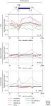
Packaging of active genes in chromosome 4, pericentric heterochromatin, and euchromatin. The plots show log2 enrichment (y-axis) for RNA Pol II (green), H3K36me3 (pink), H3K9me2 (yellow), H3K9me3 (purple), SU(VAR)3-9 (blue), POF (brown), and HP1a (red) for a scaled metagene and 2 kb flanking region created by averaging data for all active genes in a given compartment. Fourth chromosome (top) and pericentric (middle) genes show a similar depletion in silencing marks at the TSS; these marks reappear over the body of the gene only in the case of the fourth chromosome. As expected, euchromatic genes do not show association with any of the silencing marks (bottom). (Adapted from Riddle et al. 2012.)
Figure 9.
Figure 9.
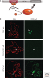
The piRNA system may drive silencing of some TEs through heterochromatin formation. (A) Depletion of HP1a (shown here), or of Piwi, results in overexpression of a subset of the TEs in the female germline. (B) This suggests that Piwi can promote transcriptional gene silencing, directly or indirectly, by targeting HP1a deposition at these TEs, presumably by a piRNA recognition event. (Adapted from Wang and Elgin 2011.)

Similar articles

Cited by

References

    1. Ahmad K, Golic KG 1996. Somatic reversion of chromosomal position effects in Drosophila melanogaster. Genetics 144: 657–70 - PMC - PubMed
    1. Ahmad K, Henikoff S 2001. Modulation of a transcription factor counteracts heterochromatin gene silencing in Drosophila. Cell 104: 839–847 - PubMed
    1. Allshire R, Ekwall K 2014. Epigenetics in Saccharomyces pombe. Cold Spring Harb Perspect Biol 10.1101/cshperspect.a018770 - DOI - PMC - PubMed
    1. Aravin AA, Lagos-Quintana M, Yalcin A, Zavolan M, Marks D, Snyder B, Gaasterland T, Meyer J, Tuschl T 2003. The small RNA profile during Drosophila melanogaster development. Dev Cell 5: 337–350 - PubMed
    1. Ashburner M, Golic KG, Hawley RS 2005. Chromosomes and position effect variegation. In Drosophila: A laboratory handbook, 2nd ed., Chaps. 4, 28 Cold Spring Harbor Laboratory Press, Cold Spring Harbor, NY

Publication types

MeSH terms

LinkOut - more resources