. 2013 Oct 15;126(Pt 20):4647-58.
doi: 10.1242/jcs.126573. Epub 2013 Jul 31.

Rab40b regulates trafficking of MMP2 and MMP9 during invadopodia formation and invasion of breast cancer cells

Affiliations

Rab40b regulates trafficking of MMP2 and MMP9 during invadopodia formation and invasion of breast cancer cells

Abitha Jacob et al. J Cell Sci. .

Abstract

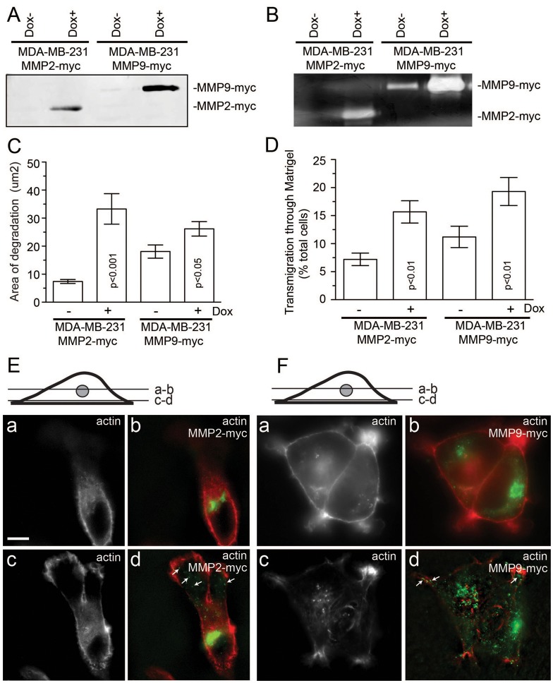
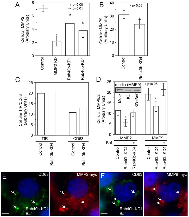
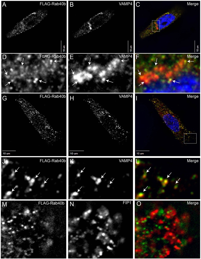
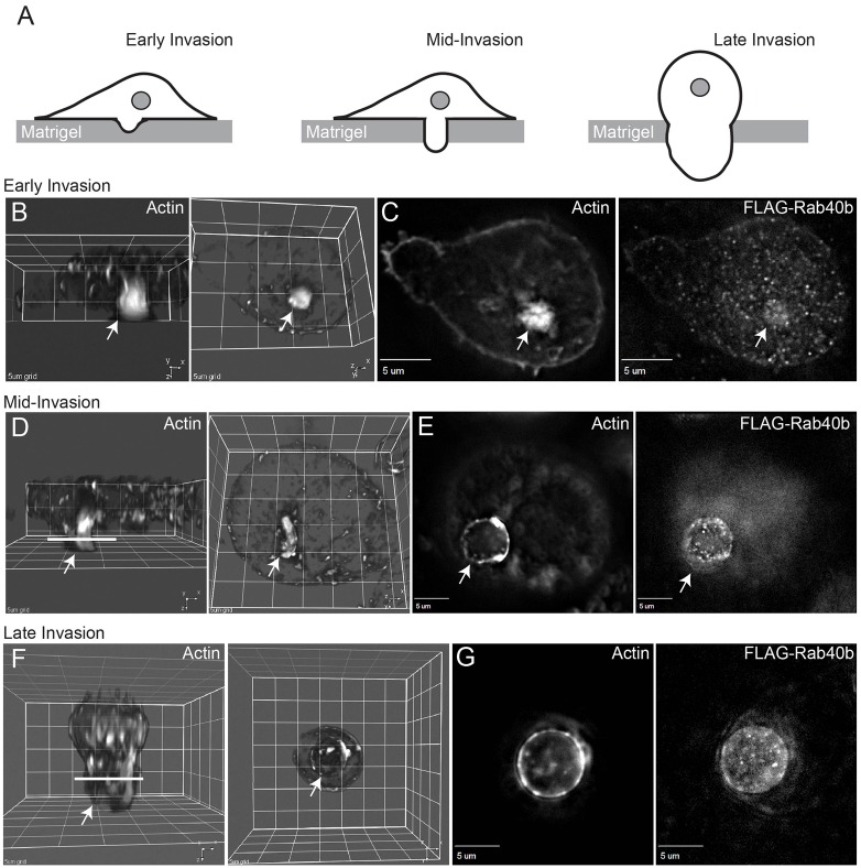
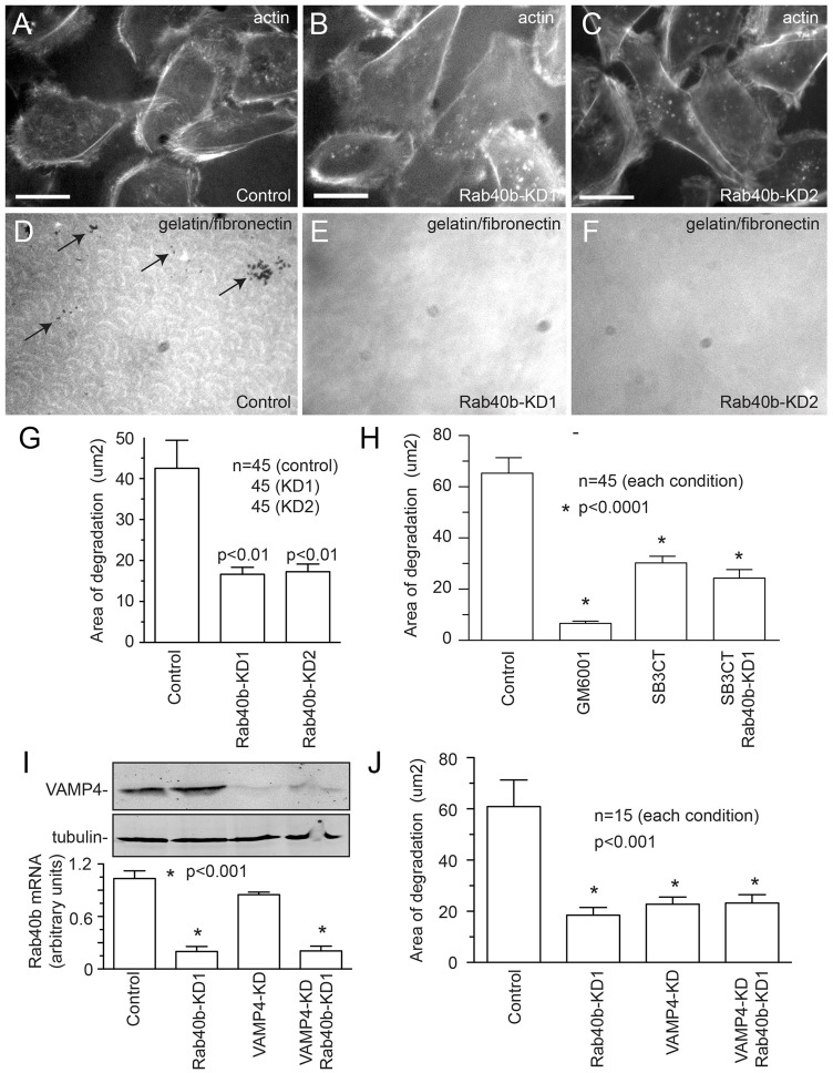
Invadopodia-dependent degradation of the basement membrane plays a major role during metastasis of breast cancer cells. Basement membrane degradation is mediated by targeted secretion of various matrix metalloproteinases (MMPs). Specifically, MMP2 and MMP9 (MMP2/9) possess the ability to hydrolyze components of the basement membrane and regulate various aspects of tumor growth and metastasis. However, the membrane transport machinery that mediates targeting of MMP2/9 to the invadopodia during cancer cell invasion remains to be defined. Because Rab GTPases are key regulators of membrane transport, we screened a human Rab siRNA library and identified Rab40b GTPase as a protein required for secretion of MMP2/9. We also have shown that Rab40b functions during at least two distinct steps of MMP2/9 transport. Here, we demonstrate that Rab40b is required for MMP2/9 sorting into VAMP4-containing secretory vesicles. We also show that Rab40b regulates transport of MMP2/9 secretory vesicles during invadopodia formation and is required for invadopodia-dependent extracellular matrix degradation. Finally, we demonstrate that Rab40b is also required for breast cancer cell invasion in vitro. On the basis of these findings, we propose that Rab40b mediates trafficking of MMP2/9 during invadopodia formation and metastasis of breast cancer cells.

Keywords: Invadopodia; Invasion; MMP2; MMP9; Rab40b; Secretion.


Figures

Fig. 1.
Fig. 1.
Characterization of MDA-MB-231 cell lines expressing tet-inducible MMP2–Myc or MMP9–Myc. (A,B) MDA-MB-231 cells expressing dox-inducible MMP2–Myc or MMP9–Myc were incubated in Opti-MEM for 24 hours in the absence or presence of 1 µg/ml doxycycline. Opti-MEM medium was then collected and the amount of secreted MMP2–Myc or MMP9–Myc was analyzed by immunoblotting (A) or zymography (B). (C) MDA-MB-231 cells expressing dox-inducible either MMP2–Myc or MMP9–Myc were plated on gelatin and fibronectin-HiLyte Fluor488-coated coverslips and incubated in the presence or absence of 1 µg/ml doxycycline. After incubation for 20 hours, cells were fixed and invadopodia-associated ECM degradation was analyzed by in situ zymography (for more details see the Materials and Methods). Data shown are the means and s.e. of three independent experiments. (D) MDA-MB-231 cells expressing dox-inducible MMP2–Myc or MMP9–Myc were plated on matrigel-coated 8-µm-pore filters and incubated in the presence or absence of 1 µg/ml doxycycline. The ability of cells to invade through Matrigel matrix was analyzed using Crystal Violet staining. The data shown are the means and s.d. from three independent experiments. (E,F) MDA-MB-231 cells expressing MMP2–Myc (E) or MMP9–Myc (F) were fixed and stained with Rhodamine-phalloidin and mouse anti-Myc antibodies. Panels a and b show optical sections at the TGN level, whereas panels c and d show optical sections at the coverslip level. Arrows indicate cytosolic organelles containing MMP2–Myc or MMP9–Myc. Scale bar: 5 µm.
Fig. 2.
Fig. 2.
MMP2 and MMP9 secretion-regulating proteins identified from siRNA screen. ELISA-based quantification of the secreted MMP2–Myc (A) and MMP9–Myc (B) in cells treated with various Rab siRNAs. For more details see the Materials and Methods. Data shown are the means and s.d. of three independent experiments. All the values above top grey line or below bottom grey line are significantly different to the control at P<0.01.
Fig. 3.
Fig. 3.
Rab40b knockdown decreases MMP2 and MMP9 secretion in MDA-MB-231 cells. (A) Schematic representation of Rab40b domain structure. (B) The efficiency of Rab40b knockdown as determined by qPCR. (C,D) MDA-MB-231 cells expressing MMP2–Myc (C) or MMP9–Myc (D) were transfected with four different Rab40b siRNAs. Two days later, equal number of cells were plated in six-well dishes and incubated with 1 ml of medium for 24 hours. Medium was then collected and the effect of Rab40b knockdown on secretion of MMP2–Myc and MMP9–Myc analyzed by western blotting. Data shown are the means and s.d. of three independent experiments. (E) MDA-MB-231 cells were transfected with Rab40b siRNA#2. Two days later, equal number of cells were plated in six-well dishes and incubated with 1 ml of Opti-MEM for 24 hours. Opti-MEM was then collected and the effect of Rab40b knockdown on secretion of endogenous MMP2 and MMP9 was analyzed by zymography. Fetal bovine serum (rich in secreted MMP2/9) in the first lane was used as a positive control. Opti-MEM collected from a six-well dish without cells was used as negative control. (F) Mock-, Rab40b-siRNA- or VAMP4-siRNA-treated MDA-MB-231 cells were harvested and analyzed by flow cytometry to measure the levels of endogenous plasma membrane MT1-MMP. Data shown are the means and s.d. of three independent experiments. (G) Mock- or Rab40b-siRNA-treated MDA-MB-231 cells were plated in six-well dishes and stimulated with 1 mg/ml of LPS. After incubation for 16 hours, medium was collected and the levels of secreted IL-6 analyzed using ELISA. The data shown are the means and s.d. of three independent experiments.
Fig. 4.
Fig. 4.
Rab40b increases lysosomal degradation of MMP2 and MMP9. (A–C) Mock- or Rab40b-siRNA-treated MDA-MB-231 cells were harvested and analyzed by flow cytometry to measure the levels of intracellular MMP2–Myc (A), MMP9–Myc (B), transferrin receptor (C) and CD63 (C). Data shown in A and B are the means and s.d. of three independent experiments. Data shown in C are the means of two independent experiments. (D) MDA-MB-231 cells stably expressing MMP2–Myc or MMP9–Myc were transfected with Rab40b siRNA. Two days later, cells were incubated in the presence of absence of bafilomycin for 12 hours and medium was collected to measure the amounts of secreted MMP2–Myc and MMP9–Myc (inset). Cells were then analyzed by flow cytometry to measure the levels of intracellular MMP2–Myc and MMP9–Myc. Data shown are the means and s.d. of three independent experiments. (E,F) MDA-MB-231 cells stably expressing MMP2–Myc (E) or MMP9–Myc (F) were transfected with Rab40b siRNA. Two days later, cells were treated with bafilomycin for 12 hours, then fixed and co-stained with anti-Myc (red) or anti-CD63 (green) antibodies. Nuclei are stained blue with DAPI. Scale bar: 5 µm.
Fig. 5.
Fig. 5.
FLAG–Rab40b colocalizes with VAMP4-containing secretory vesicles. MDA-MB-231 cells were transfected with FLAG–Rab40b and plated on collagen-coated glass coverlips. Cells were then fixed and stained with anti-FLAG (A,C,D,F,G,I,J,L,M,O), anti-VAMP4 (B,C,E,F,H,I,K,L) and anti-FIP1 (N,O) antibodies. In D–F, arrows indicate VAMP4 and Rab40b-containing vesicles at the edges of the TGN. In J–L, arrows indicate peripheral organelles containing VAMP4 and Rab40b. In C and I, boxed region marks area shown as higher magnification images in D–F and J–L. Nuclei in C,F,I are stained blue with DAPI.
Fig. 6.
Fig. 6.
Localization of Rab40b-containing organelles during cell invasion in vitro. MDA-MB-231 cells stably expressing FLAG–Rab40b were seeded on Matrigel-coated filters containing 8 µm pores. Cells were incubated for either 24 hours (B,D) or 36 hours (F). Cells were then fixed and stained with Rhodamine-phalloidin or anti-FLAG antibodies. Drawings in A depict the invasion stages imaged in panels B,D,F. Arrows in all images indicate invadopodia or pseudopodia (probably derived from invadopodia). B,D,F are 3D rendering of images shown in C,E,G and show cells from Z-Y (left panels) and X-Y (right panels) planes. Lines in D,F indicate the level of the optical sections depicted in E,G.
Fig. 7.
Fig. 7.
Rab40b is required for MDA-MB-231 cell invasion in vitro. (A,B) Mock- or siRNA-treated MDA-MB-231 cells were plated on matrigel-coated (A) or uncoated (B) 8-µm-pore filters. Cells were then incubated for 8 hours (B) or 16 hours (A) and the extent of cell migration to the bottom side of the filter was analyzed by Crystal Violet staining (see the Materials and Methods). Data shown are the means and s.d. of three independent experiments; *P>0.05. (C–H) MDA-MB-231 cells were plated on gelatin and fibronectin-HiLyte Fluor488-coated coverslips (D,E,G and H). After incubation for 20 hours, cells were fixed and stained with Rhodamine-phalloidin (C,E,F,H). Arrows indicate invadopodia. Scale bars: 5 µm (C–E), 1 µm (F–H). (I–K) MDA-MB-231 cells were plated on gelatin-coated coverslips. After incubation for 20 hours, cells were fixed and stained with Rhodamine-phalloidin (I,K) and rabbit anti-Tks5 antibodies (J,K). Scale bar: 1 µm.
Fig. 8.
Fig. 8.
Rab40b is required for invadopodia-associated ECM degradation. (A–F) Untreated or Rab40b-siRNA-treated MDA-MB-231 cells were plated on gelatin and fibronectin HiLyte Fluor488-coated coverslips (D–F). After 20 hours of incubation, cells were fixed and stained with Rhodamine-phalloidin (A–C). Scale bars: 25 µm. (G) Quantification of ECM degradation in untreated and Rab40b-siRNA-treated MDA-MB-231 cells. Data shown are the means and s.e. of three independent experiments. n is the total number of cells analyzed. (H) Quantification of ECM degradation in MDA-MB-231 cells treated with either GM6001 (broad-spectrum MMP inhibitor) or SB3CT (MMP2/MMP9 inhibitor). Where indicated, cells were also treated with Rab40b siRNA. Data shown are the means and s.e. of three independent experiments. n is the total number of cells analyzed. (I) Mock-, Rab40b-siRNA- or VAMP4 siRNA-treated cells were harvested and the levels of mRNA encoding Rab40b analyzed by qPCR (graph). The data shown are the means and s.d. The levels of VAMP4 were analyzed by western blotting (top panels) with tubulin used as a loading control. (J) Quantification of ECM degradation in mock-, Rab40b-siRNA- or VAMP4-siRNA-treated MDA-MB-231 cells. Data shown are the means and s.e. of three independent experiments. n is the total number of cells analyzed.

Similar articles

Cited by

References

    1. Artym V. V., Zhang Y., Seillier-Moiseiwitsch F., Yamada K. M., Mueller S. C. (2006). Dynamic interactions of cortactin and membrane type 1 matrix metalloproteinase at invadopodia: defining the stages of invadopodia formation and function. Cancer Res. 66, 3034–3043 10.1158/0008-5472.CAN-05-2177 - DOI - PubMed
    1. Blouw B., Seals D. F., Pass I., Diaz B., Courtneidge S. A. (2008). A role for the podosome/invadopodia scaffold protein Tks5 in tumor growth in vivo. Eur. J. Cell Biol. 87, 555–567 10.1016/j.ejcb.2008.02.008 - DOI - PMC - PubMed
    1. Bourguignon L. Y., Gunja-Smith Z., Iida N. (1998). CD44v3,8�–10 is involved in cytoskeleton-mediated tumor cell migration and matrix metalloproteinase (MMP-9) association in metastatic breast cancer cells. J. Cell Physiol. 176, 206–215 - PubMed
    1. Bravo-Cordero J. J., Marrero-Diaz R., Megías D., Genís L., García-Grande A., García M. A., Arroyo A. G., Montoya M. C. (2007). MT1-MMP proinvasive activity is regulated by a novel Rab8-dependent exocytic pathway. EMBO J. 26, 1499–1510 10.1038/sj.emboj.7601606 - DOI - PMC - PubMed
    1. Caswell P. T., Spence H. J., Parsons M., White D. P., Clark K., Cheng K. W., Mills G. B., Humphries M. J., Messent A. J., Anderson K. I. et al.(2007). Rab25 associates with alpha5beta1 integrin to promote invasive migration in 3D microenvironments. Dev. Cell 13, 496–510 10.1016/j.devcel.2007.08.012 - DOI - PubMed

Publication types