

Estradiol and tamoxifen regulate NRF-1 and mitochondrial function in mouse mammary gland and uterus
- PMID: 23892277
- PMCID: PMC3772954
- DOI: 10.1530/JME-13-0051
Estradiol and tamoxifen regulate NRF-1 and mitochondrial function in mouse mammary gland and uterus
Abstract
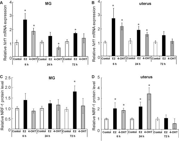
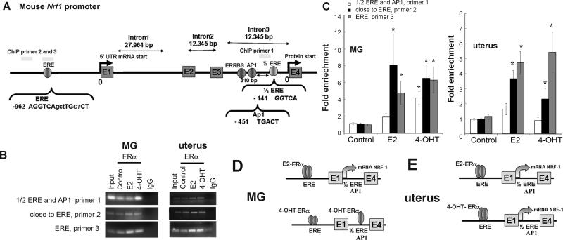
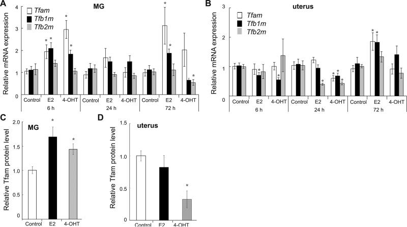
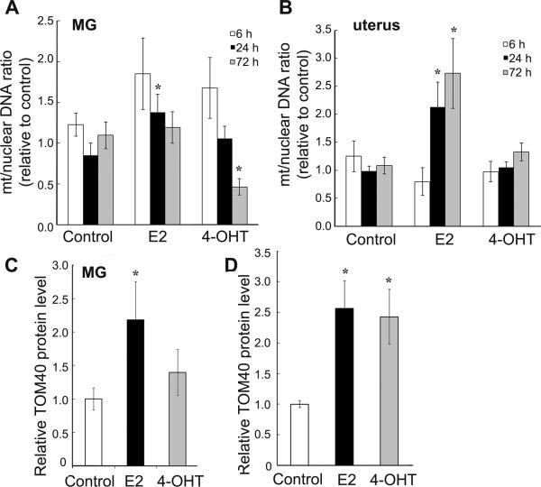
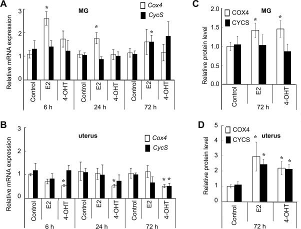
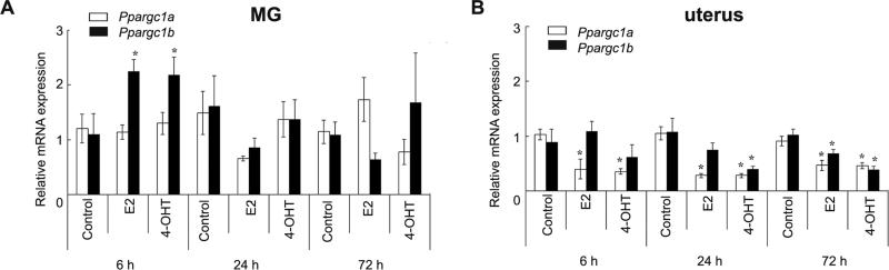
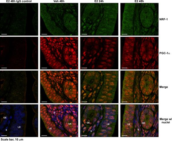
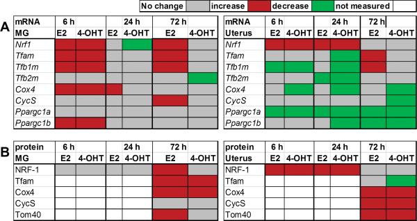
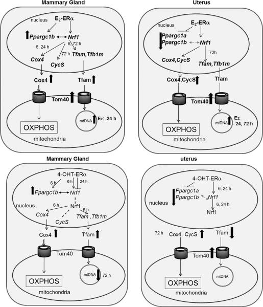
Nuclear respiratory factor-1 (NRF-1) stimulates the transcription of nuclear-encoded genes that regulate mitochondrial (mt) genome transcription and biogenesis. We reported that estradiol (E2) and 4-hydroxytamoxifen (4-OHT) stimulate NRF-1 transcription in an estrogen receptor α (ERα)- and ERβ-dependent manner in human breast cancer cells. The aim of this study was to determine whether E2 and 4-OHT increase NRF-1 in vivo. Here, we report that E2 and 4-OHT increase NRF-1 expression in mammary gland (MG) and uterus of ovariectomized C57BL/6 mice in a time-dependent manner. E2 increased NRF-1 protein in the uterus and MG; however, in MG, 4-OHT increased Nrf1 mRNA but not protein. Chromatin immunoprecipitation assays revealed increased in vivo recruitment of ERα to the Nrf1 promoter and intron 3 in MG and uterus 6 h after E2 and 4-OHT treatment, commensurate with increased NRF-1 expression. E2- and 4-OHT-induced increases in NRF-1 and its target genes Tfam, Tfb1m, and Tfb2m were coordinated in MG but not in uterus due to uterine-selective inhibition of the expression of the NRF-1 coactivators Ppargc1a and Ppargc1b by E2 and 4-OHT. E2 transiently increased NRF-1 and PGC-1α nuclear staining while reducing PGC-1α in uterus. E2, not 4-OHT, activates mt biogenesis in MG and uterus in a time-dependent manner. E2 increased mt outer membrane Tomm40 protein levels in MG and uterus whereas 4-OHT increased Tomm40 only in uterus. These data support the hypothesis of tissue-selective regulation of NRF-1 and its downstream targets by E2 and 4-OHT in vivo.
Keywords: estrogen receptor; mitochondria; mouse; nuclear respiratory factor-1.
Figures
Similar articles
-
Tamoxifen increases nuclear respiratory factor 1 transcription by activating estrogen receptor beta and AP-1 recruitment to adjacent promoter binding sites.FASEB J. 2011 Apr;25(4):1402-16. doi: 10.1096/fj.10-169029. Epub 2011 Jan 13. FASEB J. 2011. PMID: 21233487 Free PMC article.
-
Control of mitochondrial transcription specificity factors (TFB1M and TFB2M) by nuclear respiratory factors (NRF-1 and NRF-2) and PGC-1 family coactivators.Mol Cell Biol. 2005 Feb;25(4):1354-66. doi: 10.1128/MCB.25.4.1354-1366.2005. Mol Cell Biol. 2005. PMID: 15684387 Free PMC article.
-
Estradiol stimulates transcription of nuclear respiratory factor-1 and increases mitochondrial biogenesis.Mol Endocrinol. 2008 Mar;22(3):609-22. doi: 10.1210/me.2007-0029. Epub 2007 Nov 29. Mol Endocrinol. 2008. PMID: 18048642 Free PMC article.
-
Nuclear control of respiratory gene expression in mammalian cells.J Cell Biochem. 2006 Mar 1;97(4):673-83. doi: 10.1002/jcb.20743. J Cell Biochem. 2006. PMID: 16329141 Review.
-
Estrogenic control of mitochondrial function.Redox Biol. 2020 Apr;31:101435. doi: 10.1016/j.redox.2020.101435. Epub 2020 Jan 23. Redox Biol. 2020. PMID: 32001259 Free PMC article. Review.
Cited by
-
PGC-1α induced browning promotes involution and inhibits lactation in mammary glands.Cell Mol Life Sci. 2019 Dec;76(24):5011-5025. doi: 10.1007/s00018-019-03160-y. Epub 2019 Jun 1. Cell Mol Life Sci. 2019. PMID: 31154462 Free PMC article.
-
Membrane estrogen receptor 1 is required for normal reproduction in male and female mice.J Endocrinol Reprod. 2017 Jun;21(1):1-14. J Endocrinol Reprod. 2017. PMID: 34321782 Free PMC article.
-
Age, APOE and sex: Triad of risk of Alzheimer's disease.J Steroid Biochem Mol Biol. 2016 Jun;160:134-47. doi: 10.1016/j.jsbmb.2016.03.012. Epub 2016 Mar 8. J Steroid Biochem Mol Biol. 2016. PMID: 26969397 Free PMC article. Review.
-
Systematically Analyzing the Pathogenic Variations for Acute Intermittent Porphyria.Front Pharmacol. 2019 Sep 13;10:1018. doi: 10.3389/fphar.2019.01018. eCollection 2019. Front Pharmacol. 2019. PMID: 31572191 Free PMC article.
-
Metabolic and Epigenetic Regulation by Estrogen in Adipocytes.Front Endocrinol (Lausanne). 2022 Feb 22;13:828780. doi: 10.3389/fendo.2022.828780. eCollection 2022. Front Endocrinol (Lausanne). 2022. PMID: 35273571 Free PMC article. Review.
References
-
- Aboghe DH, Yoshioka M, Phaneuf D, St-Amand J. Regulation of gene expression by estrogen in mammary gland of wild type and estrogen receptor alpha knockout mice. The Journal of Steroid Biochemistry and Molecular Biology. 2009;113:116–126. - PubMed
-
- Balsitis SJ, Sage J, Duensing S, Münger K, Jacks T, Lambert PF. Recapitulation of the Effects of the Human Papillomavirus Type 16 E7 Oncogene on Mouse Epithelium by Somatic Rb Deletion and Detection of pRb-Independent Effects of E7 In Vivo. Molecular and Cellular Biology. 2003;23:9094–9103. - PMC - PubMed
-
- Barros Rodrigo PA, Gustafsson J-Å. Estrogen Receptors and the Metabolic Network. Cell Metabolism. 2011;14:289–299. - PubMed
-
- Bauerly KA, Storms DH, Harris CB, Hajizadeh S, Sun MY, Cheung CP, Satre MA, Fascetti AJ, Tchaparian E, Rucker RB. Pyrroloquinoline quinone nutritional status alters lysine metabolism and modulates mitochondrial DNA content in the mouse and rat. Biochimica et Biophysica Acta (BBA) - General Subjects. 2006;1760:1741–1748. - PubMed
-
- Billon-Galés A, Fontaine C, Filipe C, Douin-Echinard V, Fouque M-J, Flouriot G, Gourdy P, Lenfant F, Laurell H, Krust A, et al. The transactivating function 1 of estrogen receptor alpha is dispensable for the vasculoprotective actions of 17beta-estradiol. Proceedings of the National Academy of Sciences. 2009;106:2053–2058. - PMC - PubMed
Publication types
MeSH terms
Substances
Grants and funding
LinkOut - more resources
Full Text Sources
Other Literature Sources
