. 2013 Sep;57(9):4449-62.
doi: 10.1128/AAC.02284-12. Epub 2013 Jul 8.

KpnEF, a new member of the Klebsiella pneumoniae cell envelope stress response regulon, is an SMR-type efflux pump involved in broad-spectrum antimicrobial resistance

Affiliations

KpnEF, a new member of the Klebsiella pneumoniae cell envelope stress response regulon, is an SMR-type efflux pump involved in broad-spectrum antimicrobial resistance

Vijaya Bharathi Srinivasan et al. Antimicrob Agents Chemother. 2013 Sep.

Abstract

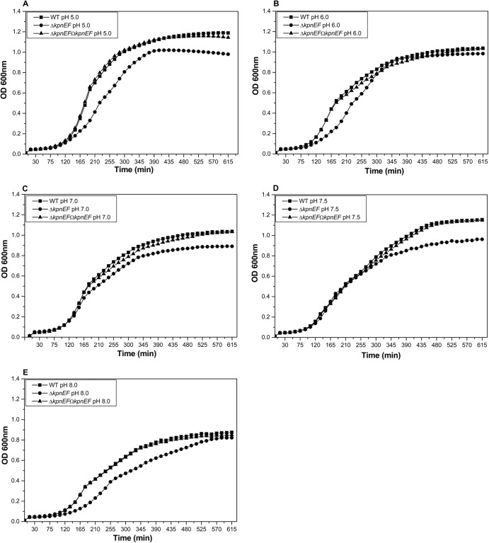
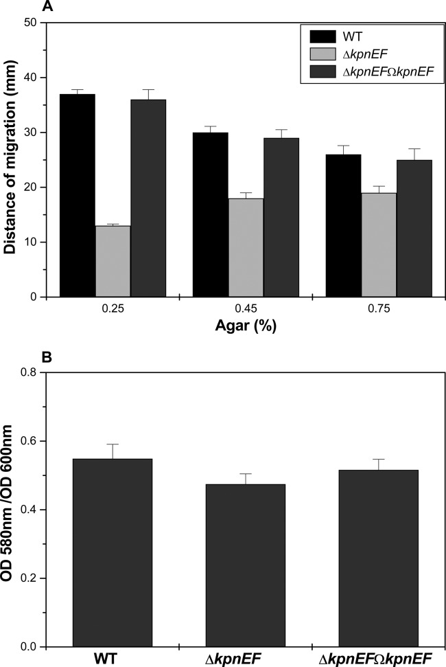
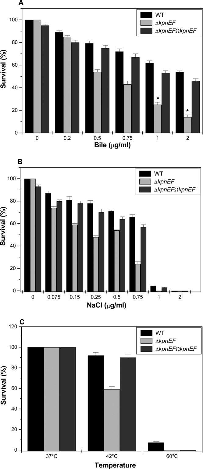
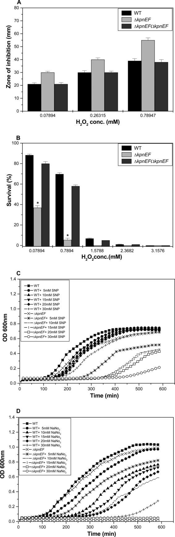
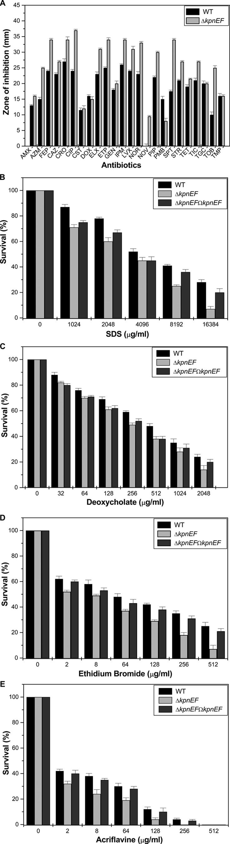
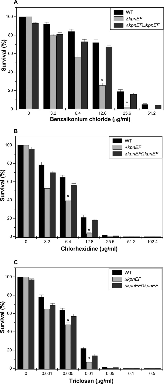
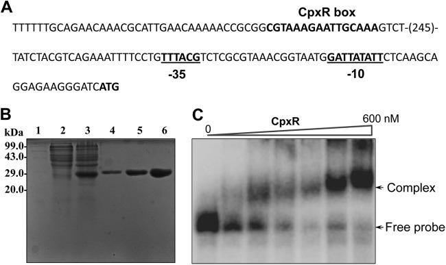
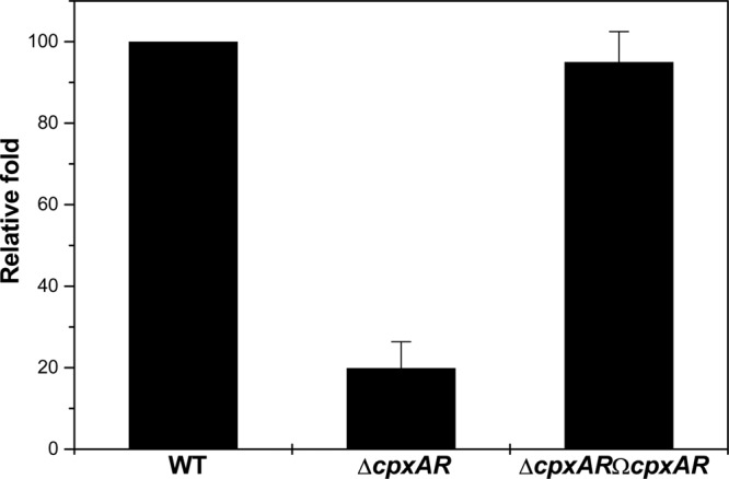
Klebsiella pneumoniae has been frequently associated with nosocomial infections. Efflux systems are ubiquitous transporters that also function in drug resistance. Genome analysis of K. pneumoniae strain NTUH-K2044 revealed the presence of ∼15 putative drug efflux systems. We discuss here for the first time the characterization of a putative SMR-type efflux pump, an ebrAB homolog (denoted here as kpnEF) with respect to Klebsiella physiology and the multidrug-resistant phenotype. Analysis of hypermucoviscosity revealed direct involvement of kpnEF in capsule synthesis. The ΔkpnEF mutant displayed higher sensitivity to hyperosmotic (∼2.8-fold) and high bile (∼4.0-fold) concentrations. Mutation in kpnEF resulted in increased susceptibility to cefepime, ceftriaxone, colistin, erythromycin, rifampin, tetracycline, and streptomycin; mutated strains changed from being resistant to being susceptible, and the resistance was restored upon complementation. The ΔkpnEF mutant displayed enhanced sensitivity toward structurally related compounds such as sodium dodecyl sulfate, deoxycholate, and dyes, including clinically relevant disinfectants such as benzalkonium chloride, chlorhexidine, and triclosan. The prevalence of kpnEF in clinical strains broadens the diversity of antibiotic resistance in K. pneumoniae. Experimental evidence of CpxR binding to the efflux pump promoter and quantification of its expression in a cpxAR mutant background demonstrated kpnEF to be a member of the Cpx regulon. This study helps to elucidate the unprecedented biological functions of the SMR-type efflux pump in Klebsiella spp.


Figures

Fig 1
Fig 1
Effect of kpnEF mutations on general growth characteristics. The effect on bacterial growth was monitored in WT, ΔkpnEF, and ΔkpnEFΩkpnEF strains in LB medium at pH 3.0, 5.0, 6.0, 7.0, 7.5, 8.0, and 12.0. The patterns for the representative pH values 5.0 (A), 6.0 (B), 7.0 (C), 7.5 (D), and 8.0 (E) are shown. After 10 h of growth, the kpnEF mutant exhibited 1.2-fold, 1.06-fold, 1.16-fold, 1.2-fold, and 1.06-fold stunted growth in LB medium at pH 5.0, 6.0, 7.0, 7.5, and 8.0, respectively, which was restored upon complementation. The data presented reflect triplicate determinations.
Fig 2
Fig 2
Alterations in motility behavior and biofilm formation. (A) The average diameters of swimming halos from three different experiments are plotted, along with the standard deviations. The P values for the differences between WT, ΔkpnEF, and ΔkpnEFΩkpnEF strains were all <0.01. The results are given in millimeters, considering K. pneumoniae NTUH-K2044 as the WT. (B) The K. pneumoniae ΔkpnEF mutant is defective in biofilm formation on glass tubes. After 24 h of incubation, biofilm formation was measured by staining with 0.5% crystal violet, and the OD580 was determined for the WT strain and for the ΔkpnEF mutant. The data are means of measurements performed three times. Significant differences with respect to NTUH-K2044 are indicated by an asterisk (*, P < 0.01).
Fig 3
Fig 3
Impact on gastrointestinal stress response. (A) Sensitivities of WT, ΔkpnEF, and ΔkpnEFΩkpnEF strains to different concentrations (0.2, 0.5, 0.75, 1.0, and 2.0%) of bile. The survival ability of the WT strain in 0.5% bile was 1.4-fold (±0.025) greater, in 0.75% bile it was 1.7-fold (±0.035) greater, in 1% bile it was 2.5-fold (±0.024) greater, and in 2% bile it was 4.0-fold (±0.05) greater compared to the ΔkpnEF strain. The complemented ΔkpnEFΩkpnEF strain displayed the same phenotype as the WT. (B) Sensitivities of WT, ΔkpnEF, and ΔkpnEFΩkpnEF strains to different concentrations (0.075, 0.15, 0.25, 0.5, 0.75, 1.0, and 2.0 M) of NaCl. The survival ability of the WT strain in 0.25 M NaCl was ∼1.629-fold (±0.025) greater, in 0.5 M NaCl it was ∼1.32- fold(±0.012) greater, and in 0.75 M NaCl it was ∼2.75-fold (±0.23) greater compared to the ΔkpnEF strain. (C) Survival of the WT, ΔkpnEF, and ΔkpnEFΩkpnEF strains at 37, 42, and 60°C after 1 h of incubation. The percentage of resistance to different stress agents was calculated compared to the numbers of viable cells in LB medium alone. Significant differences with respect to the WT strain are indicated by an asterisk.
Fig 4
Fig 4
Role in oxidative and nitrosative stress tolerance. (A) Oxidative stress response of WT, ΔkpnEF, and ΔkpnEFΩkpnEF strains. The survival abilities of WT and ΔkpnEF strains to combat different levels of H2O2 stress (0.07894, 0.26315, and 0.78947 mM) were measured by disc diffusion assay. The kpnEF mutant displayed greater sensitivity to 0.78947 mM H2O2 (inhibition zone = 55 ± 2.0 mm) than the WT (39 ± 1.2 mm). The data presented reflect triplicate determinations. (B) Survival of WT, ΔkpnEF, and ΔkpnEFΩkpnEF strains upon exposure to oxidative stress at 0.07894, 0.7894, 1.5788, 2.3682, and 3.1576 mM H2O2. After 1 h of treatment with 0.07894 mM H2O2, only 36.9% of the ΔkpnEF cells survived compared to 89% of the WT cells. The differences between the mutant and its parental WT strain are statistically significant (P < 0.05) for all H2O2 concentrations; the standard errors of the mean from three independent assays are also shown. Significant differences with respect to the WT strain are indicated by an asterisk. (C) Effect of different concentrations of SNP on the growth profiles of the WT and ΔkpnEF strains. The growth kinetics of the ΔkpnEF strain were ∼1.5-fold (P = 0.0039) lower, 1.6-fold (P = 0.0055) lower, 1.62-fold (P = 0.0076) lower, 1.7-fold (P = 0.0095) lower, or 3.2-fold (P = 0.0196) lower than for the WT cells in the presence of 5, 10, 15, 20, or 30 mM concentrations of the NO donor, SNP. (D) Growth patterns of WT and ΔkpnEF strains in the presence of acidified nitrite. The growth kinetics for the ΔkpnEF strain were ∼1.18-fold (P = 0.000424) lower, 1.3-fold (P = 0.00659) lower, 13.75-fold (P = 0.0002) lower, 15.2-fold (P = 0.00013) lower, or 20-fold (P = 0.000202) lower than for WT cells in the presence of 5, 10, 15, 20, or 30 mM concentrations of the acidified nitrite.
Fig 5
Fig 5
Contributions of the KpnEF efflux pump in K. pneumoniae antimicrobial resistance. (A) A Kirby-Bauer disc diffusion assay was performed with different antibiotics using commercial discs. Antibiotics, abbreviations, and concentrations: amoxicillin, AMX (30 μg/ml); azithromycin, AZM (15 μg/ml); cefepime, FEP (30 μg/ml); ceftazidime, CAZ (30 μg/ml); ceftriaxone, CRO (30 μg/ml); ciprofloxacin, CIP (5 μg/ml); colistin, CST (10 μg/ml); doxycycline, DOX (30 μg/ml); enrofloxacin, ELX (10 μg/ml); ertapenem, ETP (10 μg/ml); gentamicin, GEN (10 μg/ml); imipenem, IPM (10 μg/ml); levofloxacin, LVX (5 μg/ml); norfloxacin, NOR (10 μg/ml); novobiocin, NOV (30 μg/ml); piperacillin, PIP (100 μg/ml); polymyxin B, PMB (300 μg/ml); spectinomycin, SPT (100 μg/ml); streptomycin, STR (10 μg/ml); tetracycline, TET (30 μg/ml); ticarcillin, TIC (75 μg/ml); tigecycline, TGC (15 μg/ml); tobramycin, TOB (10 μg/ml); trimethoprim, TMP (5 μg/ml). The data for representative drugs are shown here. (B) Sensitivities of WT, ΔkpnEF, and ΔkpnEFΩkpnEF strains to different concentrations of SDS. The survival ability of the WT strain in SDS at 1,024 μg/ml was ∼1.22-fold greater, in SDS at 2,048 μg/ml it was ∼1.3-fold greater, in SDS at 4,096 μg/ml it was ∼1.15-fold greater, in SDS at 8,192 μg/ml it was ∼1.64-fold greater, and in SDS at 16,834 μg/ml it was 4-fold greater compared to the ΔkpnEF strain. (C) Susceptibilities of WT, ΔkpnEF, and ΔkpnEFΩkpnEF strains to different concentrations of deoxycholate. The survival ability of the WT strain in deoxycholate at 128 μg/ml was ∼1.13-fold greater, in deoxycholate at 256 μg/ml it was ∼1.2-fold greater, in deoxycholate at 512 μg/ml it was ∼1.26-fold greater, in deoxycholate at 1,024 μg/ml it was ∼1.25-fold greater, and in deoxycholate at 2,048 μg/ml it was 1.71-fold greater compared to the ΔkpnEF strain. (D) Sensitivities of WT, ΔkpnEF, and ΔkpnEFΩkpnEF strains to different concentrations of EtBr. The survival ability of the WT strain in EtBr at 8 μg/ml was ∼1.18-fold greater, in EtBr at 64 μg/ml it was ∼1.29-fold greater, in EtBr at 128 μg/ml it was ∼1.44-fold greater, in EtBr at 256 μg/ml it was ∼1.94-fold greater, and in EtBr at 512 μg/ml it was 3.57-fold greater compared to the ΔkpnEF strain. (E) Susceptibilities of WT, ΔkpnEF, and ΔkpnEFΩkpnEF strains to different concentrations of acriflavine. The survival ability of the WT strain in EtBr at 8 μg/ml was ∼1.58-fold greater, in EtBr at 64 μg/ml it was ∼1.57-fold greater, and in EtBr at 128 μg/ml it was ∼3-fold greater compared to the ΔkpnEF strain. The percent survival was calculated by comparison of the viable cells in the WT. The data are means of measurements made in triplicate and performed three times. *, Significant difference (P < 0.05, Student t test).
Fig 6
Fig 6
Contributions of kpnEF in disinfectant resistance. (A) Sensitivities of WT, ΔkpnEF, and ΔkpnEFΩkpnEF strains to different concentrations of benzalkonium chloride. The survival ability of the WT strain in benzalkonium chloride at 3.2 μg/ml was ∼1.15-fold greater, in benzalkonium chloride at 6.4 μg/ml it was ∼1.49-fold greater, in benzalkonium chloride at 12.8 μg/ml it was ∼2.81-fold greater, and in benzalkonium chloride at 25.6 μg/ml it was ∼8.05-fold greater compared to the ΔkpnEF mutant. (B) Susceptibilities of WT, ΔkpnEF, and ΔkpnEFΩkpnEF strains to different concentrations of chlorhexidine. The survival ability of the WT strain in chlorhexidine at 3.2 μg/ml was ∼1.48-fold greater, in chlorhexidine at 6.4 μg/ml it was ∼1.64-fold, and in chlorhexidine at 12.8μg/ml it was ∼6.39-fold greater compared to the ΔkpnEF strain. (C) Sensitivities of WT, ΔkpnEF, and ΔkpnEFΩkpnEF strains to different concentrations of triclosan. The survival ability of the WT strain in triclosan at 0.001 μg/ml was ∼1.20-fold greater, in triclosan at 0.005 μg/ml it was ∼1.32-fold greater, and in triclosan at 0.01 μg/ml it was ∼3.16-fold greater compared to the ΔkpnEF strain. The percent survival was calculated by comparison of viable cells in the WT. The data are means of measurements made in triplicate and performed three times. *, Significant difference (P < 0.05, Student t test).
Fig 7
Fig 7
Fluorimetric efflux assay. Accumulation studies were performed using EtBr (A) and ciprofloxacin (B) with K. pneumoniae and the ΔkpnEF mutant. The efflux of EtBr in mutant and WT cells was monitored continuously by measuring fluorescence emission at 600 nm upon excitation at 530 nm. After 5 min in a fluorimeter, cells loaded with EtBr were energized by the addition of glucose, and the efflux of EtBr was monitored. After 10 min, 100 μM CCCP was added as indicated to abolish active transport, and fluorescence emission was monitored further. The fluorescence was measured using a spectrofluorimeter (Hitachi). For ciprofloxacin, excitation was set at 275 nm and emission was set at 440 nm. Each data point represents the mean plus the standard deviation of three independent experiments.
Fig 8
Fig 8
CpxR regulates KpnEF in K. pneumoniae. (A) Promoter region analysis of kpnEF. The numbers in parentheses represent the numbers of nucleotides in the sequence. The −35 and −10 regions in the promoter are underlined. The putative CpxR binding site is indicated in boldface. (B) SDS-PAGE profile of pET-cpxR. Lane 1, medium-size marker; lane 2, pET-cpxR/BL21DE3 uninduced; lane 3, pET-cpxR/BL21DE3, induced; lanes 4 to 6, purified CpxR fractions E1, E2, and E3, respectively. Protein samples after induction were subjected to SDS-PAGE (15% gel), followed by Coomassie brilliant blue staining. (C) Gel shift assays demonstrating the binding of CpxR to the promoter of kpnEF in K. pneumoniae in a concentration-dependent manner. Lane 1 shows the free probe, and lanes 2 to 7 show increasing concentrations of CpxR protein (60 to 600 nM), respectively. Protein-bound DNA complexes and free probe are indicated by arrows. The gels are representative of at least four independent experiments.
Fig 9
Fig 9
Relative transcriptional level of kpnEF in the ΔcpxAR mutant. The relative transcriptional levels of kpnEF in ΔcpxAR and ΔcpxARΩcpxAR strains determined using real-time RT-PCR are shown compared to the WT strain. The WT expression level is represented as 1-fold. Each bar represents the average value of three independent experiments. Error bars indicate the standard deviations.

Similar articles

Cited by

References

    1. Alekshun MN, Levy SB. 2007. Molecular mechanisms of antibacterial multidrug resistance. Cell 128:1037–1050 - PubMed
    1. Piddock LJ. 2006. Multidrug-resistance efflux pumps: not just for resistance. Nat. Rev. Microbiol. 4:629–636 - PubMed
    1. Piddock LJ. 2006. Clinically relevant chromosomally encoded multidrug resistance efflux pumps in bacteria. Clin. Microbiol. Rev. 19:382–402 - PMC - PubMed
    1. Paulsen IT. 2003. Multidrug efflux pumps and resistance: regulation and evolution. Curr. Opin. Microbiol. 6:446–451 - PubMed
    1. Putman M, Van Veen HW, Konings WN. 2000. Molecular properties of bacterial multidrug transporters. Microbiol. Mol. Biol. Rev. 64:672–693 - PMC - PubMed

Publication types

MeSH terms

LinkOut - more resources