. 2013 Oct;62(10):3404-17.
doi: 10.2337/db12-1650. Epub 2013 Jul 8.

Sirt3 regulates metabolic flexibility of skeletal muscle through reversible enzymatic deacetylation

Affiliations

Sirt3 regulates metabolic flexibility of skeletal muscle through reversible enzymatic deacetylation

Enxuan Jing et al. Diabetes. 2013 Oct.

Abstract

Sirt3 is an NAD(+)-dependent deacetylase that regulates mitochondrial function by targeting metabolic enzymes and proteins. In fasting mice, Sirt3 expression is decreased in skeletal muscle resulting in increased mitochondrial protein acetylation. Deletion of Sirt3 led to impaired glucose oxidation in muscle, which was associated with decreased pyruvate dehydrogenase (PDH) activity, accumulation of pyruvate and lactate metabolites, and an inability of insulin to suppress fatty acid oxidation. Antibody-based acetyl-peptide enrichment and mass spectrometry of mitochondrial lysates from WT and Sirt3 KO skeletal muscle revealed that a major target of Sirt3 deacetylation is the E1α subunit of PDH (PDH E1α). Sirt3 knockout in vivo and Sirt3 knockdown in myoblasts in vitro induced hyperacetylation of the PDH E1α subunit, altering its phosphorylation leading to suppressed PDH enzymatic activity. The inhibition of PDH activity resulting from reduced levels of Sirt3 induces a switch of skeletal muscle substrate utilization from carbohydrate oxidation toward lactate production and fatty acid utilization even in the fed state, contributing to a loss of metabolic flexibility. Thus, Sirt3 plays an important role in skeletal muscle mitochondrial substrate choice and metabolic flexibility in part by regulating PDH function through deacetylation.


Figures
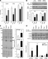
FIG. 1.
FIG. 1.
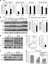
Skeletal muscle Sirt3 expression and mitochondrial protein acetylation are regulated by fasting. Wild-type 8-week-old male C57Bl/6 mice were fed ad libitum, fasted for 24 h, or fasted and then refed for 16 h. After each treatment, RNA and protein were extracted and analyzed by real-time quantitative PCR (A) or Western blotting (B) for assessment of Sirt3 expression in quadriceps (Quad), EDL, soleus (Sol), and gastrocnemius (Gast) muscle groups (n = 3–5, #P < 0.05 vs. fed, ANOVA). C: Mixed hindlimb muscles (gastrocnemius and soleus) were collected from mice in the fed or fasted state. Muscle mitochondria were isolated in the presence of protease and deacetylase inhibitors as described in research design and methods. Mitochondrial protein lysates from each animal were subjected to SDS-PAGE and Western blotting using an antibody against AcK. The intensity of specified bands was quantified with ImageJ software (n = 3, *P < 0.05, Student t test). D: PDH E1α acetylation in muscle of fed or fasted mice was measured by immunoprecipitation (IP) of mitochondrial lysates using anti-AcK antibody. Immunoprecipitates were subjected to Western blotting analysis (IB) using anti–PDH E1α antibody. The same muscle mitochondrial lysates were directly subjected to SDS-PAGE electrophoresis and Western blotting using antibodies against phosphorylated serine sites p-232, p-293, and p-300 of the PDH E1α subunit and total protein of PDH E1α. Autoradiography of Western blots was quantified with ImageJ software (n = 3, *P < 0.05, Student t test). GAPDH, glyceraldehyde-3-phosphate dehydrogenase.
FIG. 2.
FIG. 2.
Sirt3 deletion induces synergistic switch from glucose oxidation toward fatty acid oxidation and suppresses PDH activity by inducing PDH E1α hyperacetylation and hyperphosphorylation in fed Sirt3 KO skeletal muscle. A: Basal and insulin-stimulated (1 mU/mL) glucose oxidation, glycogen synthesis, glycolysis, and palmitate oxidation were measured in EDL muscles isolated from randomly fed 8- to 16-week-old WT and Sirt3 KO mice (n = 9; §P < 0.05 vs. basal, #P < 0.05 vs. WT, ANOVA). B: Basal and insulin-stimulated (10 mU/mL) phosphorylation of insulin/IGF-1 receptor, Akt, and ERK in tissue lysates of EDL muscles from fed and fasted 8- to 16-week-old WT and Sirt3 KO mice. C: Insulin-stimulated (1 mU/mL) glucose and palmitate oxidation were measured as described in research design and methods in diaphragms isolated from randomly fed 8- to 16-week-old WT and Sirt3 KO mice (n = 9; *P < 0.05, Student t test). D: Mitochondrial lysates were subjected to Western blot analysis using antibodies against p-232, p-293, and p-300 serine phosphorylation sites on PDH E1α, total PDH E1α, and Sirt3 (densitometry in Supplementary Fig. 2F). E: PDH activity was measured in 20 mg mitochondrial lysate from hindlimb of fed WT and Sirt3 KO mice using a PDH activity microplate kit as described in research design and methods. The PDH activities were calculated by linear regression of the steady-state kinetics and quantified (n = 4–5, *P < 0.05, Student t test). F: Western blot analysis was performed on tissue lysates from gastrocnemius muscle from fed and fasted WT and Sirt3 KO for PDHK4, Sirt3, and glyceraldehyde-3-phosphate dehydrogenase. G: Native PDH activity (PDHa) was measured in gastrocnemius muscle homogenate from WT fed, Sirt3 KO fed, and fasted C57Bl/6 mice by collection of 14CO2 release from 14C-pyruvate. Prior to PDH assay, aliquots of homogenates were incubated in the presence of 50 mmol/L NaF with or without deacetylase inhibitors (1 mmol/L nicotinamide and 1 mol/L trichostatin A) as described in research design and methods (n = 6–7; #P < 0.05 vs. WT plus deacetylase inhibitors, ANOVA). OD, optical density.
FIG. 3.
FIG. 3.
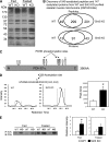
Discovery of PDH E1α subunit as a target of Sirt3 in skeletal muscle. A: Muscle mitochondrial protein lysates from WT and Sirt3 KO mice in either the fed or fasted state were subjected to Western blotting analysis using anti-AcK antibody. B: Proteomic analysis using anti-AcK antibody–based acetylated peptide enrichment and mass spectrometry discovered a total of 549 acetylated peptides present in WT and Sirt3 KO skeletal muscle mitochondria. These peptides represented a total of 147 proteins. Venn diagrams show overlapping and distinctive patterns of distribution of acetylated peptides and proteins between WT and Sirt3 KO skeletal muscle. C: Schematic diagram of lysine acetylation sites and previously reported serine phosphorylation sites on PDH E1α. D: A representative MS1 chromatogram of the triply charged precursor peak at m/z 772.73033+ (324-MVNSNLASVEELacKEIDVEVR-343) of PDH E1α was highly increased in Sirt3 KO skeletal muscle mitochondria compared with WT (n = 4; P < 0.05, Student t test). E: Skeletal muscle mitochondrial lysates from WT and Sirt3 KO mice in both fed and fasted states were immunoprecipitated (IP) with AcK antibody–bound beads. Immunoprecipitates of the mitochondrial lysate were subjected to Western blotting (IB) using an anti–PDH E1α antibody. Densitometry of either fed or fasted animals was calculated (n = 4; *P < 0.05, Student t test).
FIG. 4.
FIG. 4.
Sirt3 deletion induces a substrate switch and derangement of metabolites in skeletal muscle. Quadriceps muscles from fed male WT and Sirt3 KO mice were collected and subjected to metabolomic analysis as described in research design and methods. Relative levels of organic acids and Kreb cycle intermediates are shown in A, levels of acylcarnitines in B, and amino acid levels in C (n = 4–5; *P < 0.05, Student t test). Asx, asparagine and aspartic acid; Cit, citrulline; Glx, glutamine and glutamic acid; Orn, ornithine.
FIG. 5.
FIG. 5.
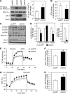
Sirt3 knockdown in C2C12 myoblasts impairs PDH activity despite decreased phosphorylation of PDH E1α and leads to a substrate switch toward fatty acid utilization. A: Total mitochondrial protein lysates from shGFP control and shSirt3 myoblasts were immunoprecipitated (IP) with AcK (AcLys) antibody and subjected to Western blot analysis (IB) using an anti–PDH E1α antibody. The same mitochondrial lysates were directly subjected to Western blot analysis using antibodies against PDH E1α, Sirt3, and voltage-dependent anion channel (VDAC) as a mitochondrial loading control. Densitometry of PDH E1α from AcK immunoprecipitates was normalized to total PDH E1α (n = 4 separate experiments; *P < 0.05, Student t test). B: Total PDH activity was assessed in confluent control and shSirt3 myoblasts using PDH activity microplate assay kit and normalized to total protein from detergent extraction (n = 5 separate experiments; †P < 0.05, paired t test). C: Phosphorylation of PDH E1α and total Sirt3 levels were determined by Western blot analysis of whole cell lysates from confluent shGFP and shSirt3 C2C12 myoblasts. D: Densitometry of Western blots from C (n = 3 separate experiments, *P < 0.05, Student t test). E: ECAR was measured in shSirt3 and control myoblasts using a Seahorse flux analyzer after incubation in glucose-free Seahorse running media for 1 h at 37°C. A representative tracing of basal and glucose-stimulated ECARs recorded before and after addition of 25 mmol/L glucose (final concentration) is shown. At the end of the glucose metabolism period, 2-deoxyglucose was injected to give a final concentration of 25 mmol/L. F: AUC calculation of glucose-stimulated ECAR from E. G: A representative tracing of palmitate OCR measured in control and Sirt3 knockdown cells after incubation with substrate-free buffer for 1 h at 37°C. Basal and palmitate-BSA–stimulated OCRs were recorded and plotted as a percentage over basal OCR. Finally, 50 μmol/L etomoxir was injected. H: AUC of palmitate-stimulated OCR from G (n = 3; *P < 0.05, Student t test). mOD, milli optical density.
FIG. 6.
FIG. 6.
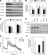
Expression of a K336Q and K336R mutant of PDH E1α in C2C12 myoblasts decreases phosphorylation but does not affect PDH activity. A: Phosphorylation of PDH E1α levels determined by Western blot analysis of whole cell lysates from confluent C2C12 myoblasts expressing WT PDH E1α, K336Q, or K336R mutant of PDH E1α. B: Densitometry of Western blots from A (n = 3 separate experiments, #P < 0.05, ANOVA). C: Total PDH activity was assessed in confluent WT, K336Q, or K336R myoblasts using detergent extracts either with PIs 2 and 3 (Sigma) or 20 mmol/L DCA added to the PDH activity assay as described in research design and methods (n = 2–4 separate experiments). D: Western blot analysis of detergent extracts from C either with PI (Phos Inhib) or with DCA after 3 h incubation at room temperature in buffer 1 of the PDH activity assay kit. E: A representative example of Seahorse analysis of glucose-induced ECAR with PDH E1α WT, K336Q, and K336R mutants. F: AUC quantification after addition of glucose showed no statistical difference between mutants (n = 4 separate experiments). 2-DG, 2-deoxyglucose.
FIG. 7.
FIG. 7.
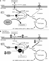
Model for the role of Sirt3 in control of skeletal muscle substrate metabolism. Sirt3 regulates PDH E1α subunit deacetylation and activates PDH activity. A: In the fed state, Sirt3 skeletal muscle expression is abundant and leads to deacetylation of PDH E1α. This is associated with dephosphorylation of PDH allowing for maximal enzyme activation, enhanced glucose utilization, and increased flux of pyruvate to acetyl-CoA used by the tricarboxylic acid (TCA) cycle and electron transport chain (ETC) to generate ATP. B: In contrast, decreased Sirt3 expression in muscle by fasting or genetic deletion leads to PDH E1α hyperacetylation and decreased PDH complex activity, which is correlated with increased PDH E1α phosphorylation in vivo. The activity of PDH controls the substrate influx to the TCA cycle from glycolysis. In the case of Sirt3 deletion, inactivation of the PDH caused by hyperacetylation leads to metabolic inflexibility as evidenced by an inability to fully oxidize glucose, a shunt of excess pyruvate toward lactate production, and increased lipid oxidation even in the fed state. CPT, carnitine palmitoyl transferase; FFA, free fatty acid; Mito, mitochondrial; OxPhos, oxidative phosphorylation.

Similar articles

Cited by

References

    1. Kim JK, Zisman A, Fillmore JJ, et al. Glucose toxicity and the development of diabetes in mice with muscle-specific inactivation of GLUT4. J Clin Invest 2001;108:153–160 - PMC - PubMed
    1. Fueger PT, Hess HS, Bracy DP, et al. Regulation of insulin-stimulated muscle glucose uptake in the conscious mouse: role of glucose transport is dependent on glucose phosphorylation capacity. Endocrinology 2004;145:4912–4916 - PubMed
    1. Storlien L, Oakes ND, Kelley DE. Metabolic flexibility. Proc Nutr Soc 2004;63:363–368 - PubMed
    1. van Loon LJ. Use of intramuscular triacylglycerol as a substrate source during exercise in humans. J Appl Physiol 2004;97:1170–1187 - PubMed
    1. Befroy DE, Petersen KF, Dufour S, et al. Impaired mitochondrial substrate oxidation in muscle of insulin-resistant offspring of type 2 diabetic patients. Diabetes 2007;56:1376–1381 - PMC - PubMed

Publication types