. 2013 Jun 20;153(7):1435-47.
doi: 10.1016/j.cell.2013.05.042.

XBP-1 is a cell-nonautonomous regulator of stress resistance and longevity

Affiliations

XBP-1 is a cell-nonautonomous regulator of stress resistance and longevity

Rebecca C Taylor et al. Cell. .

Abstract

The ability to ensure proteostasis is critical for maintaining proper cell function and organismal viability but is mitigated by aging. We analyzed the role of the endoplasmic reticulum unfolded protein response (UPR(ER)) in aging of C. elegans and found that age-onset loss of ER proteostasis could be reversed by expression of a constitutively active form of XBP-1, XBP-1s. Neuronally derived XBP-1s was sufficient to rescue stress resistance, increase longevity, and activate the UPR(ER) in distal, non-neuronal cell types through a cell-nonautonomous mechanism. Loss of UPR(ER) signaling components in distal cells blocked cell-nonautonomous signaling from the nervous system, thereby blocking increased longevity of the entire animal. Reduction of small clear vesicle (SCV) release blocked nonautonomous signaling downstream of xbp-1s, suggesting that the release of neurotransmitters is required for this intertissue signaling event. Our findings point toward a secreted ER stress signal (SERSS) that promotes ER stress resistance and longevity.


Figures

Figure 1
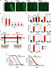
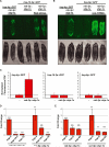
Figure 1. The Ability to Activate the UPRER Declines with Age
(A) hsp-4p::GFP UPRER reporter worms were treated at days 1, 4, 7, and 10 of adulthood with 25 ng/μl tunicamycin in M9 buffer, or buffer only, for 4 hr, and GFP fluorescence assessed. (B) hsp-4p::GFP UPRER reporter worms were treated as above at days 1, 2, 3, 4, 7, and 10 of adulthood, and GFP expression measured by fluorimetry. Fold inductions relative to untreated animals are shown, with error bars indicating mean + standard error of the mean (SEM) (n = 3). A Student's t test was used to assess statistical significance of induction at each age: *p < 0.05, n/s = not significant. (C) N2 animals were treated as above at day 1 and day 5 of adulthood. Transcript levels of UPR regulators and targets in tunicamycin-treated (red) and untreated (black) animals were measured by quantitative RT-PCR, with error bars indicating mean ± standard deviation (SD). A Student's t test was used to assess significance: ***p < 0.005, n/s = not significant. (D) Schematic describing the measurement of age-dependent ER stress resistance. Animals are transferred to plates containing tunicamycin at either day 1 or day 7 of adulthood. (E) N2 (black) and xbp-1(zc12) (red) animals were transferred at day 1 and day 7 of adulthood to plates containing 50 μg/ml tunicamycin, and survival monitored (N2 day 1, median lifespan 7 days; xbp-1(zc12) day 1, median lifespan 5 days; p < 0.0001. N2 day 7, median lifespan 4 days; xbp-1(zc12) day 7, median lifespan 4 days; p = 0.9211. See Table S1). See also Figure S1 and Table S2.
Figure 2
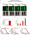
Figure 2. Expression of xbp-1s under the sur-5p Promoter Constitutively Activates the UPRER and Prevents a Decline in ER Stress Resistance
(A) Schematic of the exon/intron boundaries of the 23 bp intron removed from the xbp-1 sequence to activate the transcription factor. (B) Fluorescent micrographs of hsp-4p::GFP UPRER reporter worms expressing an integrated sur-5p::xbp-1s transgene at days 1, 4, 7, and 10 of adulthood. (C) A COPAS Biosort was used to measure length (TOF) and GFP fluorescence of hsp-4p::GFP (−) and hsp-4p::GFP; sur-5p::xbp-1s (sur-5p::xbp-1s) animals at day 1 of adulthood. Mean GFP fluorescence/ TOF and standard deviation of each genotype were calculated and normalized to control hsp-4p::GFP animals. (D) Transcript levels of UPR targets in sur-5p::xbp-1s animals at day 1 of adulthood were measured by quantitative RT-PCR. Results are shown relative to transcript levels in N2 worms, with error bars indicating mean ± SD. A Student's t test was used to assess significance: ***p < 0.005, **p < 0.01, n/s = not significant. (E) N2 (black) and sur-5p::xbp-1s (red) animals were transferred at day 1 and day 7 of adulthood to plates containing 50 mg/ml tunicamycin, and survival monitored (sur-5p::xbp-1s day 1, median lifespan 7 days; N2 day 1, median lifespan 7 days; p = 0.0078. sur-5p::xbp-1s day 7, median lifespan 7 days; N2 day 7, median lifespan 3 days; p < 0.0001. See Table S1). (F) Survival of sur-5p::xbp-1s (red) and N2 (black) worms on E. coli OP50 at 20° C (sur-5p::xbp-1s: median lifespan 17 days; N2: black; median lifespan 19 days; p = 0.0925. See Table S3). See also Figure S2 and Table S2.
Figure 3
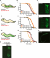
Figure 3. Neuronal and Intestinal Expression of xbp-1s Increases Lifespan, and Neuronal xbp-1s Can Induce Cell-Nonautonomous Activation of the UPRER
(A) Schematic of a tissue-specific xbp-1s expression system driving xbp-1s expression in neurons, intestine, and body-wall muscle cells. (B) Survival of N2 (black) and transgenic strains on E. coli OP50 at 20° C: (i) gly-19p::xbp-1s (line 1 [red], median lifespan 22 days; p = 0.0013; line 2 [orange], median lifespan 24 days; p < 0.0001), (ii) rab-3p::xbp-1s (line 1 [red], median lifespan 22 days; p < 0.0001; line 2 [orange], median lifespan 22 days; p < 0.0001), and (iii) unc-54p::xbp-1s (line 1 [red], median lifespan 16 days; p = 0.0003; line 2 [orange], median lifespan 14 days; p < 0.0001). See Table S3. (C) Fluorescent micrographs of hsp-4p::GFP UPRER reporter worms expressing (i) gly-19p::xbp-1s, (ii) rab-3p::xbp-1s, and (iii) unc-54p::xbp-1s transgenes at day 1 of adulthood (i and ii) and L2/L3 (iii). (D) Fluorescent micrograph of hsp-4p::GFP UPRER reporter worms expressing rgef-1p::xbp-1s. See also Figure S3.
Figure 4
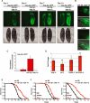
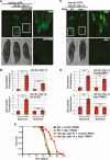
Figure 4. Expression of xbp-1s in Neurons Activates the UPRER in the Intestine and Prevents a Decline in Stress Resistance
(A) Fluorescent micrographs of hsp-4p::GFP UPRER reporter worms expressing an integrated rab-3p::xbp-1s transgene at days 1, 4, 7, and 10 of adulthood. (B) Tail region of hsp-4p::GFP UPRER reporter worms expressing an integrated rab-3p::xbp-1s transgene showing neurons at 633 magnification. (C) Length (TOF) and GFP fluorescence of hsp-4p::GFP (−) and hsp-4p::GFP; rab-3p::xbp-1s (rab-3p::xbp-1s) animals were measured using a COPAS Biosort at day 1 of adulthood. Mean GFP fluorescence/TOF and standard deviation of each genotype were calculated and normalized to control hsp-4p::GFP animals. (D) Transcript levels of UPR targets in rab-3p::xbp-1s animals were measured at day 1 of adulthood by quantitative RT-PCR. Results are shown relative to transcript levels in N2 worms, with error bars indicating mean ± SD. A Student's t test was used to assess significance: ***p < 0.005, *p < 0.05, n/s = not significant. (E) Survival of rab-3p::xbp-1s (red) and N2 (black) worms on E. coli OP50 at 20° C (rab-3p::xbp-1s: median lifespan 25 days; N2: median lifespan 19 days; p < 0.0001. See Table S3). (F) rab-3p::xbp-1s (red) and N2 (black) animals were transferred at day 1 and day 7 of adulthood to plates containing 50 mg/ml tunicamycin, and survival monitored (rab-3p::xbp-1s day 1, median lifespan 9 days; N2 day 1, median lifespan 7 days; p = 0.0078; rab-3p::xbp-1s day 7, median lifespan 7 days; N2 day 7, median lifespan 4 days; p < 0.0001. See Table S1). See also Figure S4 and Table S2.
Figure 5
Figure 5. Neuronal xbp-1s Specifically Activates the UPRER Cell Nonautonomously
(A) Fluorescent micrograph of hsp-4p::GFP UPRER and hsp-16.2p::GFP HSR reporter worms with and without the rab-3p::xbp-1s transgene at day 1 of adulthood. Fluorescence in the pharyngeal area of rab-3p::xbp-1s animals is due to the myo-2p::tdTomato injection marker. As controls, separate populations of hsp-16.2p::GFP and hsp-16.2p::GFP; rab-3p::xbp-1s worms were incubated at 34° C for 6 hr. (B) Fluorescent micrograph of hsp-4p::GFP UPRER and hsp-6p::GFP UPRmt reporter worms, with and without the rab-3p::xbp-1s transgene at day 1 of adulthood. Fluorescence in the pharyngeal area of rab-3p::xbp-1s animals is due to the myo-2p::tdTomato injection marker. As controls, separate populations of hsp-6p::GFP and hsp-6p::GFP; rab-3p::xbp-1s animals were treated with cco-1 RNAi from hatch. (C) A COPAS Biosort was used to measure length (TOF) and GFP fluorescence of hsp-4p::GFP, hsp-6p::GFP, and hsp-16.2p::GFP animals, with and without the rab-3p::xbp-1s transgene, at day 1 of adulthood. Mean GFP fluorescence/TOF and standard deviation of each genotype were calculated, with rab-3p::xbp-1s animals normalized to reporter controls. (D) Transcript levels of HSR targets in rab-3p::xbp-1s and sur-5p::xbp-1s animals were measured by quantitative RT-PCR. Results are shown relative to transcript levels in N2 worms, with error bars indicating mean ± SD. A Student's t test was used to assess significance: ***p < 0.005, **p < 0.01, n/s = not significant. (E) Transcript levels of UPRmt targets in rab-3p::xbp-1s and sur-5p::xbp-1s animals were measured by quantitative RT-PCR. Results are shown relative to transcript levels in N2 worms, with error bars indicating mean ± SD. A Student's t test was used to assess significance: ***p < 0.005, **p < 0.01, n/s = not significant. See also Figure S5.
Figure 6
Figure 6. Cell-Nonautonomous Activation of the UPRER Requires ire-1 and xbp-1
(A) Fluorescent micrograph of hsp-4p::GFP and hsp-4p::GFP; ire-1(v33) worms expressing rab-3p::xbp-1s at day 1 of adulthood. (B) Using a COPAS Biosort, fluorescence in the nerve ring and intestine were approximated by measuring GFP fluorescence in equidistant sections through the proximal 25% (nerve ring) and distal 75% (intestine) of hsp-4p::GFP (−) and hsp-4p::GFP; rab-3p::xbp-1s or hsp-4p::GFP; rab-3p::xbp-1s; ire-1(v33) (+) worms. Total fluorescence in each region was divided by number of sections to give average fluorescence per section, which was then averaged among animals and normalized to control hsp-4p::GFP worms. Error bars indicate mean ± SD. A Student's t test was used to assess significance: ***p < 0.005, n/s = not significant. See Figure S6C. (C) Fluorescent micrograph of hsp-4p::GFP; rab-3p::xbp-1s worms grown on empty vector and xbp-1 RNAi. (D) Fluorescence in the nerve ring and intestine were approximated as above in hsp-4p::GFP animals on control empty vector RNAi (−) and hsp-4p::GFP; rab-3p::xbp-1s animals on control or xbp-1 RNAi (+). Error bars indicate mean ± SD. A Student's t test was used to assess significance: ***p < 0.005, n/s = not significant. See Figure S6D. (E) Survival of rab-3p::xbp-1s and N2 worms grown on empty vector control or xbp-1 RNAi (rab-3p::xbp-1s on xbp-1 [red], median lifespan 18 days; rab-3p::xbp-1s on empty vector [orange], median lifespan 22 days; p < 0.0001. See Table S3). See also Figure S6.
Figure 7
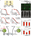
Figure 7. Cell-Nonautonomous Activation of the UPRER Requires unc-13
(A) Schematic of the roles of UNC-13 and UNC-31 in neurosecretory vesicle release. UNC-13 is required for the release of SCVs containing small-molecule neurotransmitters. UNC-31 is involved in the release of DCVs primarily containing neuropeptides. (B) (i) Fluorescent micrograph of hsp-4p::GFP, hsp-4p::GFP; rab-3p::xbp-1s, hsp-4p::GFP; rab-3p::xbp-1s; unc-13(e450), and hsp-4p::GFP; rab-3p::xbp-1s; unc-31(e928) worms at day 1 of adulthood. (ii) Using a COPAS Biosort, fluorescence in the nerve ring and intestine of hsp-4p::GFP; rab-3p::xbp-1s animals in a wild-type (WT), unc-13(e450), or unc-31(e928) background was approximated by measuring GFP fluorescence in equidistant sections through the proximal 25% (nerve ring) and distal 75% (intestine) of worms. Total fluorescence in each region was divided by number of sections to give average fluorescence per section, which was then averaged among animals and normalized to hsp-4p::GFP controls. Error bars indicate mean ± SD. A Student's t test was used to assess significance: ***p < 0.005, n/s = not significant. See Figure S6E. (C) Survival of (left panel) rab-3p::xbp-1s and N2 and (right panel) rab-3::xbp-1s; unc-13(e450) and unc-13(e450) animals was assessed at 15° C, following a single treatment with FUDR at t = 0 (left panel: rab-3p::xbp-1s [red], median lifespan 28 days; N2 [black], median lifespan 24 days; p < 0.0001; right panel: rab-3::xbp-1s; unc-13(e450) [red], median lifespan 34 days; unc-13(e450) [black], median lifespan 43 days; p < 0.0001. See Table S3). (D) (Left panel) rab-3p::xbp-1s and N2 and (right panel) rab-3::xbp-1s; unc-13(e450) and unc-13(e450) animals were transferred at day 1 and day 7 of adulthood to plates containing 50 mg/ml tunicamycin, and survival monitored (left panel: rab-3p::xbp-1s; unc-13(e450) day 1 [red], median lifespan 9 days; unc-13(e450) day 1 [black], median lifespan 8 days; p = 0.1295; right panel: rab-3p::xbp-1s; unc-13(e450) day 7 [red], median lifespan 6 days; unc-13(e450) day 7 [black], median lifespan 5 days; p = 0.0908. See Table S1). (E) Model for the communication of UPRER activation between neurons and distal cells. ER stress in neurons induces splicing of xbp-1 and activation of target gene transcription that leads to UNC-13-mediated release of a secreted ER stress signal (SERSS) from SCVs. This signal induces the UPRER in distal intestinal cells through activation of endogenous IRE-1 and XBP-1, leading to target gene transcription and increased stress resistance and longevity. See also Figure S7.

Comment in

Similar articles

Cited by

References

    1. Acosta-Alvear D, Zhou Y, Blais A, Tsikitis M, Lents NH, Arias C, Lennon CJ, Kluger Y, Dynlacht BD. XBP1 controls diverse cell type- and condition-specific transcriptional regulatory networks. Mol. Cell. 2007;27:53–66. - PubMed
    1. Akerfelt M, Morimoto RI, Sistonen L. Heat shock factors: integrators of cell stress, development and lifespan. Nat. Rev. Mol. Cell Biol. 2010;11:545–555. - PMC - PubMed
    1. Alcedo J, Kenyon C. Regulation of C. elegans longevity by specific gustatory and olfactory neurons. Neuron. 2004;41:45–55. - PubMed
    1. Apfeld J, Kenyon C. Regulation of lifespan by sensory perception in Caenorhabditis elegans. Nature. 1999;402:804–809. - PubMed
    1. Aragón T, van Anken E, Pincus D, Serafimova IM, Korennykh AV, Rubio CA, Walter P. Messenger RNA targeting to endoplasmic reticulum stress signalling sites. Nature. 2009;457:736–740. - PMC - PubMed

Publication types

Substances