A bacterial export system for generating extracellular amyloid aggregates
- PMID: 23787895
- PMCID: PMC3963027
- DOI: 10.1038/nprot.2013.081
A bacterial export system for generating extracellular amyloid aggregates
Abstract
Here we describe a protocol for the generation of amyloid aggregates of target amyloidogenic proteins using a bacteria-based system called curli-dependent amyloid generator (C-DAG). C-DAG relies on the natural ability of Escherichia coli cells to elaborate surface-associated amyloid fibers known as curli. An N-terminal signal sequence directs the secretion of the major curli subunit CsgA. The transfer of this signal sequence to the N terminus of heterologous amyloidogenic proteins similarly directs their export to the cell surface, where they assemble as amyloid fibrils. Notably, protein secretion through the curli export pathway facilitates acquisition of the amyloid fold specifically for proteins that have an inherent amyloid-forming propensity. Thus, C-DAG provides a cell-based alternative to widely used in vitro assays for studying amyloid aggregation, and it circumvents the need for protein purification. In particular, C-DAG provides a simple method for identifying amyloidogenic proteins and for distinguishing between amyloidogenic and non-amyloidogenic variants of a particular protein. Once the appropriate vectors have been constructed, results can be obtained within 1 week.
Figures


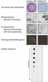
When grown on solid medium supplemented with Congo Red, E. coli cells producing CsgAss-NM form colonies that stain red, whereas cells producing CsgAss-M form pale colonies (reprinted, with permission, from Sivanathan & Hochschild, 2012).
E. coli cells secreting CsgAss-NM produce fibrillar aggregates that can be visualized by transmission electron microscopy, whereas cells secreting CsgAss-M do not produce fibrillar aggregates.
The fibrillar aggregates generated by cells secreting CsgAss-NM are immunolabeled by an anti-Sup35 antibody. No fibrillar aggregates are detected for cells secreting CsgAss-M (reprinted, with permission from Sivanathan & Hochschild, 2012).
E. coli cells secreting CsgAss-NM produce material that manifests apple-green birefringence when viewed by bright-field microscopy between crossed polarizers, whereas cells secreting CsgAss-M do not. Cell samples are taken from colonies formed on solid medium supplemented with Congo Red.
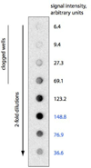
SDS-resistant aggregates are detected using the filter retention assay for samples of cells secreting CsgAss-NM, but not for cell samples secreting CsgAss-M.

Similar articles
-
Curli biogenesis: order out of disorder.Biochim Biophys Acta. 2014 Aug;1843(8):1551-8. doi: 10.1016/j.bbamcr.2013.09.010. Epub 2013 Sep 27. Biochim Biophys Acta. 2014. PMID: 24080089 Free PMC article. Review.
-
Generating extracellular amyloid aggregates using E. coli cells.Genes Dev. 2012 Dec 1;26(23):2659-67. doi: 10.1101/gad.205310.112. Epub 2012 Nov 19. Genes Dev. 2012. PMID: 23166018 Free PMC article.
-
Inhibition of curli assembly and Escherichia coli biofilm formation by the human systemic amyloid precursor transthyretin.Proc Natl Acad Sci U S A. 2017 Nov 14;114(46):12184-12189. doi: 10.1073/pnas.1708805114. Epub 2017 Oct 30. Proc Natl Acad Sci U S A. 2017. PMID: 29087319 Free PMC article.
-
Structure-Function Analysis of the Curli Accessory Protein CsgE Defines Surfaces Essential for Coordinating Amyloid Fiber Formation.mBio. 2018 Jul 17;9(4):e01349-18. doi: 10.1128/mBio.01349-18. mBio. 2018. PMID: 30018113 Free PMC article.
-
Curli provide the template for understanding controlled amyloid propagation.Prion. 2008 Apr-Jun;2(2):57-60. doi: 10.4161/pri.2.2.6746. Epub 2008 Apr 5. Prion. 2008. PMID: 19098444 Free PMC article. Review.
Cited by
-
Curli biogenesis: order out of disorder.Biochim Biophys Acta. 2014 Aug;1843(8):1551-8. doi: 10.1016/j.bbamcr.2013.09.010. Epub 2013 Sep 27. Biochim Biophys Acta. 2014. PMID: 24080089 Free PMC article. Review.
-
Antibiofilm activity of flavonoids on staphylococcal biofilms through targeting BAP amyloids.Sci Rep. 2020 Nov 3;10(1):18968. doi: 10.1038/s41598-020-75929-2. Sci Rep. 2020. PMID: 33144670 Free PMC article.
-
M60-like metalloprotease domain of the Escherichia coli YghJ protein forms amyloid fibrils.PLoS One. 2018 Jan 30;13(1):e0191317. doi: 10.1371/journal.pone.0191317. eCollection 2018. PLoS One. 2018. PMID: 29381728 Free PMC article.
-
Gut microbiota produces biofilm-associated amyloids with potential for neurodegeneration.Nat Commun. 2024 May 16;15(1):4150. doi: 10.1038/s41467-024-48309-x. Nat Commun. 2024. PMID: 38755164 Free PMC article.
-
Enabling stop codon read-through translation in bacteria as a probe for amyloid aggregation.Sci Rep. 2017 Sep 19;7(1):11908. doi: 10.1038/s41598-017-12174-0. Sci Rep. 2017. PMID: 28928456 Free PMC article.
References
Publication types
MeSH terms
Substances
Grants and funding
LinkOut - more resources
Full Text Sources
Other Literature Sources

