. 2013 Jul 15;191(2):764-72.
doi: 10.4049/jimmunol.1300312. Epub 2013 Jun 14.

Orchestration of CD4 T cell epitope preferences after multipeptide immunization

Affiliations

Orchestration of CD4 T cell epitope preferences after multipeptide immunization

Jacqueline Tung et al. J Immunol. .

Abstract

A detailed understanding of the molecular and cellular mechanisms that underlie epitope preferences in T cell priming is important for vaccines designed to elicit a broad T cell response. Protein vaccinations generally elicit CD4 T cell responses that are skewed toward a small fraction of epitopes, a phenomenon known as immunodominance. This characteristic of T cell responses, which limits the diversity of CD4 T cell recognition, is generally attributed to intracellular Ag processing. However, we recently discovered that immunodominance hierarchies persist even after vaccination with synthetic peptides. In this study, we probed the regulatory mechanisms that cause diminished CD4 T cell responses to subdominant peptides after such multipeptide immunization in mice. We have found that the delivery of subdominant and dominant epitopes on separate dendritic cells rescues expansion of less favored CD4 T cells. Furthermore, through the use of genetic models and inhibitors, we have found that selective losses in CD4 T cell responses are mediated by an IFN-γ-induced pathway, involving IDO, and that regulatory T cell activities may also regulate preferences in CD4 T cell specificity. We propose that after multipeptide immunization, the expansion and differentiation of dominant T cells initiate complex regulatory events that determine the final peptide specificity of the elicited CD4 T cell response.


Figures

Figure 1
Figure 1. Expansion of CD4 T cells specific for cryptic peptides fails to progress when there are ongoing CD4 T cell responses to dominant peptides
BALB/c mice were immunized with peptide/IFA emulsions containing OVA peptide alone (filled) or in combination with dominant MalE and Myo peptides (open). CD4 T cells were purified from pooled popliteal LNs of 2 mice/group and were restimulated with OVA peptide (A), MalE peptide (B), or Myo peptide (C) in IFN-γ Elispot assays, with the peptide used for restimulation indicated above each panel. (D) CD4 T cells were restimulated with the indicated peptides at day 6 post immunization and total number of cytokine-producing cells for the indicated peptide are shown for IFN-γ (left) or IL-2 (right). Data are represented as mean and s.d. between 4 independent experiments; p-values were calculated using 1-way ANOVA.
Figure 2
Figure 2. Use of DC-based priming regimen to evaluate the requirement for simultaneous presentation of subdominant and dominant peptides by the same DC
(A–C) BALB/c mice were immunized with DC loaded with OVA peptide alone (filled) or in combination with dominant MalE and Myo peptides (open). CD4 T cells were purified from pooled popliteal LNs of 3–5 mice/group and were restimulated with OVA peptide (A), MalE peptide (B), or Myo peptide (C) in IL-2 Elispot assays at the indicated time points post immunization, with the peptide used for restimulation indicated above each panel. (D) Mice were immunized with DC loaded with either OVA peptide alone (unfilled bar), OVA in combination with dominant peptides (filled bar), or OVA on separate DC (hatched bar). CD4 T cells were purified from pooled popliteal LNs of 3–5 mice/group at day 9 and restimulated with OVA peptide in IL-2 Elispot assays. Data is represented as the mean and s.e.m. between 4–5 independent experiments; statistical analysis was conducted using 1-way ANOVA.
Figure 3
Figure 3. Cell surface expression of MHCII and ICAM-1 on DC is not further reduced after multiple peptide immunization
BALB/c mice (4–5 mice/group) were immunized with CFSE-labeled DC loaded with no antigen, OVA peptide alone, or OVA peptide in combination with dominant MalE and Myo peptides. (A) Expression of ICAM-1 and I-Ad on gated CFSE+ cells in pooled dLNs. (B) Absolute number of CFSE bright cells per LN represented as the mean and s.e.m. between four independent experiments.
Figure 4
Figure 4. Multiple peptide immunization does not selectively increase Treg expansion
Foxp3GFP mice were immunized with peptide/IFA emulsions containing OVA peptide alone or in combination with dominant MalE and Myo peptides (+Competitors). (A) Frequency of Foxp3+ CD4 T cells in unfractionated popliteal LNs from a representative mouse of two is shown for each group over time. (B) Absolute number of Tregs per popliteal LN was quantified from the experiment in A. Data are representative of three independent experiments. Error bars represent the mean and s.d. between two individual mice per group; statistical analysis was conducted using 2-way ANOVA.
Figure 5
Figure 5. Treg activity is enhanced by the presence of dominant peptides
(A) Tregs were flow-sorted from pooled popliteal LNs of Foxp3GFP mice (2 mice/group) that were immunized 6 days prior. Tregs were co-cultured with irradiated APCs and CFSE-labeled target T cells (Thy1.1) in the presence of 1μg/ml anti-CD3 for 72 hrs. Shown is the change in the frequency of Thy1.1+ responder CD4 T cells that have proliferated in cocultures with Tregs sorted from the OVA +Competitors group compared to cocultures with Tregs sorted from the OVA alone group. Data from eight experiments are shown. (B) Foxp3DTR mice (2–3 mice/group) were immunized with peptide/IFA emulsions containing OVA peptide competitor peptides. Mice alone or OVA peptide in combination with MalE, NP261-274, and NP310-325 were administered DT or PBS control at days 3 and 4 post immunization. At day 10, CD4 T cells were purified from pooled inguinal and popliteal LNs and restimulated with OVA peptide. Data from four independent experiments are shown, with each experiment represented by a different symbol (B; left panel). The % of the OVA-specific response maintained in the presence of the competitor peptides relative to no competition with and without DT treatment is displayed in the bar graph (B; right panel).
Figure 6
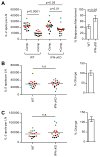
Figure 6. IFN-γ mediates the loss of cryptic CD4 T cell responses after multiple peptide immunization
WT BALB/c and IFN-γKO mice (3–5 mice/group) were immunized with peptide/IFA emulsions containing OVA peptide alone (−Comp.) or OVA peptide in combination with dominant MalE and Myo peptides (+Comp.). At day 10 post immunization, CD4 T cells were purified from popliteal LNs of individual mice and were restimulated with OVA peptide (A), MalE peptide (B), or Myo peptide (C) in IL-2 Elispot assays. The right panel of (A) shows the % of the OVA peptide-specific response maintained when in competition among the four experiments in WT or IFN-γKO mice. The right panels of (B) and (C) show the relative response to dominant peptides of IFN-γKO compared to WT mice. Data from 4 independent experiments were overlaid (each represented by a different color). p-values in the left panel of (A) were calculated using 1-way ANOVA followed by Bonferroni post test. p-values in the right panel of (A) and the left panel of (B) and (C) were calculated from Student’s t test.
Figure 7
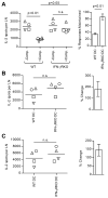
Figure 7. IFN-γ responsiveness in DC is necessary for the suppression of subdominant CD4 T cell responses
In four independent experiments (represented by different symbols), WT BALB/c mice (3–5 pooled mice/group) were immunized with WT or IFN-γRKO DC loaded with OVA peptide alone (−Comp.) or OVA peptide in combination with dominant MalE and Myo peptides (+Comp.). CD4 T cells were purified from pooled popliteal LNs at day 9 and tested for reactivity to OVA peptide (A), MalE peptide (B), or Myo peptide (C). The left panels show the number of peptide-reactive cells per LN. In (A), the right panel shows the % of the OVA response maintained when in competition. The right panels of (B) and (C) show percent change in T cell responses to the dominant peptides in mice immunized with IFN-γRKO DCs compared to mice immunized with WT DCs. p-values were calculated using 1-way ANOVA in the left panel of (A) and Student’s t test in the right panel of (A) and for (B) and (C).
Figure 8
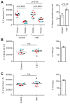
Figure 8. Involvement of the IDO pathway in regulating the selectivity of CD4 T cell responses
In three independent experiments (each represented by a different color), BALB/c mice (5 mice/group) were immunized with peptide/IFA emulsions containing OVA peptide alone (−Comp.) or OVA peptide in combination with dominant MalE and Myo peptides (+Comp.) and were orally administered 1-MT or control. At day 10 post immunization, CD4 T cells were purified from popliteal LNs of individual mice and were restimulated with OVA peptide (A), MalE peptide (B), or Myo peptide (C). The left panels show the total number of peptide-specific T cells. The right panels show the % of the OVA-specific response maintained when in competition (A) or the percent change in T cell responses in mice treated with 1-MT compared to control (B) and (C); p-values shown were calculated from 1-way ANOVA followed by Bonferroni post test in the left panel of (A) or Student’s t test in the right panel of (A) and for (B) and (C).
Figure 9
Figure 9. Ongoing CD4 T cell responses to dominant peptides induce suppressive activities in the local microenvironment that reduce the abundance of CD4 T cells specific for subdominant peptides
Peptides introduced either subcutaneously or on autologous DC initially activate all peptide-specific CD4 T cells equally whether in the presence or absence of other antigenic peptides. Within the first few days of DC arrival, CD4 T cells specific for dominant peptides expand more quickly than CD4 T cells specific for subdominant peptides. As dominant CD4 T cells expand, they differentiate into effector cells and produce IFN-γ. The presence of IFN-γ in the local environment alters the function of DCs that are co-engaging CD4 T cells specific for subdominant peptides. In response to IFN-γ, DC rapidly activate IDO, which in turn increases tryptophan catabolism and production of kynurenines that can arrest cell division of subdominant CD4 T cells and induce the generation of antigen-specific Tregs.

Similar articles

Cited by

References

    1. Scharnagl NC, Klade CS. Experimental discovery of T-cell epitopes: combining the best of classical and contemporary approaches. Expert Rev Vaccines. 2007;6:605–615. - PubMed
    1. Nielsen M, Lund O, Buus S, Lundegaard C. MHC class II epitope predictive algorithms. Immunology. 2010;130:319–328. - PMC - PubMed
    1. Wang P, Sidney J, Dow C, Mothe B, Sette A, Peters B. A systematic assessment of MHC class II peptide binding predictions and evaluation of a consensus approach. PLoS Comput Biol. 2008;4:e1000048. - PMC - PubMed
    1. Dudek NL, Perlmutter P, Aguilar MI, Croft NP, Purcell AW. Epitope discovery and their use in peptide based vaccines. Curr Pharm Des. 2010;16:3149–3157. - PubMed
    1. Sette A, Fikes J. Epitope-based vaccines: an update on epitope identification, vaccine design and delivery. Current Opinion in Immunology. 2003;15:461–470. - PubMed

Publication types

MeSH terms

Substances