

Exosomes secreted by human cells transport largely mRNA fragments that are enriched in the 3'-untranslated regions
- PMID: 23758897
- PMCID: PMC3732077
- DOI: 10.1186/1745-6150-8-12
Exosomes secreted by human cells transport largely mRNA fragments that are enriched in the 3'-untranslated regions
Abstract
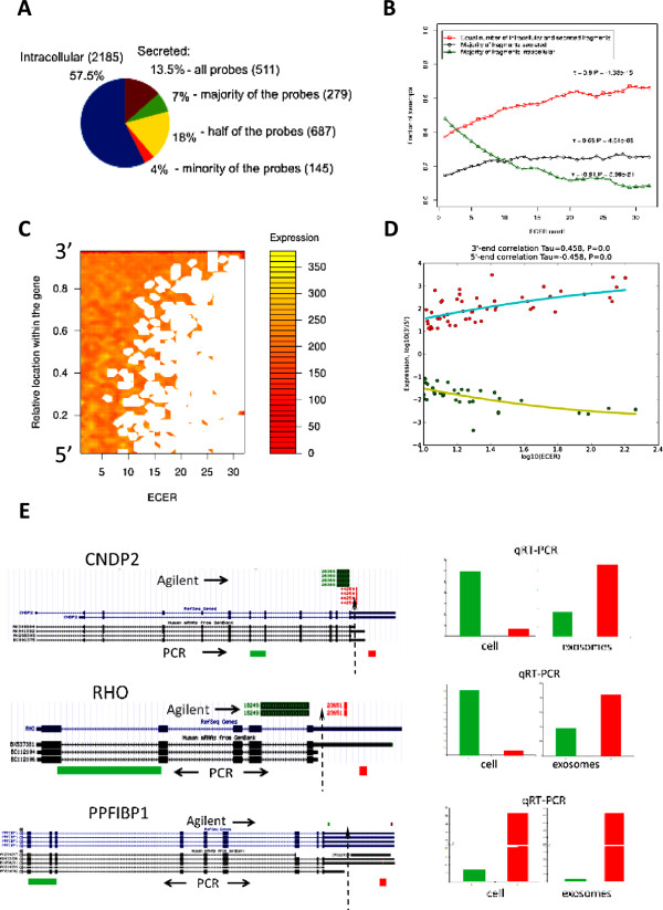
Small secreted membrane vesicles called exosomes have recently attracted a great interest after the discovery that they transfer mRNA that can be translated into protein in recipient cells. Surprisingly, we found that for the majority of exosomal mRNAs only a fraction of their corresponding probes is detectable on the expression microarrays. Exosomal mRNA fragmentation is characterized with a specific structural pattern. The closer to the 3'-end of the transcript the fragments are localized, the larger fraction among the secreted RNAs they constitute. Since the 3'-ends of transcripts contain elements conferring subcellular localization of mRNA and are rich in miRNA-binding sites, exosomal RNA may act as competing RNA to regulate stability, localization and translation activity of mRNAs in recipient cells.
Reviewers: This article was reviewed by Neil Smalheiser and Sandor Pongor.
Figures
Similar articles
-
Purification and microRNA profiling of exosomes derived from blood and culture media.J Vis Exp. 2013 Jun 14;(76):e50294. doi: 10.3791/50294. J Vis Exp. 2013. PMID: 23792786 Free PMC article.
-
Small RNAs derived from tRNAs and rRNAs are highly enriched in exosomes from both old and new world Leishmania providing evidence for conserved exosomal RNA Packaging.BMC Genomics. 2015 Mar 5;16(1):151. doi: 10.1186/s12864-015-1260-7. BMC Genomics. 2015. PMID: 25764986 Free PMC article.
-
Exosome microRNA signatures in patients with complex regional pain syndrome undergoing plasma exchange.J Transl Med. 2019 Mar 14;17(1):81. doi: 10.1186/s12967-019-1833-3. J Transl Med. 2019. PMID: 30871575 Free PMC article.
-
Implications of polyadenylation in health and disease.Nucleus. 2014;5(6):508-19. doi: 10.4161/nucl.36360. Epub 2014 Oct 31. Nucleus. 2014. PMID: 25484187 Free PMC article. Review.
-
To localize or not to localize: mRNA fate is in 3'UTR ends.Trends Cell Biol. 2009 Sep;19(9):465-74. doi: 10.1016/j.tcb.2009.06.001. Epub 2009 Aug 26. Trends Cell Biol. 2009. PMID: 19716303 Review.
Cited by
-
Prostasomes as a source of diagnostic biomarkers for prostate cancer.J Clin Invest. 2016 Apr 1;126(4):1144-51. doi: 10.1172/JCI81128. Epub 2016 Apr 1. J Clin Invest. 2016. PMID: 27035806 Free PMC article. Review.
-
Alarming Cargo: The Role of Exosomes in Trauma-Induced Inflammation.Biomolecules. 2021 Mar 31;11(4):522. doi: 10.3390/biom11040522. Biomolecules. 2021. PMID: 33807302 Free PMC article. Review.
-
Extracellular Vesicles from Animal Milk: Great Potentialities and Critical Issues.Animals (Basel). 2022 Nov 22;12(23):3231. doi: 10.3390/ani12233231. Animals (Basel). 2022. PMID: 36496752 Free PMC article. Review.
-
3D cell culture stimulates the secretion of in vivo like extracellular vesicles.Sci Rep. 2019 Sep 10;9(1):13012. doi: 10.1038/s41598-019-49671-3. Sci Rep. 2019. PMID: 31506601 Free PMC article.
-
Role of cell-free network communication in alcohol-associated disorders and liver metastasis.World J Gastroenterol. 2021 Nov 7;27(41):7080-7099. doi: 10.3748/wjg.v27.i41.7080. World J Gastroenterol. 2021. PMID: 34887629 Free PMC article. Review.
References
-
- Johnstone RM, Adam M, Hammond JR, Orr L, Turbide C. Vesicle formation during reticulocyte maturation. Association of plasma membrane activities with released vesicles (exosomes) J Biol Chem. 1987;262:9412–9420. - PubMed
-
- Skog J, Würdinger T, van Rijn S, Meijer DH, Gainche L, Sena-Esteves M, Curry WT, Carter BS, Krichevsky AM, Breakefield XO. Glioblastoma microvesicles transport RNA and proteins that promote tumour growth and provide diagnostic biomarkers. Nat Cell Biol. 2008;10:1470–1476. doi: 10.1038/ncb1800. - DOI - PMC - PubMed
MeSH terms
Substances
LinkOut - more resources
Full Text Sources
Other Literature Sources
