Review
. 2013 Jun;140(12):2535-47.
doi: 10.1242/dev.091777.

Stem cell metabolism in tissue development and aging

Affiliations
Review

Stem cell metabolism in tissue development and aging

Ng Shyh-Chang et al. Development. 2013 Jun.

Abstract

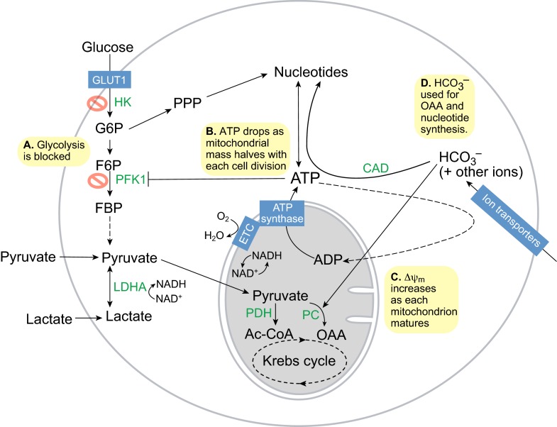
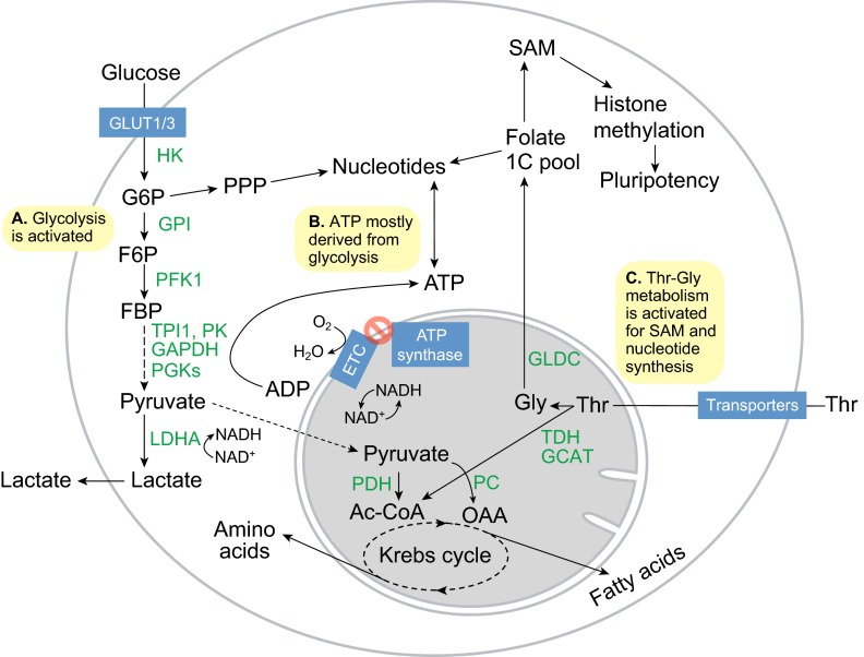
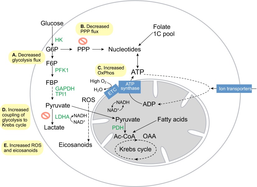
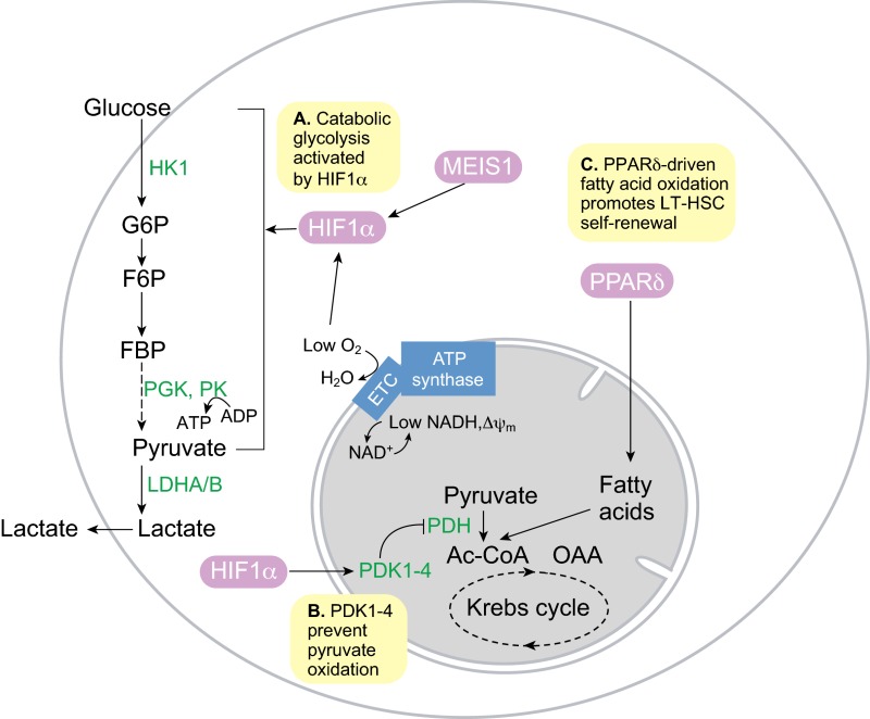
Recent advances in metabolomics and computational analysis have deepened our appreciation for the role of specific metabolic pathways in dictating cell fate. Once thought to be a mere consequence of the state of a cell, metabolism is now known to play a pivotal role in dictating whether a cell proliferates, differentiates or remains quiescent. Here, we review recent studies of metabolism in stem cells that have revealed a shift in the balance between glycolysis, mitochondrial oxidative phosphorylation and oxidative stress during the maturation of adult stem cells, and during the reprogramming of somatic cells to pluripotency. These insights promise to inform strategies for the directed differentiation of stem cells and to offer the potential for novel metabolic or pharmacological therapies to enhance regeneration and the treatment of degenerative disease.

Keywords: Blastocyst metabolism; Hematopoietic progenitors; Metabolomics; Pluripotent stem cells; Reprogramming.


Figures

Fig. 1.
Fig. 1.
Metabolism in totipotent stem cells. (A) Glycolysis is impaired due to the low activities of the rate-limiting enzymes hexokinase (HK) and phosphofructokinase 1 (PFK1). Totipotent stem cells use pyruvate as their major energy and carbon source instead, via pyruvate dehydrogenase (PDH) to generate acetyl-CoA (Ac-CoA) and via pyruvate carboxylase (PC) to generate oxaloacetate (OAA) for anaplerosis or gluconeogenesis. (B) ATP synthesis is dependent on mitochondrial oxidative phosphorylation driven by the electron transport chain (ETC) and ATP synthase. However, as mitochondrial replication has not yet initiated, the halving of mitochondrial mass with each round of mitosis leads to a drop in ATP levels during embryo cleavage. (C) Simultaneously, each mitochondrion matures and the inner mitochondrial membrane potential (ΔΨm) increases steadily, thus turning the exponential drop in ATP into a linear drop. (D) Bicarbonate (HCO3) is needed to buffer the pH and also provides a carbon source to OAA in the Krebs cycle for anaplerosis via PC or to nucleotide synthesis for DNA and RNA via carbamoyl phosphate synthetase (CAD). F6P, fructose-6-phosphate; FBP, fructose-1,6-bisphosphate; G6P, glucose-6-phosphate; LDHA, lactate dehydrogenase A; PPP, pentose phosphate pathway.
Fig. 2.
Fig. 2.
Metabolism in pluripotent stem cells. (A) Glucose flux increases with the increase in GLUT1/3 expression, and the hexokinase (HK) and phosphofructokinase 1 (PFK1) enzymes become activated to sharply increase glycolytic flux. As a result, flux into the pentose phosphate pathway (PPP) for nucleotide synthesis increases. (B) ATP synthesis is more dependent on the reactions carried out by glycolytic phosphoglycerate kinases (PGKs) and pyruvate kinases (PKs), and is decoupled from O2 consumption by the mitochondrial electron transport chain (ETC). (C) Activation of threonine dehydrogenase (TDH), glycine C-acetyltransferase (GCAT) and glycine decarboxylase (GLDC) promotes Thr-Gly catabolism to feed the folate one-carbon (1C) pool, which in turn fuels S-adenosylmethionine (SAM) and nucleotide synthesis to maintain pluripotency and proliferation. Ac-CoA, acetyl coenzyme A; F6P, fructose-6-phosphate; FBP, fructose-1,6-bisphosphate; G6P, glucose-6-phosphate; GAPDH, glyceraldehyde-3-phosphate dehydrogenase; GPI, glucose-6-phosphate isomerase; LDHA, lactate dehydrogenase A; OAA, oxaloacetate; PC, pyruvate carboxylase; PDH, pyruvate dehydrogenase complex; PK, pyruvate kinase; TPI1, triosephosphate isomerase.
Fig. 3.
Fig. 3.
Metabolism in differentiating embryonic stem cells. (A) Glycolytic flux and lactate production drop rapidly upon embryonic stem cell differentiation. (B) Flux into the pentose phosphate pathway (PPP) decreases as a result. (C) O2 consumption increases sharply as the electron transport chain (ETC) again becomes coupled to ATP synthase to fulfill the needs of cell differentiation. (D) Glycolysis also becomes more coupled to the Krebs cycle, as pyruvate is transported more efficiently into mitochondria. (E) Increased ETC activity leads to increased reactive oxygen species (ROS) and eicosanoid signaling, which promote cell differentiation. Ac-CoA, acetyl coenzyme A; HK, hexokinase; F6P, fructose-6-phosphate; FBP, fructose-1,6-bisphosphate; G6P, glucose-6-phosphate; GAPDH, glyceraldehyde-3-phosphate dehydrogenase; LDHA, lactate dehydrogenase A; OAA, oxaloacetate; Ox Phos, oxidative phosphorylation; PDH, pyruvate dehydrogenase complex; PFK1, phosphofructokinase 1; TPI1, triosephosphate isomerase.
Fig. 4.
Fig. 4.
Metabolism in quiescent long-term hematopoietic stem cells. (A) The hematopoietic stem cell (HSC) transcription factor MEIS1 and low O2 levels combine to activate hypoxia-inducible factor 1α (HIF1α) activity, which in turn promotes glycolysis in quiescent long-term HSCs (LT-HSCs). (B) HIF1α-dependent pyruvate dehydrogenase kinases (PDK1-4) prevent pyruvate oxidation by suppressing pyruvate dehydrogenase complex (PDH). (C) Peroxisome proliferator activator receptor δ (PPARδ)-driven fatty acid oxidation in the mitochondria is required for LT-HSC self-renewal and quiescence. Inhibition of fatty acid oxidation leads to LT-HSC proliferation and differentiation. Ac-CoA, acetyl coenzyme A; ETC, electron transport chain; F6P, fructose-6-phosphate; FBP, fructose-1,6-bisphosphate; G6P, glucose-6-phosphate; HK1, hexokinase 1; LDHA, lactate dehydrogenase A/B; PGK, phosphoglycerate kinase; PK, pyruvate kinase.
Fig. 5.
Fig. 5.
Metabolism in proliferative hematopoietic stem and progenitor cells. (A) Anabolic glycolysis is driven in part by the myeloid transcription factor PU.1 and the Akt kinase. (B) Increased reactive oxygen species (ROS) production during oxidative phosphorylation (OxPhos), fueled by PTPMT1-driven pyruvate oxidation, might lead to increased synthesis of eicosanoids, e.g. prostaglandin E2 (PGE2), which promote hematopoiesis. (C) Insulin-PI3K-Akt signaling activates glycolysis, promotes ROS production by repressing the FOXO-mediated stress response, and promotes mitochondrial biogenesis by activating mTOR signaling. This leads to hematopoietic stem cell (HSC) proliferation, differentiation and aging. AMPK, AMP-activated protein kinase; Ac-CoA, acetyl coenzyme A; ETC, electron transport chain; F6P, fructose-6-phosphate; FBP, fructose-1,6-bisphosphate; G6P, glucose-6-phosphate; HK2/3, hexokinase 2/3; LDHA, lactate dehydrogenase A; LKB1, serine/threonine protein kinase 11; OAA, oxaloacetate; PDH, pyruvate dehydrogenase complex; PGK, phosphoglycerate kinase; PK, pyruvate kinase.
Fig. 6.
Fig. 6.
Metabolism in neural stem cells and progenitors. (A) Neural stem cells (NSCs) remain quiescent in a hypoxic niche with low O2. NSCs require FOXO3 to suppress reactive oxygen specis (ROS). (B) Neural progenitors, which exist under normoxia, upregulate both glycolysis and oxidative phosphorylation (OxPhos). In normoxia, FOXO3 is repressed and leads to increased ROS, which prime NSCs for differentiation. Activation of acetyl-CoA (Ac-CoA) carboxylase (ACC) and fatty acid synthase (FASN) increase fatty acid synthesis from Ac-CoA to fuel phospholipid membrane synthesis.
Fig. 7.
Fig. 7.
Metabolism in mesenchymal stem cells and progenitors. (A) Bone marrow mesenchymal stem cells remain quiescent in a hypoxic niche and use glycolysis. (B) Pre-adipocytes upregulate oxidative phosphorylation (OxPhos), and reactive oxygen species (ROS) production from the electron transport chain (ETC) complex III is highly active, to prime adipocyte differentiation. (C) Adipocytes upregulate glycolysis and ATP citrate lyase (ACL), which leads to increased cytosolic acetyl-CoA (Ac-CoA) synthesis and, hence, an increase in histone H3 acetylation (H3ac) and in lipid synthesis. H3ac, in turn, leads to activation of the carbohydrate-responsive element-binding protein (ChREBP) transcription factor to promote GLUT4-mediated glucose uptake and glycolysis in order to generate the acetyl-CoA needed. (D) In osteoblasts, which give rise to bone, OxPhos and O2 consumption are upregulated, but ROS is suppressed via superoxide dismutase (SOD) and catalase (CAT). (E) Glycolysis is further upregulated during chondrogenesis in chondroblasts, which give rise to cartilage.
Fig. 8.
Fig. 8.
Metabolism in skeletal myoblasts and myotubes. (A) Myoblasts use GLUT1 and GLUT3 for glucose uptake. Glycolysis mediated by phosphoglycerate kinase 1 (PGK1) is necessary for myoblast self-renewal. The low activity pyruvate kinase isoform M2 (PKM2) then facilitates accumulation of glycolytic intermediates for anabolic metabolism in myoblasts. For example, the pentose phosphate pathway (PPP), which is mediated by hexose-6-phosphate dehydrogenase (H6PD), shunts glucose-6-phosphate (G6P) into ribose and NADPH synthesis. ATP citrate lyase (ACL), which generates acetyl-CoA (Ac-CoA) from citrate, is also necessary for myoblast self-renewal. (B) Upon cell fusion and differentiation into myotubes, glucose uptake and glycolysis are increased by GLUT4, which is sensitive to regulation by insulin-PI3K-Akt signaling. Mitochondrial oxidative phosphorylation (OxPhos) also increases, partly owing to the switch to the high activity pyruvate kinase isoform M1 (PKM1).

Similar articles

Cited by

References

    1. Alexander P. B., Wang J., McKnight S. L. (2011). Targeted killing of a mammalian cell based upon its specialized metabolic state. Proc. Natl. Acad. Sci. USA 108, 15828-15833 - PMC - PubMed
    1. Ambros V., Horvitz H. R. (1984). Heterochronic mutants of the nematode Caenorhabditis elegans. Science 226, 409-416 - PubMed
    1. Ang Y.-S., Tsai S.-Y., Lee D.-F., Monk J., Su J., Ratnakumar K., Ding J., Ge Y., Darr H., Chang B., et al. (2011). Wdr5 mediates self-renewal and reprogramming via the embryonic stem cell core transcriptional network. Cell 145, 183-197 - PMC - PubMed
    1. Arantes-Oliveira N., Apfeld J., Dillin A., Kenyon C. (2002). Regulation of life-span by germ-line stem cells in Caenorhabditis elegans. Science 295, 502-505 - PubMed
    1. Azuara V., Perry P., Sauer S., Spivakov M., Jørgensen H. F., John R. M., Gouti M., Casanova M., Warnes G., Merkenschlager M., et al. (2006). Chromatin signatures of pluripotent cell lines. Nat. Cell Biol. 8, 532-538 - PubMed

Publication types