

Stages of pTDP-43 pathology in amyotrophic lateral sclerosis
- PMID: 23686809
- PMCID: PMC3785076
- DOI: 10.1002/ana.23937
Stages of pTDP-43 pathology in amyotrophic lateral sclerosis
Abstract
Objective: To see whether the distribution patterns of phosphorylated 43kDa TAR DNA-binding protein (pTDP-43) intraneuronal inclusions in amyotrophic lateral sclerosis (ALS) permit recognition of neuropathological stages.
Methods: pTDP-43 immunohistochemistry was performed on 70 μm sections from ALS autopsy cases (N = 76) classified by clinical phenotype and genetic background.
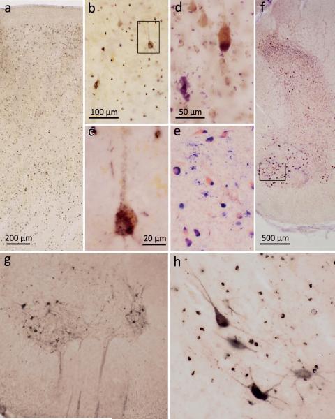
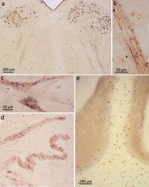
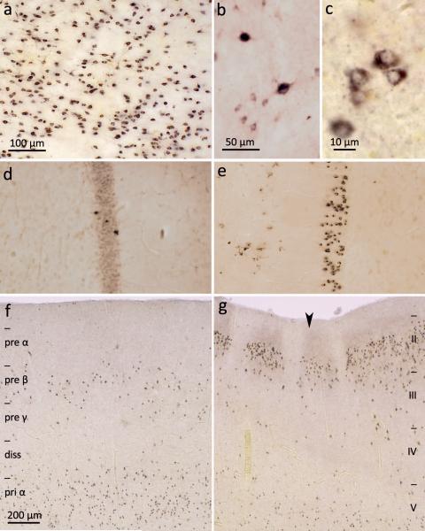
Results: ALS cases with the lowest burden of pTDP-43 pathology were characterized by lesions in the agranular motor cortex, brainstem motor nuclei of cranial nerves V, VII, and X-XII, and spinal cord α-motoneurons (stage 1). Increasing burdens of pathology showed involvement of the prefrontal neocortex (middle frontal gyrus), brainstem reticular formation, precerebellar nuclei, and the red nucleus (stage 2). In stage 3, pTDP-43 pathology involved the prefrontal (gyrus rectus and orbital gyri) and then postcentral neocortex and striatum. Cases with the greatest burden of pTDP-43 lesions showed pTDP-43 inclusions in anteromedial portions of the temporal lobe, including the hippocampus (stage 4). At all stages, these lesions were accompanied by pTDP-43 oligodendroglial aggregates. Ten cases with C9orf72 repeat expansion displayed the same sequential spreading pattern as nonexpansion cases but a greater regional burden of lesions, indicating a more fulminant dissemination of pTDP-43 pathology.
Interpretation: pTDP-43 pathology in ALS possibly disseminates in a sequential pattern that permits recognition of 4 neuropathological stages consistent with the hypothesis that pTDP-43 pathology is propagated along axonal pathways. Moreover, the finding that pTDP-43 pathology develops in the prefrontal cortex as part of an ongoing disease process could account for the development of executive cognitive deficits in ALS.
© 2013 American Neurological Association.
Figures
Similar articles
-
Sequential distribution of pTDP-43 pathology in behavioral variant frontotemporal dementia (bvFTD).Acta Neuropathol. 2014 Mar;127(3):423-439. doi: 10.1007/s00401-013-1238-y. Epub 2014 Jan 10. Acta Neuropathol. 2014. PMID: 24407427 Free PMC article.
-
TDP-43 pathology and neuronal loss in amyotrophic lateral sclerosis spinal cord.Acta Neuropathol. 2014 Sep;128(3):423-37. doi: 10.1007/s00401-014-1299-6. Epub 2014 Jun 12. Acta Neuropathol. 2014. PMID: 24916269 Free PMC article.
-
Sense-encoded poly-GR dipeptide repeat proteins correlate to neurodegeneration and uniquely co-localize with TDP-43 in dendrites of repeat-expanded C9orf72 amyotrophic lateral sclerosis.Acta Neuropathol. 2018 Mar;135(3):459-474. doi: 10.1007/s00401-017-1793-8. Epub 2017 Dec 1. Acta Neuropathol. 2018. PMID: 29196813 Free PMC article.
-
The multisystem degeneration amyotrophic lateral sclerosis - neuropathological staging and clinical translation.Arch Ital Biol. 2017 Dec 1;155(4):118-130. doi: 10.12871/00039829201746. Arch Ital Biol. 2017. PMID: 29405032 Review.
-
Prevalence of brain and spinal cord inclusions, including dipeptide repeat proteins, in patients with the C9ORF72 hexanucleotide repeat expansion: a systematic neuropathological review.Neuropathol Appl Neurobiol. 2016 Oct;42(6):547-60. doi: 10.1111/nan.12284. Neuropathol Appl Neurobiol. 2016. PMID: 26373655 Review.
Cited by
-
Neuropeptide FF (NPFF)-positive nerve cells of the human cerebral cortex and white matter in controls, selected neurodegenerative diseases, and schizophrenia.Acta Neuropathol Commun. 2024 Jun 28;12(1):108. doi: 10.1186/s40478-024-01792-1. Acta Neuropathol Commun. 2024. PMID: 38943180 Free PMC article.
-
Absence of Subcerebral Projection Neurons Is Beneficial in a Mouse Model of Amyotrophic Lateral Sclerosis.Ann Neurol. 2020 Oct;88(4):688-702. doi: 10.1002/ana.25833. Epub 2020 Jul 11. Ann Neurol. 2020. PMID: 32588450 Free PMC article.
-
Prion-like propagation as a pathogenic principle in frontotemporal dementia.J Neurochem. 2016 Aug;138 Suppl 1(Suppl Suppl 1):163-83. doi: 10.1111/jnc.13668. J Neurochem. 2016. PMID: 27502124 Free PMC article. Review.
-
The Role of VCP Mutations in the Spectrum of Amyotrophic Lateral Sclerosis-Frontotemporal Dementia.Front Neurol. 2022 Feb 22;13:841394. doi: 10.3389/fneur.2022.841394. eCollection 2022. Front Neurol. 2022. PMID: 35273561 Free PMC article. Review.
-
Cognitive impairment across ALS clinical stages in a population-based cohort.Neurology. 2019 Sep 3;93(10):e984-e994. doi: 10.1212/WNL.0000000000008063. Epub 2019 Aug 13. Neurology. 2019. PMID: 31409738 Free PMC article.
References
-
- Kiernan MC, Vucic S, Cheah BC, et al. Amyotrophic lateral sclerosis. Lancet. 2011 Mar 12;377(9769):942–55. - PubMed
-
- Neumann M, Sampathu DM, Kwong LK, et al. Ubiquitinated TDP-43 in frontotemporal lobar degeneration and amyotrophic lateral sclerosis. Science. 2006 Oct 6;314(5796):130–3. - PubMed
-
-
Braak H, Thal DR, Ghebremedhin E, Del Tredici K. Stages of the pathologic process in Alzheimer disease: age categories from 1 to 100 years. Journal of neuropathology and experimental neurology. 2011 Nov;70(11):960–969. This is the correct reference and not Braak et al. 2006.
-
-
- Braak H, Del Tredici K. Where, when, and in what form does sporadic Alzheimer’s disease begin? Current opinion in neurology. 2012 Dec;25(6):708–14. - PubMed
Publication types
MeSH terms
Substances
Grants and funding
- P30 AG010124/AG/NIA NIH HHS/United States
- K08 AG039510/AG/NIA NIH HHS/United States
- AG010124/AG/NIA NIH HHS/United States
- K08 AG033101/AG/NIA NIH HHS/United States
- AG039510/AG/NIA NIH HHS/United States
- NS044266/NS/NINDS NIH HHS/United States
- R01 NS044266/NS/NINDS NIH HHS/United States
- P01 AG032953/AG/NIA NIH HHS/United States
- P01 AG017586/AG/NIA NIH HHS/United States
- AG017586/AG/NIA NIH HHS/United States
- T32 AG000255/AG/NIA NIH HHS/United States
- T32-AG000255/AG/NIA NIH HHS/United States
- AG032953/AG/NIA NIH HHS/United States
- AG033101/AG/NIA NIH HHS/United States
LinkOut - more resources
Full Text Sources
Other Literature Sources
Medical
Miscellaneous
