. 2013:3:1809.
doi: 10.1038/srep01809.

Prominent role for T cell-derived tumour necrosis factor for sustained control of Mycobacterium tuberculosis infection

Affiliations

Prominent role for T cell-derived tumour necrosis factor for sustained control of Mycobacterium tuberculosis infection

Nasiema Allie et al. Sci Rep. 2013.

Abstract

Tumour Necrosis Factor (TNF) is critical for host control of M. tuberculosis, but the relative contribution of TNF from innate and adaptive immune responses during tuberculosis infection is unclear. Myeloid versus T-cell-derived TNF function in tuberculosis was investigated using cell type-specific TNF deletion. Mice deficient for TNF expression in macrophages/neutrophils displayed early, transient susceptibility to M. tuberculosis but recruited activated, TNF-producing CD4(+) and CD8(+) T-cells and controlled chronic infection. Strikingly, deficient TNF expression in T-cells resulted in early control but susceptibility and eventual mortality during chronic infection with increased pulmonary pathology. TNF inactivation in both myeloid and T-cells rendered mice critically susceptible to infection with a phenotype resembling complete TNF deficient mice, indicating that myeloid and T-cells are the primary TNF sources collaborating for host control of tuberculosis. Thus, while TNF from myeloid cells mediates early immune function, T-cell derived TNF is essential to sustain protection during chronic tuberculosis infection.


Figures
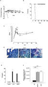
Figure 1
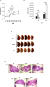
Figure 1. TNF from myeloid cells is required to control acute M. tuberculosis replication in vivo.
(A–D) WT (closed circle), M-TNF−/− (grey triangle) and TNF−/− (open circle) mice were infected by aerosol inhalation with 200–500 cfu/lung of M. tuberculosis H37Rv and body weight changes (A) and mortality (B) (n = 10 mice/group) were monitored. Changes in bodyweight values are expressed as the mean of 4–10 mice/group. Data represents one of three independent experiments. The number of viable bacteria present in lungs was assessed at the indicated time points (C). The results are expressed as the mean ± SD of 5 mice/group per time point and represent one of three independent experiments. * = p < 0.05 (WT versus M-TNF−/− mice), # = p < 0.05 (WT versus TNF−/− mice.), ϕ = p < 0.05 (M-TNF−/− versus TNF−/− mice). Ziehl-Neelsen staining of lungs at 35 days post infection (D). The results represent one of four similar experiments. (Magnification = 600×). Arrows indicate the presence of AFB. (E) WT (black), M-TNF−/− (grey) and TNF−/− (clear) bone marrow derived macrophages were stimulated either with LPS (100 ng/ml) or M. tuberculosis H37Rv (at a MOI of 2:1) for 48 hrs and TNF measured by ELISA. The results are expressed as the mean ± SD of quadruplicate values and are representative of one of three independent experiments. ND: Not detected. (F) Bone marrow derived macrophages were infected with M. tuberculosis expressing luciferase and bacterial replication measured as relative luminescence after 100 hrs. The results are expressed as the mean ± SD of quadruplicate values and represent one of three independent experiments.
Figure 2
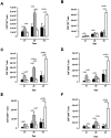
Figure 2. TNF from myeloid cells regulates cellular recruitment for granuloma formation but is dispensable for initiation and maintenance of granuloma structure.
(A; B) WT (black), M-TNF−/− (grey), and TNF−/− (clear) mice were infected with M. tuberculosis H37Rv as described under “Materials and Methods” and lung weights (A) and cell numbers (B) assessed at the indicated time points. Data represent the mean ± SD of 5 mice per time point and one of three independent experiments. * = p < 0.05 (WT versus M-TNF−/− mice), # = p < 0.05 (WT versus TNF−/− mice.), ϕ = p < 0.05 (M-TNF−/− versus TNF−/− mice). (C–D). Macroscopic lung pathology (C) in WT, M-TNF−/− and TNF−/− mice at 28 days post infection (one of three independent experiments; n = 5 mice/strain). Low magnification (20×) of pulmonary granulomas at (i) 28 days and (ii) 166 days postinfection after haemotoxylin and eosin staining (D). Arrows indicate granulomatous structures and X indicates necrosis. The results represent one of 5 similar experiments.
Figure 3
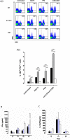
Figure 3. Transiently enhanced pulmonary infiltration of CD4+ and CD8+ activated T cells is independent of TNF produced by myeloid cells during M. tuberculosis infection.
WT (black), M-TNF−/− (grey) and TNF−/− (clear) mice were infected via aerosol inhalation with 200–500 cfu/lung of Mycobacterium tuberculosis. Lung cell suspensions from infected mice were analyzed by flow cytometry to determine (A) CD3+CD4+ and (B) CD3+CD8+ T cell pulmonary populations at day 21, 28 and 35 post-infection and activation status analysed for markers CD44 (C and D) and CD69 (E and F) on CD4+ (C, E) and CD8+ (D, F) T cells by flow cytometry. The results are expressed as the mean ± SD of 5 mice per group and are from one experiment, representative of two independent experiments.
Figure 4
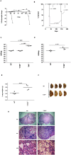
Figure 4. Enhanced M. tuberculosis specific priming of pulmonary CD4+ T cells in infected M-TNF−/− mice.
(A) Lung cell suspensions from M. tuberculosis H37Rv infected WT, M-TNF−/−, and TNF−/− mice harvested at 21 days post infection were restimulated with ESAT 6, PPD or antibodies to CD3 and CD28 for 72 hrs and intracellular IFNγ produced by CD4+T cells assessed by flow cytometry. Fig. 4A(i) are representative dot blots of single mice while the results in Fig. 4A(ii) are expressed as the mean ± SD of 4 mice per group and represent one of two similar experiments. (B, C) Lung concentrations of IFNγ (B) and IL-12p70 (C) were determined by ELISA in lung homogenates of M. tuberculosis H37Rv infected WT (black), M-TNF−/− (grey) and TNF−/− (open) mice at different time points. The data are expressed as the mean and ± SD of 4 mice/group for each time point and represent one of two similar experiments.
Figure 5
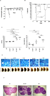
Figure 5. T-TNF−/− mice succumb to chronic M. tuberculosis infection.
(A; B) WT (closed circle), T-TNF−/− (grey diamond), and TNF−/− (clear circle) mice were infected via aerosol inhalation with 200–500 cfu/lung of M. tuberculosis H37Rv are represented (n = 10 mice/strain). Change in bodyweight values (A) are expressed as the mean of 4–10 mice/group. Differences in mortality rates (B) were analysed by the student's logrank test. (C; D). The number of viable bacteria present in lungs were assessed at 33 days (C) and 150 days (D) post-infection. The results are expressed as the mean of 4 mice/group. (E; F and G). Lung weights (E), macroscopic lung pathology (F) and haemotoxylin and eosin staining of lung sections (G) from WT-, and T-TNF−/− mice infected with M. tuberculosis. Images of left lobes of WT and T-TNF−/− mice at 150 days post infection are presented (n = 4 mice/group). The data is from one experiment representative of three independent experiments.
Figure 6
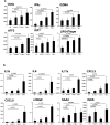
Figure 6. Inflammatory gene expression during acute M. tuberculosis infection in cell-specific TNF−/− mice.
(A; B) Analysis of selected inflammatory gene expression was performed by quantitative RT-PCR on day 28 after M. tuberculosis infection and normalized to b-actin. (n = 3–4 mice per point). Genes include CD8a, IFNγ, granzyme A,Irgm1/LRG47, IRF7 and ATF3 (Fig. 6A), the proinflammatory cytokines and chemokines IL-1b, IL-6, IL-17a, CXCL3 or CXCL5 and s100a9, the acute phase reactant, SAA and iNOS (Fig. 6B).
Figure 7
Figure 7. MT-TNF−/− mice are highly susceptible and succumb to acute M. tuberculosis infection.
(A; B) WT (closed circle), M-TNF−/− (grey triangle), T-TNF−/− (grey diamond), MT-TNF−/− mice (clear square) and TNF−/− (clear circle) mice infected as described were monitored for change in bodyweight (A; expressed as the mean of 4–10 mice/group) and survival (B; n = 10 mice/group) Data is from one experiment representative of three independent experiments. (C; D) The lung weights (C) and number of viable bacteria present in lungs (D) were assessed at 39 days post-infection. The results are expressed as the mean ± SD of 4–5 mice/group and are from one experiment, representative of two independent experiments. (E; F) Pulmonary bacilli burdens on day 39 were confirmed by Ziehl-Neelsen staining (E; magnification 600×). Arrows indicate the presence of single AFB within macrophages. Images of left lobes illustrating macroscopic pulmonary pathology in WT-, M-TNF−/−, T-TNF−/−, MT-TNF−/− and TNF−/− mice. (n = 5 mice/group). Images are representative of one of three independent experiments. Haemotoxylin and eosin staining show enlarged, necrotic nodules in the lungs of MT-TNF−/− and TNF−/− mice at day 39 post infection (F). Images are representative of one of three independent experiments.

Similar articles

Cited by

References

    1. Moreno C. et al. Lipoarabinomannan from Mycobacterium tuberculosis induces the production of tumour necrosis factor from human and murine macrophages. Clin Exp Immunol 76, 240–245 (1989). - PMC - PubMed
    1. Roach T. I., Barton C. H., Chatterjee D. & Blackwell J. M. Macrophage activation: lipoarabinomannan from avirulent and virulent strains of Mycobacterium tuberculosis differentially induces the early genes c-fos, KC, JE, and tumor necrosis factor-alpha. J Immunol 150, 1886–1896 (1993). - PubMed
    1. Falcone V., Bassey E. B., Toniolo A., Conaldi P. G. & Collins F. M. Differential release of tumor necrosis factor-alpha from murine peritoneal macrophages stimulated with virulent and avirulent species of mycobacteria. FEMS Immunol Med Microbiol 8, 225–232 (1994). - PubMed
    1. Oswald I. P., Dozois C. M., Fournout S., Petit J. F. & Lemaire G. Tumor necrosis factor is required for the priming of peritoneal macrophages by trehalose dimycolate. Eur Cytokine Netw 10, 533–540 (1999). - PubMed
    1. Flesch I. E. & Kaufmann S. H. Activation of tuberculostatic macrophage functions by gamma interferon, interleukin-4, and tumor necrosis factor. Infect Immun 58, 2675–2677 (1990). - PMC - PubMed

Publication types

MeSH terms