RIG-I detects triphosphorylated RNA of Listeria monocytogenes during infection in non-immune cells
- PMID: 23653683
- PMCID: PMC3639904
- DOI: 10.1371/journal.pone.0062872
RIG-I detects triphosphorylated RNA of Listeria monocytogenes during infection in non-immune cells
Abstract
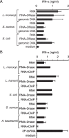
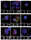
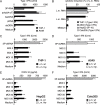
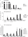
The innate immune system senses pathogens by pattern recognition receptors in different cell compartments. In the endosome, bacteria are generally recognized by TLRs; facultative intracellular bacteria such as Listeria, however, can escape the endosome. Once in the cytosol, they become accessible to cytosolic pattern recognition receptors, which recognize components of the bacterial cell wall, metabolites or bacterial nucleic acids and initiate an immune response in the host cell. Current knowledge has been focused on the type I IFN response to Listeria DNA or Listeria-derived second messenger c-di-AMP via the signaling adaptor STING. Our study focused on the recognition of Listeria RNA in the cytosol. With the aid of a novel labeling technique, we have been able to visualize immediate cytosolic delivery of Listeria RNA upon infection. Infection with Listeria as well as transfection of bacterial RNA induced a type-I-IFN response in human monocytes, epithelial cells or hepatocytes. However, in contrast to monocytes, the type-I-IFN response of epithelial cells and hepatocytes was not triggered by bacterial DNA, indicating a STING-independent Listeria recognition pathway. RIG-I and MAVS knock-down resulted in abolishment of the IFN response in epithelial cells, but the IFN response in monocytic cells remained unaffected. By contrast, knockdown of STING in monocytic cells reduced cytosolic Listeria-mediated type-I-IFN induction. Our results show that detection of Listeria RNA by RIG-I represents a non-redundant cytosolic immunorecognition pathway in non-immune cells lacking a functional STING dependent signaling pathway.
Conflict of interest statement
Figures


Similar articles
-
RIG-I detects infection with live Listeria by sensing secreted bacterial nucleic acids.EMBO J. 2012 Nov 5;31(21):4153-64. doi: 10.1038/emboj.2012.274. Epub 2012 Oct 12. EMBO J. 2012. PMID: 23064150 Free PMC article.
-
Secretion of c-di-AMP by Listeria monocytogenes Leads to a STING-Dependent Antibacterial Response during Enterocolitis.Infect Immun. 2020 Nov 16;88(12):e00407-20. doi: 10.1128/IAI.00407-20. Print 2020 Nov 16. Infect Immun. 2020. PMID: 33020211 Free PMC article.
-
Dengue Virus Subverts Host Innate Immunity by Targeting Adaptor Protein MAVS.J Virol. 2016 Jul 27;90(16):7219-7230. doi: 10.1128/JVI.00221-16. Print 2016 Aug 15. J Virol. 2016. PMID: 27252539 Free PMC article.
-
How Dengue Virus Circumvents Innate Immunity.Front Immunol. 2018 Dec 4;9:2860. doi: 10.3389/fimmu.2018.02860. eCollection 2018. Front Immunol. 2018. PMID: 30564245 Free PMC article. Review.
-
Crosstalk between Cytoplasmic RIG-I and STING Sensing Pathways.Trends Immunol. 2017 Mar;38(3):194-205. doi: 10.1016/j.it.2016.12.004. Epub 2017 Jan 7. Trends Immunol. 2017. PMID: 28073693 Free PMC article. Review.
Cited by
-
Innate Immune Sensing by Cells of the Adaptive Immune System.Front Immunol. 2020 May 29;11:1081. doi: 10.3389/fimmu.2020.01081. eCollection 2020. Front Immunol. 2020. PMID: 32547564 Free PMC article. Review.
-
STING-dependent type I IFN production inhibits cell-mediated immunity to Listeria monocytogenes.PLoS Pathog. 2014 Jan;10(1):e1003861. doi: 10.1371/journal.ppat.1003861. Epub 2014 Jan 2. PLoS Pathog. 2014. PMID: 24391507 Free PMC article.
-
Apoptosis, Toll-like, RIG-I-like and NOD-like Receptors Are Pathways Jointly Induced by Diverse Respiratory Bacterial and Viral Pathogens.Front Microbiol. 2017 Mar 1;8:276. doi: 10.3389/fmicb.2017.00276. eCollection 2017. Front Microbiol. 2017. PMID: 28298903 Free PMC article.
-
B. abortus RNA is the component involved in the down-modulation of MHC-I expression on human monocytes via TLR8 and the EGFR pathway.PLoS Pathog. 2017 Aug 2;13(8):e1006527. doi: 10.1371/journal.ppat.1006527. eCollection 2017 Aug. PLoS Pathog. 2017. PMID: 28767704 Free PMC article.
-
Discriminating self from non-self in nucleic acid sensing.Nat Rev Immunol. 2016 Sep;16(9):566-80. doi: 10.1038/nri.2016.78. Epub 2016 Jul 25. Nat Rev Immunol. 2016. PMID: 27455396 Free PMC article. Review.
References
-
- Chen G, Shaw MH, Kim YG, Nunez G (2009) NOD-like receptors: role in innate immunity and inflammatory disease. Annu Rev Pathol 4: 365–398. - PubMed
-
- Schlee M, Hornung V, Hartmann G (2006) siRNA and isRNA: two edges of one sword. Mol Ther 14: 463–470. - PubMed
-
- Krieg AM, Yi AK, Matson S, Waldschmidt TJ, Bishop GA, et al. (1995) CpG motifs in bacterial DNA trigger direct B-cell activation. Nature 374: 546–549. - PubMed
-
- Hemmi H, Takeuchi O, Kawai T, Kaisho T, Sato S, et al. (2000) A Toll-like receptor recognizes bacterial DNA. Nature 408: 740–745. - PubMed
-
- Coch C, Busch N, Wimmenauer V, Hartmann E, Janke M, et al. (2009) Higher activation of TLR9 in plasmacytoid dendritic cells by microbial DNA compared with self-DNA based on CpG-specific recognition of phosphodiester DNA. J Leukoc Biol 86: 663–670. - PubMed
Publication types
MeSH terms
Substances
Grants and funding
LinkOut - more resources
Full Text Sources
Other Literature Sources
Research Materials
Miscellaneous

