. 2013:4:1816.
doi: 10.1038/ncomms2828.

A functional deficiency of TERA/VCP/p97 contributes to impaired DNA repair in multiple polyglutamine diseases

Affiliations

A functional deficiency of TERA/VCP/p97 contributes to impaired DNA repair in multiple polyglutamine diseases

Kyota Fujita et al. Nat Commun. 2013.

Abstract

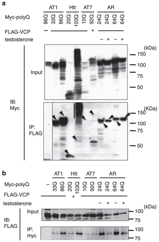
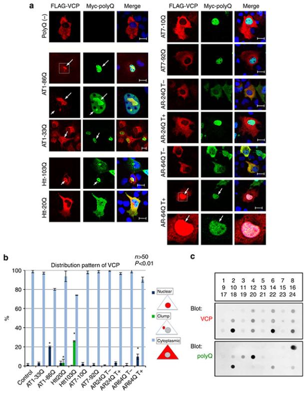
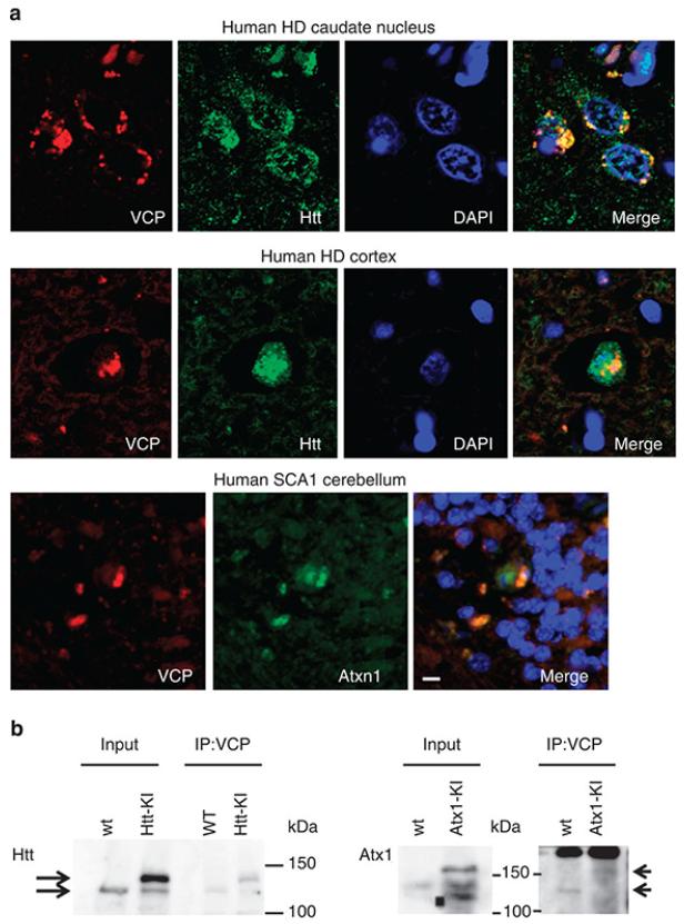
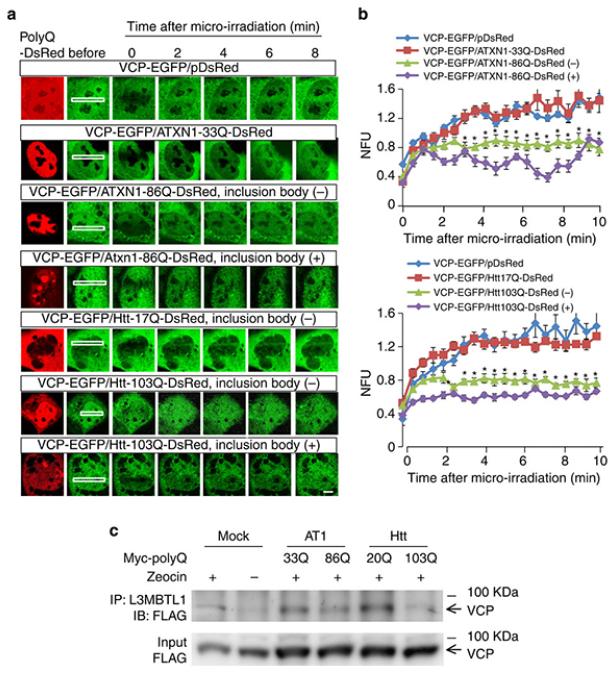
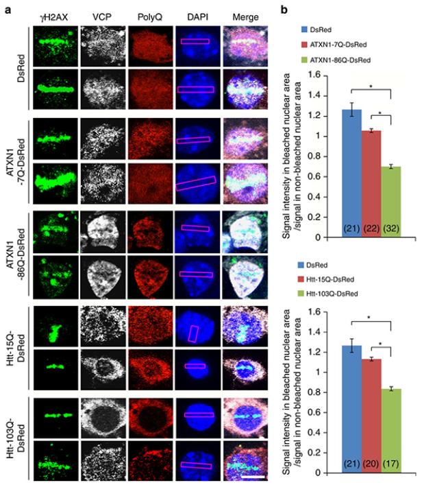
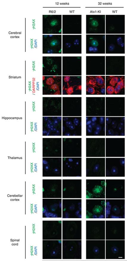
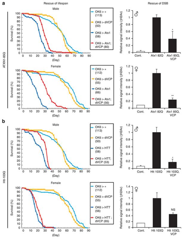
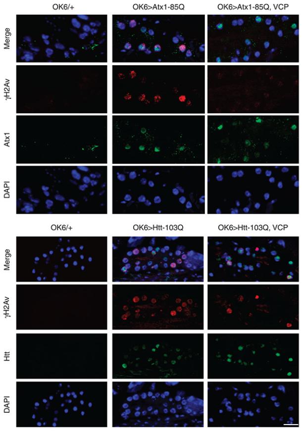
It is hypothesized that a common underlying mechanism links multiple neurodegenerative disorders. Here we show that transitional endoplasmic reticulum ATPase (TERA)/valosin-containing protein (VCP)/p97 directly binds to multiple polyglutamine disease proteins (huntingtin, ataxin-1, ataxin-7 and androgen receptor) via polyglutamine sequence. Although normal and mutant polyglutamine proteins interact with TERA/VCP/p97, only mutant proteins affect dynamism of TERA/VCP/p97. Among multiple functions of TERA/VCP/p97, we reveal that functional defect of TERA/VCP/p97 in DNA double-stranded break repair is critical for the pathology of neurons in which TERA/VCP/p97 is located dominantly in the nucleus in vivo. Mutant polyglutamine proteins impair accumulation of TERA/VCP/p97 and interaction of related double-stranded break repair proteins, finally causing the increase of unrepaired double-stranded break. Consistently, the recovery of lifespan in polyglutamine disease fly models by TERA/VCP/p97 corresponds well to the improvement of double-stranded break in neurons. Taken together, our results provide a novel common pathomechanism in multiple polyglutamine diseases that is mediated by DNA repair function of TERA/VCP/p97.


Figures

Figure 1
Figure 1. Multiple polyQ proteins interact with TERA/VCP/p97
(a) VCP coprecipitates with normal as well as mutant forms of ataxin-1 (ATXN1), huntingtin (Htt), full-length ataxin-7 (ATXN7), or androgen receptor (AR). HeLa cells were cotransfected with Myc-polyQ and FLAG-VCP protein expression vectors, and the cell lysates were immunoprecipitated with anti-FLAG antibody. Lane 1 is a negative control in which HeLa cells are transfected with Myc-ATXN1-86Q expression vector only. Coprecipitation of ATXN7 is relatively unstable. AR binds strongly to VCP in the absence of testosterone (T−) but only weakly in the presence of testosterone (T+) because of the dissociation of their intracellular distribution. (b) Normal and mutant polyQ proteins coprecipitate with VCP. ATXN7 did not obviously coprecipitate in some cases. Mutant Htt seems to bind more strongly to VCP than to normal Htt in these blots; however, the difference is not definite in other blots.
Figure 2
Figure 2. Mutant polyQ proteins sequester TERA/VCP/p97 into the inclusion body
(a) HeLa cells cotransfected with Myc-polyQ and FLAG-VCP protein expression vectors are stained with anti-FLAG and anti-Myc antibodies. The top panels show normal cytoplasmic distribution of the FLAG-VCP protein. The ATXN1-86Q and Htt-103Q proteins sequester VCP to nuclear and cytoplasmic inclusion bodies, respectively. In contrast, the ATXN1-33Q and Htt-20Q proteins do not affect the cytoplasmic distribution of VCP regardless of their interaction. Normal and mutant ATXN7 expression did not affect intracellular localization of VCP. In the presence of testosterone, mutant but not normal AR sequesters VCP into the nucleus. Scale bars represent 20 μm. (b) Quantitative analysis of the change of intracellular distribution of VCP by polyQ proteins. The distribution patterns are described in right panels. Means and s.e.m. are indicated. Asterisks indicate statistical difference from the control (p<0.01, Student’s t-test). (c) Filter binding assay shows co-aggregation of polyQ disease proteins and VCP. 1, 2: buffer, 3 empty plasmid, 4: empty plasmid+VCP, 5: ATXN1-33Q, 6: ATXN1-33Q+VCP, 7: ATXN1-86Q, 8: ATXN1-86Q+VCP, 9: Htt-20Q, 10: Htt-20Q+VCP, 11: Htt-103Q, 12: Htt-103Q+VCP, 13: ATXN7-10Q, 14: ATXN7-10Q+VCP, 15: ATXN7-92Q, 16: ATXN7-92Q+VCP, 17: AR-24Q testosterone (−), 18: AR-24Q+VCP testosterone (−), 19: AR-24Q testosterone (+), 20: AR-24Q+VCP testosterone (+), 21: AR-64Q testosterone (−), 22: AR-64Q+VCP testosterone (−), 23: AR-64Q testosterone (+), 24: AR-64Q+VCP testosterone (+).
Figure 3
Figure 3. Mutant polyQ proteins interact with TERA/VCP/p97 in the human brain
(a) Mutant polyQ proteins and VCP are colocalized in inclusion bodies of human patients with polyQ diseases. Subcellular distribution of VCP was remarkably changed in neurons possessing aggregates of polyQ disease proteins. Scale bar represents 10 μm. In control human brains, subcellular accumulation or foci of TERA/VCP/p97 was not observed (Supplementary Fig. S6). (b) Coprecipitation of mutant polyQ and VCP proteins from mutant polyQ-KI mouse brain samples.
Figure 4
Figure 4. Mutant polyQ proteins delay accumulation of TERA/VCP/p97 to DSB foci
(a) Sequential images showing accumulation of VCP-EGFP to the linear DSB. In cells expressing mutant polyQ proteins (Atxn1-86Q-DsRed and Htt-103Q-DsRed), the accumulation of VCP-EGFP was delayed in comparison to non-transfected or normal polyQ protein (Atxn1-33Q-DsRed and Htt-17Q-DsRed) expressing cells. Among the cells expressing mutant polyQ proteins, the delay in the accumulation of VCP-EGFP was more remarkable in inclusion body-positive cells than in inclusion body-negative cells. Scale bar represents 5 μm. (b) Quantitative analysis of the recovery of VCP-EGFP signals at the linear photobleach areas that correspond to accumulation of VCP-EGFP to the DSB lesion (N=6). The signals were obtained from more than four sites on the linear damage area, and the mean value was used for the quantitative analyses at each time point. Means and s.e.m. are indicated. Asterisks indicate statistical difference from the value of normal polyQ protein-expressing cells at the same time point (p<0.01, Student’s t-test). H2Ax staining confirmed that DSB was actually induced in this condition (Supplementary Figure 8). NFU: normalized fluorescence unit. (c) Mutant polyQ proteins inhibit interaction between TERA/VCP/p97 and L3MBTL1. Zeocin (400 μg/mL) was added to the culture medium of HeLa cells to induce DSBs where TERA/VCP/p97 interacts with L3MBTL1. Mutant Htt and ATXN1 both decrease the interaction between TERA/VCP/p97 and L3MBTL1.
Figure 5
Figure 5. Delayed dynamics of TERA/VCP/p97 recruitment to DSBs by mutant polyQ proteins in primary cortical neurons
(a) The fluorescent signal of TERA/VCP/p97 at DSBs was decreased in Atxn1-86Q-DsRed or Htt-103Q-DsRed expressing mouse primary cortical neurons more than the control neurons expressing DsRed, Atxn1-7Q-DsRed or Htt-15Q-DsRed at 1 hour after laser microirradiation. Signals of TERA/VCP/p97 were obtained from γH2AX-positive bleached area. Scale bar represents 5 μm. (b) The signal intensity in bleached nuclear area / signal in non-bleached nuclear area was decreased in mutant polyQ expressing neurons more than the control neurons. Means and s.e.m. are indicated. Asterisks indicate statistical difference of the value between control neurons and polyQ protein-expressing neurons (p<0.01, Student’s t-test).
Figure 6
Figure 6. DSBs are prominent in vulnerable types of neurons
Various brain regions of R6/2 (12w) and Atxn1-KI mice (32w) were stained with γH2AX antibody. The controls were age-matched littermates. DARPP32 is a marker of medium spiny neurons. Scale bar represents 5 μm.
Figure 7
Figure 7. TERA/VCP/p97 rescues lifespan shortening in mutant Htt and ATXN1 transgenic flies
Human Atxn1-85Q full-length protein (a) or Human Htt103Q-exon1 peptide (b) expression in motor neurons by OK6-driver shortens the lifespan. Coexpression of VCP elongates the lifespan of both Atxn1 and Htt transgenic flies (log rank test: p < 0.01 in Atxn1 males and females and Htt males). The numbers of flies are shown after each fly genotype. Right graphs show the signals of γH2Av indicating double strand break (DSB) DNA damages. The signals were obtained from thoracic motor neurons (n=10~30/slide, 1 slide/fly, 5 flies/genotype), subtracted with background signals, and corrected by the number of neurons. The relative values were calculated as the ratio to the signals of mutant Atxn1 or Htt transgenic flies. Means and s.e.m. are indicated. *: p<0.05, Student’s t-test, **: p<0.01, Student’s t-test.
Figure 8
Figure 8. TERA/VCP/p97 recovers DSBs in Drosophila motor neurons without affecting inclusion body formation
Drosophila thoracic motor neurons were co-stained with anti-γH2Av and anti-Atxn1(H21) or anti-Htt(N-18) antibodies. DSB detected by anti-γH2Av antibody was recovered by co-expression of VCP in SCA1 or Htt fly models, while inclusion body formation of mutant Htt or diffuse nuclear accumulation of mutant ATXN1 was not remarkably changed. Scale bar represents 10 μm.

Similar articles

Cited by

References

    1. Watts GD, et al. Inclusion body myopathy associated with Paget disease of bone and frontotemporal dementia is caused by mutant valosin-containing protein. Nat Genet. 2004;36:377–381. - PubMed
    1. Johnson JO, et al. Exome sequencing reveals VCP mutations as a cause of familial ALS. Neuron. 2010;68:857–864. - PMC - PubMed
    1. Imafuku I, et al. Polar amino acid-rich sequences bind to polyglutamine tracts. Biochem Biophys Res Commun. 1998;253:16–20. - PubMed
    1. Hirabayashi M, et al. VCP/p97 in abnormal protein aggregates, cytoplasmic vacuoles, and cell death, phenotypes relevant to neurodegeneration. Cell Death Differ. 2001;8:977–984. - PubMed
    1. Woodman PG. p97, a protein coping with multiple identities. J Cell Sci. 2003;116:4283–4290. - PubMed

Publication types

MeSH terms