. 2013 May 29;32(11):1543-55.
doi: 10.1038/emboj.2013.102. Epub 2013 May 3.

cAMP/PKA signalling reinforces the LATS-YAP pathway to fully suppress YAP in response to actin cytoskeletal changes

Affiliations

cAMP/PKA signalling reinforces the LATS-YAP pathway to fully suppress YAP in response to actin cytoskeletal changes

Minchul Kim et al. EMBO J. .

Abstract

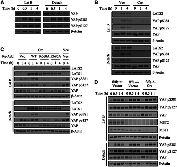
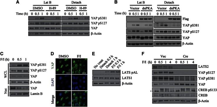
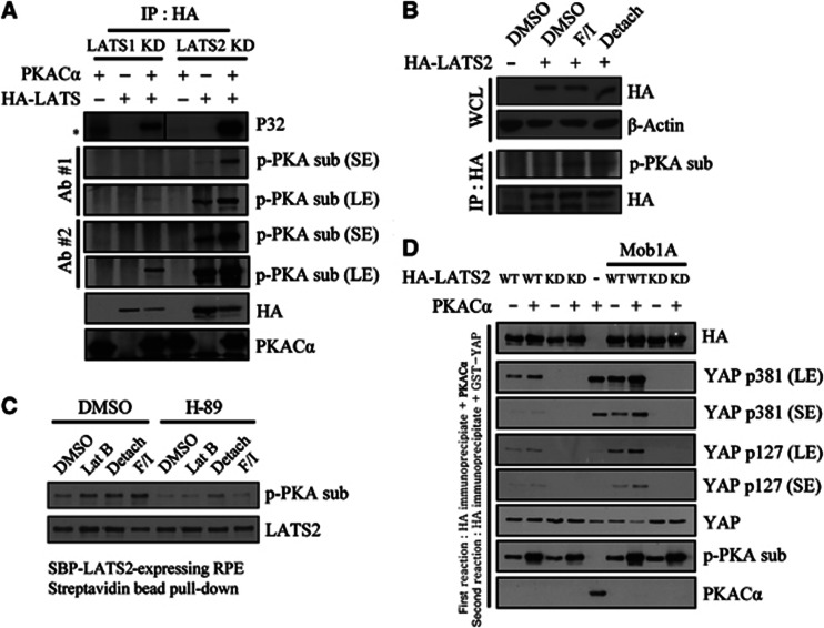
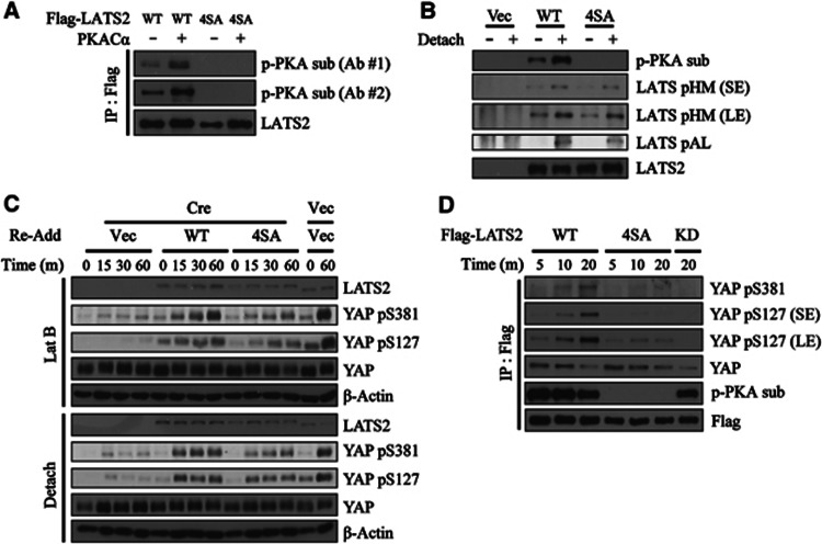
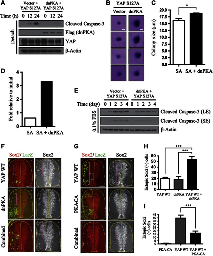
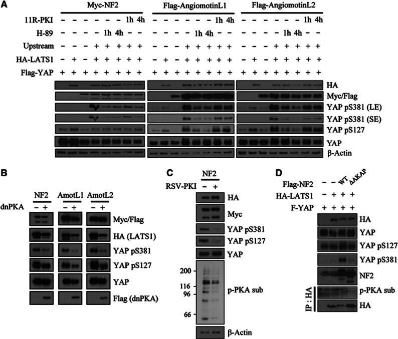
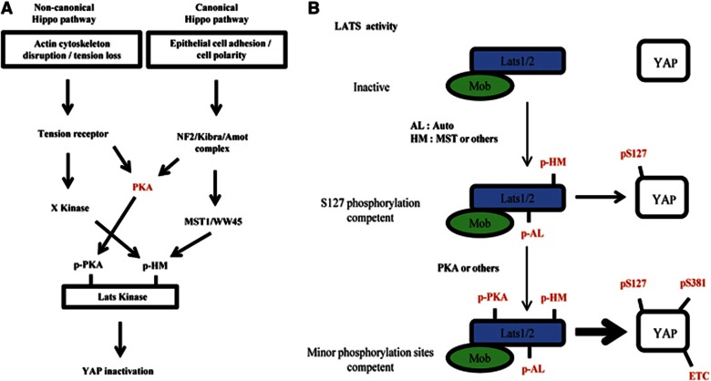
Actin cytoskeletal damage induces inactivation of the oncoprotein YAP (Yes-associated protein). It is known that the serine/threonine kinase LATS (large tumour suppressor) inactivates YAP by phosphorylating its Ser127 and Ser381 residues. However, the events downstream of actin cytoskeletal changes that are involved in the regulation of the LATS-YAP pathway and the mechanism by which LATS differentially phosphorylates YAP on Ser127 and Ser381 in vivo have remained elusive. Here, we show that cyclic AMP (cAMP)-dependent protein kinase (PKA) phosphorylates LATS and thereby enhances its activity sufficiently to phosphorylate YAP on Ser381. We also found that PKA activity is involved in all contexts previously reported to trigger the LATS-YAP pathway, including actin cytoskeletal damage, G-protein-coupled receptor activation, and engagement of the Hippo pathway. Inhibition of PKA and overexpression of YAP cooperate to transform normal cells and amplify neural progenitor pools in developing chick embryos. We also implicate neurofibromin 2 as an AKAP (A-kinase-anchoring protein) scaffold protein that facilitates the function of the cAMP/PKA-LATS-YAP pathway. Our study thus incorporates PKA as novel component of the Hippo pathway.


Conflict of interest statement

The authors declare that they have no conflict of interest.

Figures

Figure 1
Figure 1
The LATS/Mob1 complex is essential for induction of YAP Ser127 and Ser381 phosphorylation by cytoskeletal damage. (A) Phosphorylation at Ser127 and Ser381 by actin cytoskeletal damage. NIH3T3 cells were treated with 5 μM latrunculin B or seeded onto poly-HEMA-coated dishes and incubated for the indicated times. (B) Indispensability of LATS1/2 for YAP phosphorylation. Lats1−/−;Lats2fl/fl MEFs were transduced with either empty or Cre retroviruses. Selected cells were treated as indicated. (C) Indispensability of intact LATS/Mob1 complex for YAP phosphorylation. SV40 LT-immortalized Lats1−/−;Lats2fl/fl MEFs were complemented with either LATS1 WT, LATS1 R660A or R696A mutants. After Cre infection, cells were treated as indicated. Lanes 13 and 14 were infected with empty virus in place of Cre to measure LATS2 deletion efficiency. (D) Dispensability of MST1/2 for YAP phosphorylation. Mst1fl/fl;Mst2+/+vector, Mst1fl/fl;Mst2−/− vector, and Mst1fl/fl;Mst2−/− Cre MEFs were infected and treated as indicated.
Figure 2
Figure 2
Requirement of cAMP/PKA signalling for induction of YAP Ser381 phosphorylation by cytoskeletal damage. (A) Effect of H-89 on YAP phosphorylation by cytoskeletal damage. NIH3T3 cells were pre-treated with 20 μM H-89 for 1 h followed by addition of latrunculin B or seeding onto poly-HEMA-coated dishes. (B) Effect of dnPKA on YAP phosphorylation by cytoskeletal damage. NIH3T3 cells were infected with Flag–dnPKA retroviruses. Selected cells were treated with latrunculin B or seeded onto poly-HEMA-coated dishes. (C, D) Effect of PKA agonist on YAP phosphorylation. NIH3T3 cells were stimulated with 20 μM forskolin and 500 μM IBMX for the indicated times. YAP phosphorylation (C) and localization (C, D) were determined. WCL, whole cell lysate; Nuc, nuclear lysate. Scale bar, 10 μm. (E) LATS activation by PKA agonist or cell detachment. LATS activation loop (AL) phosphorylation was determined in cells treated with the indicated stimuli. (F) Requirement of LATS1/2 for YAP phosphorylation by PKA agonist. Lats1−/−;Lats2fl/fl MEFs were transduced with either empty or Cre retroviruses. Selected cells were treated with forskolin/IBMX. *, nonspecific signal.
Figure 3
Figure 3
Phosphorylation of LATS1/2 by PKA enhances LATS kinase activity in cell-free systems and in intact cells. (A) PKA phosphorylates LATS in cell-free system. HA–LATS1 KD (kinase dead) or HA–LATS2 KD was immunoprecipitated from transfected 293T cells. Immunoprecipitated beads were incubated with purified PKACα and radiolabelled ATP. Reaction products were analysed isotopically or by western blotting with phospho-PKA substrate antibodies. *, nonspecific signal. (B) PKA phosphorylates LATS in intact cells. HA–LATS2-transfected NIH3T3 cells were treated with the indicated stimuli for 1 h. After immunoprecipitation with anti-HA antibody, phosphorylation by PKA was examined using a phospho-PKA substrate antibody. WCL, whole cell lysate. (C) RPE cells stably expressing SBP-LATS2 were pre-treated with 20 μM H-89 for 1 h, followed by an additional 1-h treatment with the indicated stimuli. LATS2 was pulled down using Streptavidin agarose bead and assayed as in panel (B). (D) LATS2 pre-incubated with PKA has increased kinase activity. HA–LATS2 WT or KD mutant was immunoprecipitated from 293T cells and reacted with PKACα. PKACα was extensively washed out, followed by incubation with 1 μg GST–YAP (full-length) protein and 200 μM unlabelled ATP. Reaction products were analysed by SDS–PAGE and immunoblotting. In lanes 6–9, HA–Mob1A was co-transfected to increase overall kinase activity. In lane 5, GST–YAP was reacted with PKACα to examine possible background phosphorylation of YAP by PKA. SE, short exposure; LE, long exposure.
Figure 4
Figure 4
Identification of PKA target sites on LATS2 and their contribution to cytoskeletal damage-induced YAP phosphorylation. (A) LATS2 4SA mutant is not phosphorylated by PKA in vitro. Flag–LATS2 WT or LATS2 4SA were prepared by immunoprecipitation from transfected 293T cells. Flag immunoprecipiates were incubated with cold ATP and PKACα. Reaction products were analysed by two antibodies against phosphorylated PKA substrate. (B) LATS2 4SA mutant is not phosphorylated by cell detachment. NIH3T3 cells were transfected with empty vector, Flag–LATS2 WT, or LATS2 4SA. Transfected cells were suspended for 1 h followed by Flag immunoprecipiation. Flag immunoprocipiates were fractionated by SDS–PAGE and analysed with indicated phospho-specific antibodies. SE, short exposure; LE, long exposure. (C) LATS2 4SA-reconstituted cells attenuate YAP phosphorylation. SV40 LT-immortalized Lats1−/−;Lats2fl/fl MEFs were complemented with either LATS2 WT or LATS2 4SA mutant. After Cre infection, cells were treated as indicated. Lanes 13 and 14 were infected with empty virus in place of Cre to measure LATS2 deletion efficiency. The different mobility of human LATS2 (complemented products) and murine Lats2 (endogenous product before deletion) ensures efficient excision of Lats2 in lanes 1–12. (D) Reduced kinase activity of LATS2 4SA mutant. 293T cells were transfected with indicated Flag-tagged LATS2 constructs. LATS2 was immunoprecipiated by Flag antibody followed by time-course kinase assay as indicated.
Figure 5
Figure 5
Functional cooperation between PKA and YAP. (A) YAP S127A and dnPKA cooperate to resist anoikis. NIH3T3 cells expressing Flag–YAP S127A alone or Flag–YAP S127A plus Flag–dnPKA were seeded onto poly-HEMA-coated dishes and incubated for the indicated times. The apoptosis index was measured by immunoblotting for cleaved caspase-3. (B) Cells from (A) were also grown in soft agar for 15 days. Colonies were stained with crystal violet and imaged with a dissecting microscope. Scale bar, 10 μm. (C) Quantification of average colony size for the results in (B). The result was quantified from three independent experiments with triplicates in each experiment. Error bar indicates s.e.m. (two-tailed Student’s t-test). *P<0.05. (D) YAP S127A and dnPKA cooperate to resist serum starvation-induced cell death. Approximately 5 × 10³ cells from (A) were seeded onto 6-well plates. One day later, media were changed to DMEM containing 0.1% FBS and cells were maintained for 6 days. Cell numbers were counted and expressed as fold induction relative to the original number of cells (5 × 10³). The graph shown is a representative result from two independent experiments. (E) Cells incubated in 0.1% FBS for the indicated number of days were analysed by immunoblotting for caspase-3 cleavage. SE, short exposure; LE, long exposure. (F) YAP WT and dnPKA cooperate to generate ectopic neural progenitors in developing chick. Immunohistochemical analyses of ectopic neural progenitor formation in chick spinal cords electroporated with YAP WT, dnPKA, or YAP WT plus dnPKA, along with LacZ (left) to mark the transfected side. Neural progenitors were detected by immunostaining for Sox2. Brackets indicate ectopic Sox2+ neural progenitor cells. +, electroporated side. Scale bar, 75 μm. (G) PKA-CA suppresses YAP-induced generation of ectopic neural progenitors. Similar procedure as in (F) using YAP WT, PKA-CA, and YAP WT plus PKA-CA. Scale bar, 75 μm. (H) The number of ectopic neural progenitors from (F) were quantified. ***P<0.001. (I) Quantification of results in (G). ***P<0.001. Error bars indicate s.e.m.’s (two-tailed Student’s t-test).
Figure 6
Figure 6
Requirement for PKA activity in mediating YAP phosphorylation induced by engagement of the canonical Hippo pathway. (A) YAP phosphorylation induced by Hippo pathway upstream components is opposed by PKA inhibition. 293T cells were transfected with Flag–YAP, HA–LATS1, and Myc–NF2 or Flag-angiomotin-like 1/2 as indicated. Forty-eight hours after transfection, cells were incubated with 20 μM H-89 or 50 μM 11R-PKI for the indicated times. SE, short exposure; LE, long exposure. (B) 293T cells were transfected as in (A) with or without dnPKA co-transfection. Lysates were analysed 48 h after transfection. (C) 293T cells were transfected as in (A) with or without RSV-PKI co-transfection. Transfection of RSV-PKI was confirmed by western blotting the whole transferred membrane against phospho-PKA substrate antibody. (D) NF2 mutant that is defective in AKAP function do not induce YAP Ser381 and PKA-mediated LATS phosphorylation. 293T cells were transfected with Flag–YAP, HA–LATS1, and Flag–NF2 WT or Flag–NF2 ΔAKAP mutant. HA–LATS1 was immunoprecipitated and analysed for phosphorylation by PKA.
Figure 7
Figure 7
Model depicting the refined Hippo pathway and step-wise LATS activation. (A) Signalling scheme of the Hippo pathway. PKA serves a common function in both non-canonical and canonical Hippo pathways. See the text for further details. (B) LATS1/2 is activated in two steps. The first step involves Mob1 binding and phosphorylation of the AL and HM. These events are sufficient for YAP Ser127 phosphorylation, which is a good LATS substrate. The second step involves further activation by PKA (or others kinases). Only this fully activated LATS can effectively phosphorylate poor substrates, including YAP Ser38.

Similar articles

Cited by

References

    1. Angus L, Moleirinho S, Herron L, Sinha A, Zhang X, Niestrata M, Dholakia K, Prystowsky MB, Harvey KF, Reynolds PA, Gunn-Moore FJ (2012) Willin/FRMD6 expression activates the Hippo signaling pathway kinases in mammals and antagonizes oncogenic YAP. Oncogene 31: 238–250 - PubMed
    1. Baldwin C, Garnis C, Zhang LW, Rosin MP, Lam WL (2005) Multiple microalterations detected at high frequency in oral cancer. Cancer Res 65: 7561–7567 - PubMed
    1. Bashyam MD, Bair R, Kim YH, Wang P, Hernandez-Boussard T, Karikari CA, Tibshirani R, Maitra A, Pollack JR (2005) Array-based comparative genomic hybridization identifies localized DNA amplifications and homozygous deletions in pancreatic cancer. Neoplasia 7: 556–562 - PMC - PubMed
    1. Baumgartner R, Poernbacher I, Buser N, Hafen E, Stocker H (2010) The WW domain protein kibra acts upstream of Hippo in Drosophila. Developmental Cell 18: 309–316 - PubMed
    1. Cai J, Zhang N, Zheng Y, de Wilde RF, Maitra A, Pan D (2010) The Hippo signaling pathway restricts the oncogenic potential of an intestinal regeneration program. Genes Dev 24: 2383–2388 - PMC - PubMed

Publication types

MeSH terms