. 2013 Jun;140(12):2632-42.
doi: 10.1242/dev.090829. Epub 2013 May 1.

VEGFA-dependent and -independent pathways synergise to drive Scl expression and initiate programming of the blood stem cell lineage in Xenopus

Affiliations

VEGFA-dependent and -independent pathways synergise to drive Scl expression and initiate programming of the blood stem cell lineage in Xenopus

Aldo Ciau-Uitz et al. Development. 2013 Jun.

Abstract

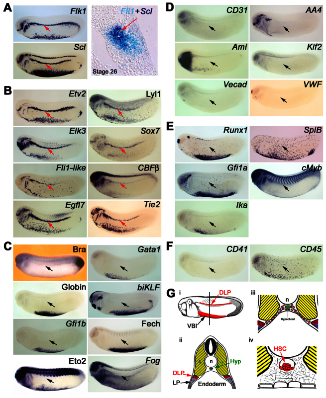
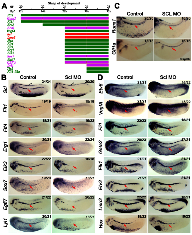
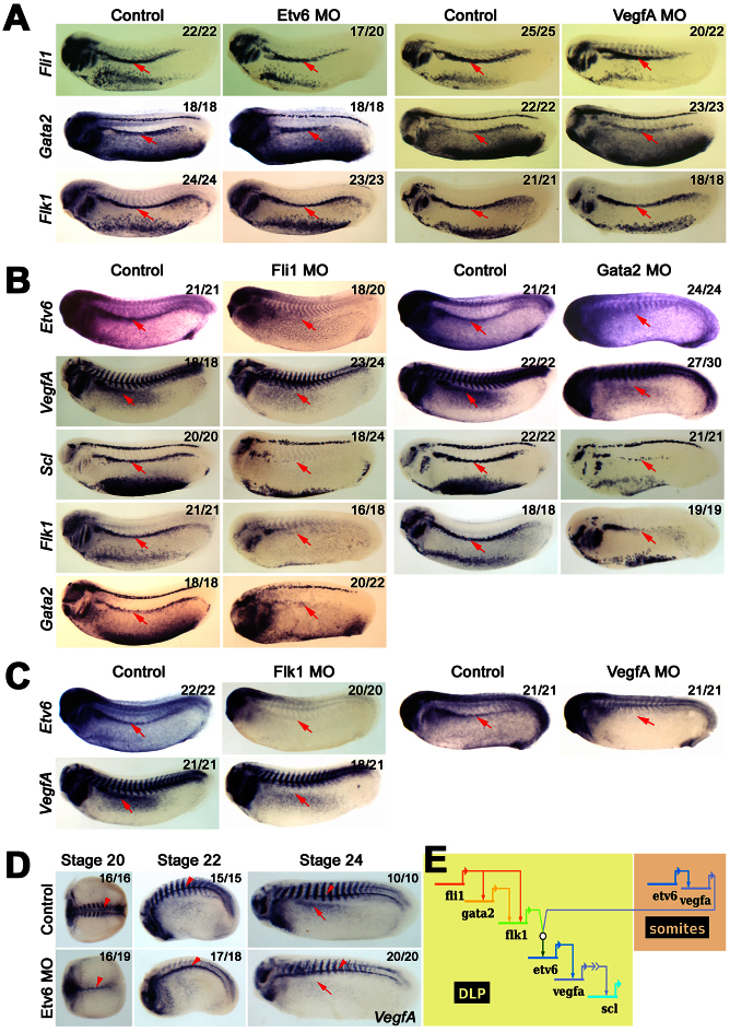
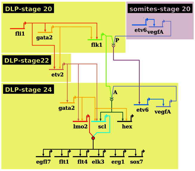
The first haematopoietic stem cells share a common origin with the dorsal aorta and derive from putative adult haemangioblasts in the dorsal lateral plate (DLP) mesoderm. Here we show that the transcription factor (TF) stem cell leukaemia (Scl/Tal1) is crucial for development of these adult haemangioblasts in Xenopus and establish the regulatory cascade controlling its expression. We show that VEGFA produced in the somites is required to initiate adult haemangioblast programming in the adjacent DLP by establishing endogenous VEGFA signalling. This response depends on expression of the VEGF receptor Flk1, driven by Fli1 and Gata2. Scl activation requires synergy between this VEGFA-controlled pathway and a VEGFA-independent pathway controlled by Fli1, Gata2 and Etv2/Etsrp/ER71, which also drives expression of the Scl partner Lmo2. Thus, the two ETS factors Fli1 and Etv6, which drives the VEGFA expression in both somites and the DLP, sit at the top of the adult haemangioblast gene regulatory network (GRN). Furthermore, Gata2 is initially activated by Fli1 but later maintained by another ETS factor, Etv2. We also establish that Flk1 and Etv2 act independently in the two pathways to Scl activation. Thus, detailed temporal, epistatic measurements of key TFs and VEGFA plus its receptor have enabled us to build a Xenopus adult haemangioblast GRN.

Keywords: ETS; Gene regulatory network; Haemangioblast; Haematopoietic stem cell; Scl; VEGF.


Figures

Fig. 1.
Fig. 1.
Characterisation of DLP mesoderm adult haemangioblasts. (A) Adult haemangioblasts localise in the DLP adjacent to the somites and are the earliest HSC progenitors detectable by co-expression of VEGFR2, Flk1, and the stem cell leukaemia gene (Scl), which marks the emergence of haemangioblasts. The panel on the right shows co-expression of the endothelial gene, Fli1, and Scl on a 10 μM section. Note that no morphological differences are observed between adult haemangioblasts and surrounding tissues. (B) Expression analysis revealing novel haematopoietic, Lyl1, Sox7 and CBFβ, as well as novel endothelial, Etv2, Elk3, Fli1-like, Egfl7 and Tie2, gene expression in adult haemangioblasts. (C) Adult haemangioblasts do not express the early mesodermal marker, Brachyury (Bra), nor the erythrocyte differentiation genes, Gata1, Globin, biKLF, Gfi1b, Ferrochelatase (Fech), Eto2 and Fog. (D) Adult haemangioblasts do not express genes associated with mature blood vessels, CD31, AA4, Ami, Klf2, Vecad and VWF. (E) Adult haemangioblasts do not express key HSC-associated genes such as Runx1, SpiB, Gfi1a, cMyb and Ikaros (Ika). (F) Expression of CD41, a gene associated with haematopoietic commitment, and CD45, which is associated with haematopoietic differentiation, are undetectable in adult haemangioblasts. The DLP is indicated by the red or black arrows. All embryos were hybridised as whole mounts and are shown in lateral view, with anterior to the left and dorsal to the top. All embryos are shown at stage 26. (G) Schematic representation of the location of the DLP adult haemangioblast population and its derivatives. (i) Representation of a stage 26 embryo showing in red the Scl expression domains, the ventral blood island and the DLP mesoderm. (ii) Cross section of a stage 26 embryo, at the level indicated by the line in panel i, showing the location of the DLP. Note that adult haemangioblasts (red tissue) lies immediately ventral to the somites (yellow tissue) but at some distance from the hypochord (green tissue), where the DA and HSCs eventually emerge. (iii) Picture showing DA/HSC progenitors migrating from the DLP to the hypochord, a process taking place from stage 28 to 31. (iv) Schematic representation of HSCs emerging in association with the ventral wall of the DA; these haematopoietic clusters are found from stage 42 to 44, 4 days after the specification of adult haemangioblasts in the DLP. Hyp, hypochord; n, notochord; s, somites; VBI, ventral blood island.
Fig. 2.
Fig. 2.
Scl marks and is essential for the emergence of adult haemangioblasts in the DLP. (A) Bar chart summarising the haematopoietic (magenta and red) and endothelial (green) expression hierarchy in the DLP reveals that adult haemangioblast programming takes place in four discrete transcriptional steps. Expression profiles were obtained by analysis of embryos subjected to whole-mount in situ hybridisation. Stages of development by morphological traits (NF) and hours post fertilisation (Hpf) are indicated at the top and are as indicated by Nieuwkoop and Faber (Nieuwkoop and Faber, 1967). (B) Expression analysis showing haematopoietic (Scl, Sox7 and Lyl1) and endothelial (Flt1, Flt4, Egr1, Elk3 and Egfl7) genes dependent on Scl for their expression in the DLP (arrows). (C) Expression analysis showing that haemogenic endothelium, as indicated by Runx1 and Gfi1a, fails to be established in the DA of Scl morphants. The DA is indicated by red arrows. (D) Expression analysis showing that the haematopoietic genes, Gata2, Lmo2 and Etv6; and the endothelial genes, Fli1, Flk1, Etv2 and Hex, in the DLP, as well as the expression of the growth factor, VegfA, are not dependent on Scl. The DLP is indicated by the red arrow. All embryos were hybridised as whole mounts and are shown in lateral view, with anterior to the left and dorsal to the top. Embryos in B and D were analysed at stage 26, whereas those in C are stage 39 embryos. Numbers of embryos represented by each panel, out of the number analysed, are indicated in the top right corner. n, neurons, VBI, ventral blood island.
Fig. 3.
Fig. 3.
Fli1 and Gata2 regulate Etv6/VEGFA signalling in the DLP to control the establishment of adult haemangioblasts. (A) Expression analysis showing that the expression of Fli1, Gata2 and Flk1 in the DLP (arrows) are not dependent on Etv6/VegfA signalling. (B) Expression analysis showing that the expression of Etv6, VegfA, Scl and Flk1 in the DLP (arrows) are dependent on both Fli1 and Gata2 transcriptional activities and that Gata2 expression is controlled by Fli1. (C) Expression analysis showing that the expression of Etv6 and VegfA in the DLP (arrows) is dependent on VEGFA signalling. (D) Expression analysis in staged embryos shows that Etv6 controls the expression of VegfA in the DLP (arrows) and in the somites (striped staining) but not in the hypochord (arrowheads). Note that VegfA expression in the somites is initially completely dependent on Etv6 but that by stage 24 some recovery is observed. (E) Diagram summarising the genetic cascade regulating Scl expression in the DLP. Relationships between genes are depicted by an arrow from the regulating gene to the regulated genes. This regulatory network shows that, although Fli1 and Gata2 are required for Etv6, VegfA and Scl expression, the establishment of endogenous VEGFA signalling and Scl expression in the DLP can only happen when VEGFA is produced in the somites, that paracrine VEGFA signalling is absolutely required for the initiation of the adult haemangioblast programme. The open circle indicates the interaction between the VEGFA receptor, Flk1, and VEGFA ligand from the somites. The arrow with chevrons indicates that VEGFA activates Scl indirectly, and that an intermediate factor controlled by VEGFA signalling is required. All embryos were hybridised as whole mounts and are shown in lateral view, with anterior to the left and dorsal to the top. All embryos are shown at stage 26, unless otherwise indicated. Numbers of embryos represented by each panel, out of the number analysed, are indicated in the top right corner.
Fig. 4.
Fig. 4.
Etv2 acts downstream of Fli1 to control Gata2 and adult haemangioblast specification. (A) Expression analysis showing that Etv2 is a major regulator of gene expression in the DLP (arrows). Note that Etv6 and VegfA expression in the DLP is not dependent on Etv2. (B) Analysis of the transcriptional regulation of Etv2 in the DLP. Expression analysis showing that Etv2 expression in the DLP (arrows) is not dependent on Etv6/VEGFA signalling but dependent on Fli1 and Gata2. Note Etv2 ectopic expression in Fli1 morphants (arrowhead). (C) Expression analysis in staged embryos showing that Gata2 expression in the DLP (arrows) is initially independent of Etv2's transcriptional regulation but is controlled by Etv2 after stage 24 of development. All embryos were hybridised as whole mounts and are shown in lateral view, with anterior to the left and dorsal to the top. All embryos are shown at stage 26, unless otherwise indicated. Numbers of embryos represented by each panel, out of the number analysed, are indicated in the top right corner. (D) Diagram summarising an endogenous cell-autonomous and VEGFA-independent genetic cascade required for Scl expression in the DLP. This cascade is initiated by Fli1 and maintained by Etv2.
Fig. 5.
Fig. 5.
The adult haemangioblast genetic regulatory network. Network summarising the genetic interactions programming adult haemangioblasts in the DLP as determined by MO knockdown. Strikingly, somitic VEGFA is absolutely essential for Flk1 receptor activation in haemangioblast precursors; this activation results in the establishment of endogenous, cell-autonomous VEGFA signalling. Importantly, Scl expression and haemangioblasts cannot be specified unless this endogenous VEGFA signalling synergises with a VEGFA-independent pathway controlled by Fli1 and Etv2. The timing and tissue of expression is denoted by the position of the gene on the network, whereas its relationships with genes expressed at the same time or later is depicted by an arrow from the regulating gene to the regulated gene. Arrows with chevrons indicate the interaction between VEGFA and its receptor, Flk1. P indicates paracrine VEGFA signalling, whereas A indicates endogenous autocrine signalling. The red circle represents the formation of a well-characterised protein complex between Scl and Lmo2.

Similar articles

Cited by

References

    1. Afouda B. A., Ciau-Uitz A., Patient R. (2005). GATA4, 5 and 6 mediate TGFbeta maintenance of endodermal gene expression in Xenopus embryos. Development 132, 763-774 - PubMed
    1. Bertwistle D., Walmsley M. E., Read E. M., Pizzey J. A., Patient R. K. (1996). GATA factors and the origins of adult and embryonic blood in Xenopus: responses to retinoic acid. Mech. Dev. 57, 199-214 - PubMed
    1. Chung Y. S., Zhang W. J., Arentson E., Kingsley P. D., Palis J., Choi K. (2002). Lineage analysis of the hemangioblast as defined by FLK1 and SCL expression. Development 129, 5511-5520 - PubMed
    1. Ciau-Uitz A., Walmsley M., Patient R. (2000). Distinct origins of adult and embryonic blood in Xenopus. Cell 102, 787-796 - PubMed
    1. Ciau-Uitz A., Liu F., Patient R. (2010a). Genetic control of hematopoietic development in Xenopus and zebrafish. Int. J. Dev. Biol. 54, 1139-1149 - PubMed

Publication types

MeSH terms