. 2013 Jul 1;73(13):3913-26.
doi: 10.1158/0008-5472.CAN-12-4318. Epub 2013 May 1.

miR-124 inhibits STAT3 signaling to enhance T cell-mediated immune clearance of glioma

Affiliations

miR-124 inhibits STAT3 signaling to enhance T cell-mediated immune clearance of glioma

Jun Wei et al. Cancer Res. .

Abstract

miRNAs (miR) have been shown to modulate critical gene transcripts involved in tumorigenesis, but their role in tumor-mediated immunosuppression is largely unknown. On the basis of miRNA gene expression in gliomas using tissue microarrays, in situ hybridization, and molecular modeling, miR-124 was identified as a lead candidate for modulating STAT3 signaling, a key pathway mediating immunosuppression in the tumor microenvironment. miR-124 is absent in all grades and pathologic types of gliomas. Upon upregulating miR-124 in glioma cancer stem cells (gCSC), the STAT3 pathway was inhibited, and miR-124 reversed gCSC-mediated immunosuppression of T-cell proliferation and induction of forkhead box P3 (Foxp3)(+) regulatory T cells (Treg). Treatment of T cells from immunosuppressed glioblastoma patients with miR-124 induced marked effector response including upregulation of interleukin (IL)-2, IFN-γ, and TNF-α. Both systemic administration of miR-124 or adoptive miR-124-transfected T-cell transfers exerted potent anti-glioma therapeutic effects in clonotypic and genetically engineered murine models of glioblastoma and enhanced effector responses in the local tumor microenvironment. These therapeutic effects were ablated in both CD4(+)- and CD8(+)-depleted mice and nude mouse systems, indicating that the therapeutic effect of miR-124 depends on the presence of a T-cell-mediated antitumor immune response. Our findings highlight the potential application of miR-124 as a novel immunotherapeutic agent for neoplasms and serve as a model for identifying miRNAs that can be exploited as immunotherapeutics.


Figures

Figure 1
Figure 1
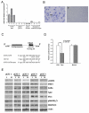
miR-124 expression is significantly reduced in GBMs and inhibits STAT3. (A) Relative expression levels of miR-124 were detected by TaqMan quantitative PCR. There was a significant difference in the relative expression levels of miR-124 between normal brain tissues, GBM specimens, and gCSCs. (B) Representative specimens of normal brain (I) demonstrating miR-124 expression (arrow) by in situ hybridization in neurons and in a GBM (II) lacking miR-124 expression (at 40x magnification). (C) Sequence of the predicted miR-124 binding site on the STAT3-3’UTR and the STAT3-3’UTR mutant that disrupted miR-124 binding. On the basis of predictive algorithms, there exists an 11-bp nucleotide binding site in the STAT3 3′-UTR that is imperfectly paired with miR-124, which is predicted to lead to STAT3 mRNA degradation. (D) Relative luciferase activity of HeLa cells after transfection with miR-124- or scramble control-expressing plasmids in conjunction with a wild type STAT3 3′-UTR or miR-124 binding site mutant reporter construct, demonstrating inhibition of STAT3. **P < 0.01. (E) Western blot analysis of the STAT3 signaling pathway gCSCs transfected with scramble control (S) compared with miR-124 (M).
Figure 2
Figure 2
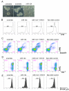
miR-124 reverses glioma-mediated immunosuppression. (A) The gCSC phenotype was markedly altered by miR-124, as photographed 48 hours after transfection. (B) T-cell proliferation was detected by flow cytometry analysis with CFSE staining after 3 days of treatment with medium alone, gCSC-scramble-transfected conditioned medium, gCSC-miR-124-transfected conditioned medium, or gCSC-miR-124 + STAT3-transfected conditioned medium. miR-124 up regulation caused a reversal of inhibition in T-cell proliferation compared with the gCSC-scramble-transfected conditioned medium. STAT3 overexpression restored gCSC inhibition on T-cell proliferation. (C) T-cell apoptosis was measured by the percentage of annexin V+ 7-AAD+ cells. Similarly, miR-124 up regulation decreased gCSC-induced T-cell apoptosis, whereas STAT3 overexpression reversed the miR-124 effect. (D) T-cells were analyzed on the basis of CD4 and FoxP3 expression levels, by flow cytometry analysis. Up regulation of miR-124 caused a decline in Treg induction compared with the scrambled gCSC control, and STAT3 addition restored the ability of the gCSCs to induce Tregs. (E) The functional suppressive activity of FoxP3+ Tregs induced in (D) was verified by autologous coculture with CD4+ T-cells. The experiments were repeated three times with similar results, and one representative set of data is shown.
Figure 3
Figure 3
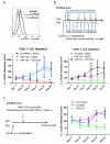
miR-124 enhances T-cell effector cytokine production in GBM patients. PBMCs were isolated from the freshly drawn blood of GBM patients undergoing resection, transfected with miR-124 or scramble control oligonucleotides, and sequentially stimulated with anti-CD3/anti-CD28 antibodies for T-cell proliferation. miR-124 administration significantly enhanced effector cytokine responses, such as IFN-γ (n=10) (A), IL-2 (n=10) (B), and TNF-α (n=7) (C), in both CD4+ T and CD8+ T-cells of PBMCs from GBM patients. miR-124 up regulation suppressed pSTAT3 activity in CD8+ T-cells (n=9) and to a lesser degree in the CD4+ T-cells (n=9) (D). Each symbol represents PBMCs from different patients without transfection or with transfection with either the scramble control or miR-124.
Figure 4
Figure 4
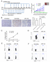
miR-124 exerts potent efficacy to suppress subcutaneous GL261 tumors in a syngeneic mouse model. (A) The treatment schema and the volumes of subcutaneous GL261 tumors in C57BL/6J mice treated intratumorally with either miR-124 or scramble control, or left untreated starting on day 6 (n=10/group/experiment). The figure is the result of a single experiment. In the miR-124 group, * denotes a P value of <0.01 compared with both the untreated and scramble control tumors. Standard deviations are shown. Arrows indicate days of treatment and tumor size measurements. The inset photo on the right is of a representative ex vivo GL261 tumor comparison between miR-124- and scramble control-treated tumor-bearing mice. (B) Ex vivo immunohistochemical analysis of gliomas, untreated (n=2), or treated with a scramble control (n=5), or miR-124 (n=3), which demonstrates a marked decrease in p-STAT3 expression in the local tumor microenvironment (P = 0.0039). (C) Splenocytes from miR-124 intratumorally treated GL261 mice (n=3) have markedly increased cytotoxicity at 48 hours after coculture with GL261 cells compared with that in splenocytes from scramble control oligonucleotide-treated tumor-bearing mice (P < 0.001). The ratios of splenocytes to GL261 cells are 10:1, 40:1, and 100:1. Error bars in the curve represent the standard deviation in the data from 3 mice. (D) Decreased tumor-infiltrating FoxP3+ Tregs in miR-124-treated GL261 mice. A single example is shown, but identical results were obtained in two other tumor bearing animals. Furthermore, miR-124 administration enhances IFN-γ and TNF-α production in CD4 T-cells (E) and CD8 T-cells (F) in the tumor local microenvironment. One set of representative FACS plots is shown. Similar results were obtained from another two tumor-bearing mice treated with miR-124 or scramble control, respectively.
Figure 5
Figure 5
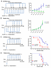
The therapeutic effect of miR-124 is lost in immune-incompetent models. (A) The treatment schema and the volumes of subcutaneous GL261 tumors in C57BL/6J mice treated intravenously with miR-124 or scramble control starting on day 7 (n=10/group/experiment). In the miR-124 group, * denotes a P value of <0.01 compared with scramble control tumors. Standard deviations are shown. The arrows indicate days of treatment and tumor size measurements. (B) The treatment schema and volumes of subcutaneous GL261 tumors in nude mice treated intratumorally with miR-124 or scramble control starting on day 7 (n=10/group/experiment). Standard deviations are shown. Arrows indicate days of treatment and tumor size measurements. (C) Treatment schema and graph of the Kaplan-Meier estimate, demonstrating better survival in C57BL/6J mice with miR-124-treated intracerebral GL261 gliomas (n=10/group/experiment) than in scrambled controls. * denotes a P value = 0.02 compared with scramble control gliomas. (D) Graph of the Kaplan-Meier estimate demonstrates miR-124’s lack of therapeutic effect in nude mice with intracerebral GL261 gliomas (n=10/group/experiment, P = 0.24).
Figure 6
Figure 6
T-cells mediate the antiglioma immune therapeutic efficacy of miR-124. (A) Histogram shows miR-124 transfection inhibited p-STAT3 activity in adoptively transferred T cells. (B) The treatment schema and volumes of subcutaneous GL261 tumors in C57BL/6J mice treated intratumorally with miR-124 or scramble control subcutaneously on day 7 (n=8/group/experiment) in the setting of in vivo CD4+ T-cells or CD8+ T-cell depletion. Arrows indicate days of treatment and tumor size measurements. *denotes a P value of <0.01 comparing the anti-CD4 or anti-CD8 depletion group with the isotype and miR-124-treated group. Standard deviations are shown. (C) The treatment schema and volumes of subcutaneous GL261 tumors in C57BL/6J mice treated intravenously with miR-124 or scramble control transfected CD3+ T-cells on day 14 (n=10/group/experiment). *denotes a P value of <0.01 comparing the miR-124 transfected T-cell treated group with the scramble control and untreated group.
Figure 7
Figure 7
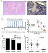
miR-124 exerts a therapeutic effect in Ntv-a mice. (A) Representative hematoxylin and eosin staining of a high-grade glioma induced in Ntv-a mice transfected with RCAS-PDGFB and RCAS-STAT3 transgenes demonstrates neovascular proliferation (arrow) and pseudopallisading necrosis (arrowhead) at 100x magnification. (B) Representative specimen from the brain of an Ntv-a mouse transfected with the RCAS-PDGFB and RCAS-STAT3 transgenes demonstrates miR-124 expression by in situ hybridization in neurons surrounding a glioma devoid of miR-124 expression (arrow) at 400x magnification. (C) Treatment schema and graph of the Kaplan-Meier estimate demonstrates improved survival in miR-124-treated Ntv-a mice transfected with the RCAS-PDGFB and RCAS-STAT3 transgenes (n=9/group) compared with scramble control and untreated mice (lipofectamine 2000 vehicle only). *denotes a P value of 0.04. (D) Summary graph demonstrates the incidence of high- and low-grade gliomas on the basis of hematoxylin and eosin staining features of necrosis and neovascular proliferation in miR-124-treated Ntv-a mice transfected with RCAS-PDGFB and RCAS-STAT3 transgenes (n=7) compared with scrambled controls (n=8) and untreated mice (n=7) (P < 0.0001). (E) An ex vivo immunohistochemical analysis of gliomas, untreated (n=7) or treated with a scramble control (n=6) or miR-124 (n=7), demonstrates a marked decrease in p-STAT3 expression in the local tumor microenvironment of the miR-124-treated group (scramble vs. miR-124: P = 0.003; untreated vs miR-124: P = 0.007; untreated vs scramble: P = 0.87). Quantification of p-STAT3 expression was obtained by averaging the number of nuclear positive p-STAT3 cells by immunohistochemistry from 10 non-overlapping high-power microscopic fields (magnification × 400) of the gliomas obtained from either untreated RCAS-PDGF-B + RCAS-STAT3 mice or mice treated with the scramble control or miR-124. Each dot represents the analysis of one mouse glioma.

Similar articles

Cited by

References

    1. Brooks WH, Markesbery WR, Gupta GD, Roszman TL. Relationship of lymphocyte invasion and survival of brain tumor patients. Ann Neurol. 1978;4:219–24. - PubMed
    1. Jacobs JF, Idema AJ, Bol KF, Grotenhuis JA, de Vries IJ, Wesseling P, et al. Prognostic significance and mechanism of Treg infiltration in human brain tumors. J Neuroimmunol. 2010;225:195–9. - PubMed
    1. Kong LY, Wu AS, Doucette T, Wei J, Priebe W, Fuller GN, et al. Intratumoral mediated immunosuppression is prognostic in genetically engineered murine models of glioma and correlates to immunotherapeutic responses. Clin Cancer Res. 2010;16:5722–33. - PMC - PubMed
    1. Palma L, Di Lorenzo N, Guidetti B. Lymphocytic infiltrates in primary glioblastomas and recidivous gliomas. Incidence, fate, and relevance to prognosis in 228 operated cases. J Neurosurg. 1978;49:854–61. - PubMed
    1. Safdari H, Hochberg FH, Richardson EP., Jr Prognostic value of round cell (lymphocyte) infiltration in malignant gliomas. Surg Neurol. 1985;23:221–6. - PubMed

Publication types

MeSH terms