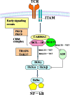
Changes in the MALT1-A20-NF-κB expression pattern may be related to T cell dysfunction in AML
- PMID: 23627638
- PMCID: PMC3641943
- DOI: 10.1186/1475-2867-13-37
Changes in the MALT1-A20-NF-κB expression pattern may be related to T cell dysfunction in AML
Abstract
To elucidate the characteristics of T-cell receptor (TCR) signal transduction in T-cells from acute myeloid leukemia (AML), the mucosa-associated-lymphoid-tissue lymphoma-translocation gene 1 (MALT1), A20, NF-κB and MALT1-V1 gene expression levels in CD3+ T cells sorted from the peripheral blood of patients with AML were analyzed by real-time PCR. A significantly lower MALT1 and A20 expression level was found in T cells from patients with AML compared with healthy controls (p = 0.045, p < 0.0001); however, the expression level of MALT1-V1 (variant 1) was significantly higher in the AML group than in the healthy control group (p = 0.006), and the expression level of NF-κB was increased in the AML group. In conclusion, the characteristics of the expression pattern of MALT1-A20-NF-κB and the distribution of MALT1 variants in T cells from AML were first characterized. Overall, low TCR-CD3 signaling is related to low MALT1 expression, which may related to T cell immunodeficiency, while the up-regulation of MALT1-V1 may play a role in overcoming the T cell activity by downregulating A20 in patients with AML, which may be related to a specific response to AML-associated antigens.
Figures


Similar articles
-
Alternative expression pattern of MALT1-A20-NF-κB in patients with rheumatoid arthritis.J Immunol Res. 2014;2014:492872. doi: 10.1155/2014/492872. Epub 2014 May 26. J Immunol Res. 2014. PMID: 24971370 Free PMC article.
-
Abnormal expression of A20 and its regulated genes in peripheral blood from patients with lymphomas.Cancer Cell Int. 2014 Apr 26;14:36. doi: 10.1186/1475-2867-14-36. eCollection 2014. Cancer Cell Int. 2014. PMID: 24790527 Free PMC article.
-
Characteristics of CARMA1-BCL10-MALT1-A20-NF-κB expression in T cell-acute lymphocytic leukemia.Eur J Med Res. 2014 Nov 11;19(1):62. doi: 10.1186/s40001-014-0062-8. Eur J Med Res. 2014. PMID: 25384343 Free PMC article.
-
MALT1--a universal soldier: multiple strategies to ensure NF-κB activation and target gene expression.FEBS J. 2015 Sep;282(17):3286-97. doi: 10.1111/febs.13325. Epub 2015 Jun 10. FEBS J. 2015. PMID: 25996250 Review.
-
Anti-apoptotic action of API2-MALT1 fusion protein involved in t(11;18)(q21;q21) MALT lymphoma.Apoptosis. 2005 Jan;10(1):25-34. doi: 10.1007/s10495-005-6059-6. Apoptosis. 2005. PMID: 15711920 Review.
Cited by
-
Changes of T-lymphocyte subpopulation and differential expression pattern of the T-bet and GATA-3 genes in diffuse large B-cell lymphoma patients after chemotherapy.Cancer Cell Int. 2014 Dec 24;14:85. doi: 10.1186/s12935-014-0085-9. eCollection 2014. Cancer Cell Int. 2014. PMID: 25705124 Free PMC article.
-
A20/Tumor Necrosis Factor α-Induced Protein 3 in Immune Cells Controls Development of Autoinflammation and Autoimmunity: Lessons from Mouse Models.Front Immunol. 2018 Feb 21;9:104. doi: 10.3389/fimmu.2018.00104. eCollection 2018. Front Immunol. 2018. PMID: 29515565 Free PMC article. Review.
-
Alternative expression pattern of MALT1-A20-NF-κB in patients with rheumatoid arthritis.J Immunol Res. 2014;2014:492872. doi: 10.1155/2014/492872. Epub 2014 May 26. J Immunol Res. 2014. PMID: 24971370 Free PMC article.
-
A20 Inhibits Intraocular Inflammation in Mice by Regulating the Function of CD4+T Cells and RPE Cells.Front Immunol. 2021 Feb 4;11:603939. doi: 10.3389/fimmu.2020.603939. eCollection 2020. Front Immunol. 2021. PMID: 33613524 Free PMC article.
-
Alteration of gene expression profile in CD3+ T-cells after downregulating MALT1.Immunotargets Ther. 2018 Nov 20;7:77-81. doi: 10.2147/ITT.S179656. eCollection 2018. Immunotargets Ther. 2018. PMID: 30538965 Free PMC article.
References
LinkOut - more resources
Full Text Sources
Other Literature Sources

