. 2013 Jul;87(13):7393-408.
doi: 10.1128/JVI.02825-12. Epub 2013 Apr 24.

Human cytomegalovirus pUL97 regulates the viral major immediate early promoter by phosphorylation-mediated disruption of histone deacetylase 1 binding

Affiliations

Human cytomegalovirus pUL97 regulates the viral major immediate early promoter by phosphorylation-mediated disruption of histone deacetylase 1 binding

Tarin M Bigley et al. J Virol. 2013 Jul.

Abstract

Human cytomegalovirus (HCMV) is a common agent of congenital infection and causes severe disease in immunocompromised patients. Current approved therapies focus on inhibiting viral DNA replication. The HCMV kinase pUL97 contributes to multiple stages of viral infection including DNA replication, controlling the cell cycle, and virion maturation. Our studies demonstrate that pUL97 also functions by influencing immediate early (IE) gene expression during the initial stages of infection. Inhibition of kinase activity using the antiviral compound maribavir or deletion of the UL97 gene resulted in decreased expression of viral immediate early genes during infection. Expression of pUL97 was sufficient to transactivate IE1 gene expression from the viral genome, which was dependent on viral kinase activity. We observed that pUL97 associates with histone deacetylase 1 (HDAC1). HDAC1 is a transcriptional corepressor that acts to silence expression of viral genes. We observed that inhibition or deletion of pUL97 kinase resulted in increased HDAC1 and decreased histone H3 lysine 9 acetylation associating with the viral major immediate early (MIE) promoter. IE expression during pUL97 inhibition or deletion was rescued following inhibition of deacetylase activity. HDAC1 associates with chromatin by protein-protein interactions. Expression of active but not inactive pUL97 kinase decreased HDAC1 interaction with the transcriptional repressor protein DAXX. Finally, using mass spectrometry, we found that HDAC1 is uniquely phosphorylated upon expression of pUL97. Our results support the conclusion that HCMV pUL97 kinase regulates viral immediate early gene expression by phosphorylation-mediated disruption of HDAC1 binding to the MIE promoter.


Figures
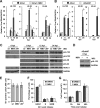
Fig 1
Fig 1
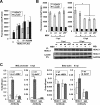
Disruption of HCMV pUL97 kinase activity alters viral gene expression at early times of infection. (A) Human foreskin fibroblasts were infected at 1 IU/cell with wild-type HCMV strain AD169 (ADwt) and treated with either DMSO vehicle or 40 μM maribavir (MBV). Viral RNA was harvested at multiple times and quantified by qRT-PCR relative to cellular GAPDH using sequence-specific primers. Error bars represent standard deviations from the means of two biological replicate experiments, and statistical significance was measured by Student's t test (*, P < 0.05). (B) Fibroblasts were infected using either ADwt or a UL97 galK substitution virus, ADdel97, at 1 IU/cell. Viral RNA was quantified as described above, and the data were evaluated as described above. (C) Western blot analysis using whole-cell lysates from infected fibroblasts treated as described above using the indicated antibodies. d97, ADdel97. (D) Western blot analysis of lysates from ADdel97-infected cells at 8 hpi with DMSO or MBV. (E) Cells were infected with ADwt, ADwt with 40 μM MBV, or ADdel97 at 0.01 IU/cell and fixed at 48 hpi, stained using an antibody against pUL123 and using DAPI for DNA, and evaluated by fluorescence microscopy. The data show the standard deviations and the means of 5 fields from each of 2 biological replicates, quantified at a magnification of ×100. (F) Fibroblasts were serum starved for 48 h and treated with 40 μM MBV or DMSO. Cells were released by the addition of serum with either 40 μM MBV or DMSO and analyzed for DNA content at 24 h using flow cytometry. The data show means and standard deviations from two biological replicates. (G) Fibroblasts (7.5 × 10⁴) were serum starved for 48 h. Cells were released from starvation by adding media containing serum and either DMSO or 40 μM MBV. Cells were dissociated from the plate and counted at the designated time points. The data show the averages of two biological replicates. Error bars are the standard deviations of biological replicates.
Fig 2
Fig 2
Characterization of entry and viral particle content following HCMV kinase disruption. (A) Fibroblasts were exposed to 1 IU/cell using ADwt, ADwt with 40 μM MBV, or ADdel97 virus and washed, and total cellular DNA was harvested at 1 hpi. Viral genomes were quantified using primers to MIEP relative to cellular DNA by qPCR; the data represent standard deviations and means of two biological replicates. (B) Cells were infected as above, fixed at 1 hpi, stained using an antibody against pUL83 and using DAPI for DNA, and evaluated by fluorescence microscopy at a magnification of ×400. (C) Viral particles from ADwt or two independent stocks of ADdel97 prepared from separate BAC transfections were isolated using centrifugation through a sucrose cushion. Equivalent infectious units (IU) of each stock were evaluated using Western blot analysis and the indicated antibodies. (D) Equivalent IUs of ADwt and ADdel97 virions were analyzed for DNA content by qPCR using the indicated primer sets. The data were normalized to DNA content from fibroblasts infected with the equivalent IU of ADwt. The data represent the standard deviations and means of two biological replicates.
Fig 3
Fig 3
Expression of kinase-active pUL97 is sufficient to enhance HCMV pUL123 expression from the viral genome. (A) Primary fibroblasts were electroporated with the ADdel97 BAC DNA and either pCGN, pCGN-UL97HA (UL97), pCGN-UL97K355MHA (UL97K355M), or pCGN-UL82HA (UL82) expression vectors. Cells were fixed at 7 days posttransfection, stained using an antibody against pUL123, and evaluated by immunofluorescence analysis. Error bars represent standard deviations from the means of six biological replicates, and statistical significance (*) was measured using analysis of variance (ANOVA). (B) U373 cells were transfected using lipid transfection and the same DNAs as described above. pUL123-positive cells were quantified at 48 h posttransfection. Error bars represent standard deviations from the means of nine biological replicates, and statistical significance (*) was measured using a one-way ANOVA. (C) Representative immunofluorescence from panel B demonstrating a pUL123-positive cell along with HA-positive cells and DAPI for DNA content. (D) Fibroblasts were pretreated for 1 h with 100 μg/ml cycloheximide (CHX) and then mock infected or infected with ADwt, ADwt with 40 μM MBV, or ADdel97 at 1 IU/cell. CHX-mediated inhibition of protein expression was confirmed by Western blotting of mock-infected or infected lysates harvested at 8 hpi using the indicated antibodies. RNA was collected at 6 hpi, and UL123 RNA was analyzed by qRT-PCR and normalized to cellular GAPDH using sequence-specific primers. Error bars represent standard deviations from the means of 2 biological replicates, and one-way ANOVA was used to measure statistical significance (*).
Fig 4
Fig 4
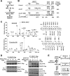
pUL97 interacts with HDAC1 and alters H3 acetylation at the MIE promoter. (A) U-2 OS cells were transfected with an empty or HDAC1-Flag expression vector along with either empty vector control or pCGN-UL97HA or pCGN-UL97K355M (355) expression vectors. Immunoprecipitation was completed using whole-cell lysates from 48 h posttransfection and an antibody against the FLAG epitope. Western blotting was completed using the indicated antibodies. (B) Fibroblasts were infected at 1 IU/cell with ADwt and treated with DMSO vehicle, 40 μM MBV, or MBV and 300 μM trichostatin A (TSA). Western blot analysis was completed using whole-cell lysates collected at the indicated times using the indicated antibodies. (C) Cells were infected with ADwt (−) or with ADdel97 (d97) (+) at 1 IU/cell and treated with either DMSO, 40 μM MBV, or MBV with 0.8 μM mocetinostat (MGC). Western blot analysis was completed using whole-cell lysate collected at 8 hpi and the indicated antibodies. (D) Cells were infected with ADwt, ADwt with 40 μM MBV, or ADdel97 virus at 1 IU/cell. Chromatin immunoprecipitation was performed at 1 and 6 hpi using an antibody against histone 3 acetylated at lysine 9 (H3K9ac) or an IgG control. Levels of the viral MIE promoter and beta-actin sequence were quantified using qPCR. Data are presented as output relative to input quantity and the standard deviations of the means of two biological replicates with statistical significance (*) calculated using a one-way ANOVA.
Fig 5
Fig 5
Association of HDAC1 with the MIE promoter increases in the absence of kinase activity. (A) To assess deacetylase activity, U-2 OS cells were transfected with an empty vector control (C) or cotransfected with HDAC1-Flag expression vector and empty control (C) or pCGN-UL97HA (97) or pCGN-UL97K355MHA (K355M) expression vectors. Immunoprecipitation was performed with an anti-FLAG antibody and used in a fluorescence-based HDAC activity assay. Activity was normalized to immunoprecipitated HDAC1 levels determined by Western blotting and chemiluminescence detection (Fig. 6A). Activity was also measured in 1% of the lysate and normalized to total protein levels. Error bars represent standard deviations from the means of five biological replicates. (B) Fibroblasts were mock infected (m), infected with ADwt (wt) virus, or infected with ADdel97 (d97) at 1 IU/cell and treated with DMSO vehicle or 40 μM MBV. Immunoprecipitation using an antibody against HDAC1 was performed from lysates collected at 1 or 6 hpi. Deacetylase activity was determined using the isolated proteins with a fluorescence-based HDAC activity assay. Activity was normalized to immunoprecipitated HDAC1 levels as determined by Western blotting and chemiluminescence detection (lower panel). Activity was also measured in 0.5% of the lysate and normalized to total protein levels. Error bars denote standard deviations from the means of two biological replicates and statistical significance by one-way ANOVA (*). (C) Fibroblasts were infected at 1 IU/cell with ADwt (wt) and treated with DMSO or 40 μM MBV at the time of infection or infected with ADwt or ADdel97 (del97) virus. Chromatin immunoprecipitation was performed with an antibody to HDAC1 or IgG control from samples collected at 6 hpi. Viral MIE promoter or the beta-actin sequence was quantified using qPCR. Data are presented as output relative to input DNA, and error bars represent the standard deviations from the means of two biological replicates with statistical significance calculated using Student's t test (*, P < 0.05).
Fig 6
Fig 6
pUL97-mediated changes in HDAC1 phosphorylation and binding to DAXX. (A) U-2 OS cells were transfected with an empty or HDAC1-Flag expression vector and cotransfected with an empty vector or pUL97HA or pUL97K355M (355) expression vector. Samples were immunoprecipitated with a Flag-specific antibody and analyzed by Western blotting using the indicated antibodies. (B) U373 cells were transfected with an HDAC1-Flag expression vector for 36 h and then mock infected (−) or infected at 3 IU/cell with ADwt in the presence or absence of 40 μM MBV. At 1 hpi, immunoprecipitation was performed with a Flag-specific antibody and analyzed by Western blotting using the indicated antibodies. The asterisk marks a potential modification of HDAC1. (C) U-2 OS cells were transfected with an HDAC1-Flag expression vector along with pUL97-HA or pUL97K355M-HA expression vector. Samples were evaluated by Western blotting using whole-cell lysates and the indicated antibodies. Modifications are marked. (D) Cells were transfected as described above, including an additional sample containing HDAC1 with empty vector or the pUL97-HA expression vector with DMSO vehicle or 40 μM MBV added at 1 h prior to transfection. Lysates were split into two aliquots and were treated with calf intestinal phosphatase (CIP) (+) or were left untreated (−). Proteins were evaluated by Western blotting using the indicated antibodies.
Fig 7
Fig 7
HDAC1 is uniquely phosphorylated in the presence of kinase-active pUL97. (A) Strategy used to identify phosphopeptides. U-2 OS cells were transfected with an HDAC1-Flag expression vector and cotransfected with an empty vector or pUL97HA, pUL97HA with MBV, or pUL97K355M expression vector. Samples were immunoprecipitated with a Flag-specific antibody, and isolated proteins were digested by trypsin, enriched for phosphopeptides using a combination of IMAC and TiO2 resins, and loaded onto an LTQ-Orbitrap mass spectrometer. Samples were analyzed using multistage activation (MSA), and the resulting spectra were evaluated using Sequest and Mascot algorithms. (B) Sites of phosphorylation identified in HDAC1 from three biological replicate experiments. The overall percentage of the total protein coverage using trypsin is noted. The numbers represent amino acid positions in HDAC1, and the ratio in parentheses represents the number of phosphorylated peptides over the total number of peptides observed. (C) Representative spectra demonstrating b and y ions with a 98-Da mass shift matching the phosphorylation at serine 85 or threonine 208 in HDAC1 as well as identified m/z of the b and y ions. (D) U-2 OS cells were transfected with HDAC1-Flag expressing vectors that were either wild type (T208) or carried an alanine (T208A), glutamic acid (T208E), or aspartic acid (T208D) substitution. Immunoprecipitation was performed with a Flag-specific antibody and analyzed using Western blotting with the indicated antibodies. U-2 OS cells were cotransfected with empty vector or a pUL97HA expressing vector and HDAC1-Flag expressing vectors containing T208, T208A, or T208E. A Flag-specific antibody was used for immunoprecipitation and analyzed by Western blotting with the indicated antibodies. (E) Model showing pUL97 interaction with and phosphorylation of HDAC1 during HCMV infection, resulting in decreased association of HDAC1 with the MIE promoter, increased histone H3-lysine 9 acetylation, and subsequent IE gene expression. Inhibition of kinase activity or gene disruption abrogates these activities, while the addition of HDAC inhibitors compensates for the absence of pUL97 kinase.

Similar articles

Cited by

References

    1. Mocarski E, Pass TSRF. 2007. Cytomegaloviruses, p 2702–2772 In Knipe DM, Howley PM, Griffin DE, Lamb RA, Martin MA, Roizman B, Straus SE. (ed), Fields virology, 5th ed Lippincott Williams & Wilkins, Philadelphia, PA
    1. Hakki M, Chou S. 2011. The biology of cytomegalovirus drug resistance. Curr. Opin. Infect. Dis. 24:605–611 - PMC - PubMed
    1. Littler E, Stuart AD, Chee MS. 1992. Human cytomegalovirus UL97 open reading frame encodes a protein that phosphorylates the antiviral nucleoside analogue ganciclovir. Nature 358:160–162 - PubMed
    1. Sullivan V, Talarico CL, Stanat SC, Davis M, Coen DM, Biron KK. 1992. A protein kinase homologue controls phosphorylation of ganciclovir in human cytomegalovirus-infected cells. Nature 359:85. - PubMed
    1. van Zeijl M, Fairhurst J, Baum EZ, Sun L, Jones TR. 1997. The human cytomegalovirus UL97 protein is phosphorylated and a component of virions. Virology 231:72–80 - PubMed

Publication types

MeSH terms

LinkOut - more resources