. 2013 May 23;50(4):540-51.
doi: 10.1016/j.molcel.2013.03.018. Epub 2013 Apr 18.

Control of protein quality and stoichiometries by N-terminal acetylation and the N-end rule pathway

Affiliations

Control of protein quality and stoichiometries by N-terminal acetylation and the N-end rule pathway

Anna Shemorry et al. Mol Cell. .

Abstract

N(α)-terminal acetylation of cellular proteins was recently discovered to create specific degradation signals termed Ac/N-degrons and targeted by the Ac/N-end rule pathway. We show that Hcn1, a subunit of the APC/C ubiquitin ligase, contains an Ac/N-degron that is repressed by Cut9, another APC/C subunit and the ligand of Hcn1. Cog1, a subunit of the Golgi-associated COG complex, is also shown to contain an Ac/N-degron. Cog2 and Cog3, direct ligands of Cog1, can repress this degron. The subunit decoy technique was used to show that the long-lived endogenous Cog1 is destabilized and destroyed via its activated (unshielded) Ac/N-degron if the total level of Cog1 increased in a cell. Hcn1 and Cog1 are the first examples of protein regulation through the physiologically relevant transitions that shield and unshield natural Ac/N-degrons. This mechanistically straightforward circuit can employ the demonstrated conditionality of Ac/N-degrons to regulate subunit stoichiometries and other aspects of protein quality control.


Figures

Figure 1
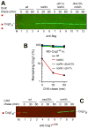
Figure 1. The Ac/N-degron of Cog1
(A) Cycloheximide (CHX)-chases were performed at 30°C with wt or doa10Δ S. cerevisiae expressing either wt Cog1, termed MD-Cog1wt, or its MK-Cog1 derivative in which Asp2 was replaced with Lys2. Both proteins were C-terminally ha-tagged. At the indicated times of chase, proteins in cell extracts were fractionated by SDS-PAGE and assayed by immunoblotting with anti-ha antibody. (B) 35S-pulse-chase with MD-Cog1wt or P-Cog1 in wt, doa10Δ, or naa10Δ (ard1Δ) S. cerevisiae, the latter strain lacking the catalytic subunit of the non-cognate (for MD-Cog1wt) NatA Nt-acetylase (Figure S2). Cog1 proteins were C-terminally tagged with 3 flag epitopes modified to contain a Met residue in each epitope, to increase 35S-Met in Cog1. (C) Same as in B but another 35S-pulse-chase. It included naa20Δ (nat3Δ) S. cerevisiae lacking the catalytic subunit of the cognate (for MD-Cog1wt) NatB Nt-acetylase (Figure S2). (D) Quantification of data in C.◆, MD-Cog1wt in wt cells. ▲, MD-Cog1wt in naa20Δ cells. ■, P-Cog1 in wt cells. (E) Anti-Cog1AcNt antibody specific for Nt-acetylated MD-Cog1wt (see Figure S4A–C) was used for immunoblotting in CHX-chase assays with MD-Cog1wt and P-Cog1 (C-terminally tagged with 3 flag epitopes) in either wt or naa20Δ (nat3Δ) S. cerevisiae. (F) Same as in E, except that membrane was reprobed with anti-flag antibody. (G) Quantification of anti-Cog1AcNt-specific immunoblotting patterns in E using a linear scale, with the level of MD-Cog1wt at time zero in wt cells taken as 100%. ◆, MD-Cog1wt in wt cells. ▲, MD-Cog1wt in naa20Δ cells. ■, P-Cog1 in wt cells. (H) Same as in G but a semi-log plot of the flag-specific Cog1 immunoblotting patterns in F. Same designations as in G. See also Figures S1, S2, S5, and Tables S1 and S2.
Figure 2
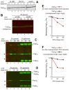
Figure 2. Stabilization of Cog1 in S. cerevisiae Lacking the Not4 E3 Ubiquitin Ligase
(A) CHX-chases with yeast expressing MD-Cog1wt C-terminally tagged with three flag epitopes. Lane M and red stars, Mr markers of 37, 50, and 100 kDa, respectively. MD-Cog1wt in wt yeast (lanes 1–3), and in not4Δ (lanes 4–6), not4Δ ubr1Δ (lanes 7–9), and not4Δ doa10Δ mutants (lanes 10–12). (B) Quantification of immunoblots in H, with the level of MD-Cog1wt in not4Δ cells at the beginning of chase taken as 100%. ■, MD-Cog1wt C in not4Δ cells (black curve). ◆, MD-Cog1wt in wt cells (red curve).▲, MD-Cog1wt in not4Δ ubr1Δ cells (green curve). X,MD-Cog1wt in not4Δ doa10Δ cells (blue curve). (C) Same as in A but an independent CHX-chase, and immunoblotting with anti-Cog1AcNt antibody specific for Nt-acetylated MD-Cog1wt. Lane M and red star, an Mr marker of 50 kDa. MD-Cog1wt in wt yeast (lanes 1–3), and in naat20Δ (nat3Δ) (lanes 4–6), and not4Δ mutants lanes 7–9). See also Figure S5.
Figure 3
Figure 3. Stabilization of Overexpressed, Short-Lived Cog1 by Coexpressed Cog2-Cog4
(A) CHX-chases with endogenous MD-Cog13hawt (C-terminally tagged with 3 ha epitopes) expressed from the chromosomal COG1 locus and the native PCOG1 promoter in wt, doa10Δ, and naa20Δ (nat3Δ) cells. (B) Same as in A but an independent CHX-chase. S. cerevisiae (in 2% glucose) expressing endogenous MD-Cog13hawt and carrying a plasmid that could express the MD-Cog13fwt decoy but only in the presence of galactose. Lanes 4–6, same as lanes 1–3 but in naa20Δ cells. (C) Stabilization of overexpressed MD-Cog1wt (C-terminally tagged with 3 flag epitopes) by coexpressed Cog2 and Cog3. Lane M and red stars, Mr markers of 37, 50, and 100 kDa, respectively. Lanes 1–3, wt S. cerevisiae in 2% glucose, expressing MD-Cog1wt from the PCUP1 promoter on a low copy plasmid and carrying a high copy plasmid that expressed, only in galactose, both Cog2 and Cog3 (C-terminally tagged with ha) from the bidirectional PGAL1/10 promoter. Lanes 4–6, same as lanes 1–3 but with cells in 2% galactose. Asterisk on the right indicates a protein crossreacting with anti-ha. (D) Same as in C but cells also carried a second high copy plasmid expressing Cog4 (C-terminally tagged with ha) from the PGAL1/10 promoter. (E) Quantification of data in C for MD-Cog1wt. ◆, MD-Cog1wt in cells that did not coexpress other COG subunits. ■, MD-Cog1wt in cells that coexpressed (in galactose) Cog2 and Cog3. (F) Quantification of data in D for MD-Cog1wt. ◆, MD-Cog1wt in cells that did not coexpress other COG subunits. ■, MD-Cog1wt in cells that coexpressed (in galactose) Cog2-Cog4. See also Figure S1.
Figure 4
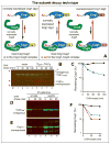
Figure 4. Subunit Decoy Technique and the Cause of Stability of Endogenous Cog1
(A) The subunit decoy technique. “CogX” denotes a Cog1-interacting COG subunit (Cog2 or Cog3) that can shield the Ac/N-degron of Cog1. “Normally expressed” refers to levels of expression from endogenous promoters and chromosomal loci. The normally expressed Cog1-tag1 bears a C-terminal tag denoted as “tag1”, whereas the otherwise identical but overexpressed Cog1-tag2 decoy bears a different C-terminal tag (“tag2”). In the absence of decoy, the bulk of (normally expressed) Cog1-tag1 would occur as a CogX-Cog1-tag1 complex in which the Ac/N-degron of Cog1 is largely sequestered. By contrast, in the presence of overexpressed Cog1-tag2 decoy, the bulk of both Cog1-tag1 and Cog1-tag2 would not be in the complex with CogX (i.e., their Ac/N-degron would be active), given relatively low levels of a (normally expressed) CogX “shielding” protein. (B) Lane M and red stars, Mr markers of 37, 50, and 100 kDa, respectively. Lanes 1–4, stability of endogenous MD-Cog113mycwt (C-terminally tagged with 13 myc epitopes) in the absence of the MD-Cog13fwt decoy (C-terminally tagged with 3 flag epitopes). CHX-chase with MD-Cog113mycwt expressed from the chromosomal COG1 locus and the native PCOG1 promoter in wt cells in the presence of vector alone. Lanes 5–8, same as lanes 1–4 but cells carried a plasmid that expressed the MD-Cog13fwt decoy from the PADH1 promoter. An asterisk denotes a protein crossreacting with anti-flag. (C) Quantification of data in B. ◆, endogenous MD-Cog113mycwt in wt cells that did not express the MD-Cog13fwt decoy. ■, endogenous MD-Cog113mycwt in wt cells that expressed MD-Cog13fwt . (D) Same as in C but with wt and naa20Δ cells expressing the endogenous MD-Cog13hawt in 2% galactose, i.e., in the presence of the coexpressed MD-Cog13fwt decoy. Immunoblotting with anti-ha, specific for MD-Cog13hawt . (E) Same as in D but also probed (in a parallel immunoblot) with anti-flag, specific for MD-Cog13fwt . (F) Quantification of data in D, E. ◆, endogenous MD-Cog13hawt in wt cells grown in 2% glucose. ■, endogenous MD-Cog13hawt in wt cells grown in 2% galactose, i.e., in the presence of the MD-Cog13fwt decoy. ▲, MD-Cog13fwt decoy. See also Figures S4 and S5.
Figure 5
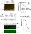
Figure 5. Hcn1 and Repression of Its Ac/N-degron by Cut9
(A) CHX-chases in wt or naa30Δ (mak3Δ) S. cerevisiae expressing the wt S. pombe Hcn1, termed ML-Hcn1wt, C-terminally tagged with 3 flag epitopes. naa30Δ cells lacked the catalytic subunit of the cognate NatC Nt-acetylase (Figure S2). Lane 1 and red stars, Mr markers of 10, 15, 20, 37, and 50 kDa, respectively. (B) 35S-pulse-chase with ML-Hcn1wt in wt and naa30Δ (mak3Δ) S. cerevisiae. Lane 7, vector alone. (C) Quantification of data in B. ◆, ML-Hcn1wt in wt cells. ■, ML-Hcn1wt in naa30Δ cells. (D) Lanes 1–4, CHX-chase with wt cells in 2% galactose (and without methionine) that expressed ML-Hcn1wt from the methionine-repressible PMET25 promoter on a low copy plasmid and carried a vector alone (no Cut9 expression). Note the metabolic instability of ML-Hcn1wt (lower panel). Lanes 5–8, same as lanes 1–4 but with a low copy plasmid (instead of control vector) expressing Cut9 from the galactose-inducible PGAL1 promoter, with both ML-Hcn1wt and Cut9 C-terminally tagged with 3 flag epitopes. Note the metabolic stabilization of ML-Hcn1wt(lower panel), including a strong increase of its level at the beginning of the chase. (E) Quantification of data in D. ◆, ML-Hcn1wt in the absence of co-expressed Cut9. ■, ML-Hcn1wt in the presence of co-expressed Cut9. See also Figure S1.
Figure 6
Figure 6. Conditionality of Ac/N-degrons
This diagram summarizes the functional understanding of the dynamics of Nt-acetylated proteins vis-à-vis the Ac/N-end rule pathway attained in the present study, in conjunction with results that initially revealed the Ac/N-end rule pathway (Hwang et al., 2010b). See also Figure S1.

Similar articles

Cited by

References

    1. Arnesen T, Van Damme P, Polevoda B, Helsens K, Evjenth R, Colaert N, Varhaug JE, Vandekerckhove J, Lillehaug JR, Sherman F, et al. Proteomics analyses reveal the evolutionary conservation and divergence of N-terminal acetyltransferases from yeast to humans. Proc Natl Acad Sci USA. 2009;106:8157–8162. - PMC - PubMed
    1. Barford D. Structure, function and mechanism of the anaphase promoting complex (APC/C) Q Rev Biophys. 2011;44:153–190. - PubMed
    1. Brandman O, Stewart-Ornstein J, Wong D, Larson A, Williams CC, Li GW, Zhou S, King D, Shen PS, Weibezahn J, et al. A ribosome-bound quality control complex triggers degradation of nascent peptides and signals translation stress. Cell. 2012;151:1042–1054. - PMC - PubMed
    1. Chen J, Archer T. Regulting SWI/SNF subunit levels via protein-protein interactions and proteasomal degradation: BAF155 and BAF170 limit expression of BAF57. Mol Cell Biol. 2005;25:9016–9027. - PMC - PubMed
    1. Collart M, Panasenko O, Nikolaev S. The Not3/5 subunit of the Ccr4-Not complex: A central regulator of gene expression that integrates signals between the cytoplasm and the nucleus in eukaryotic cells. Cell Signal. 2012;25:743–751. - PubMed

Publication types

MeSH terms

Substances