. 2013 Apr 25;153(3):678-91.
doi: 10.1016/j.cell.2013.04.001. Epub 2013 Apr 18.

Genome-wide profiling of 5-formylcytosine reveals its roles in epigenetic priming

Affiliations

Genome-wide profiling of 5-formylcytosine reveals its roles in epigenetic priming

Chun-Xiao Song et al. Cell. .

Abstract

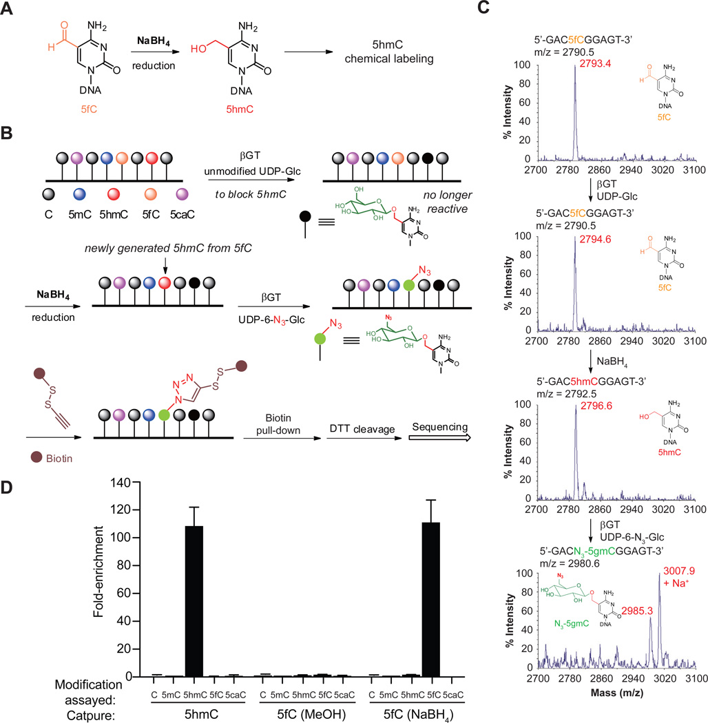
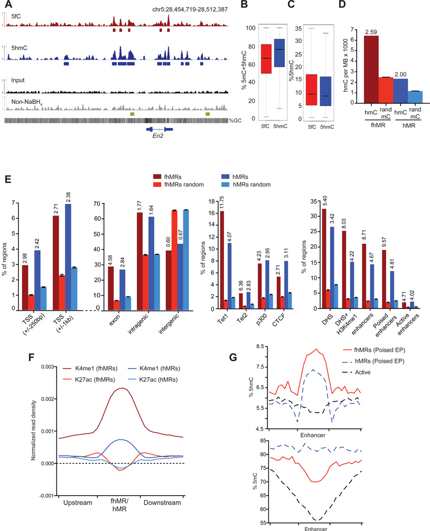
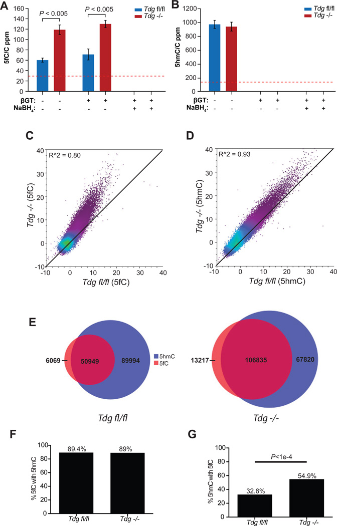
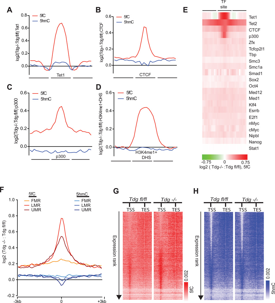
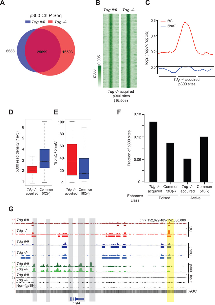
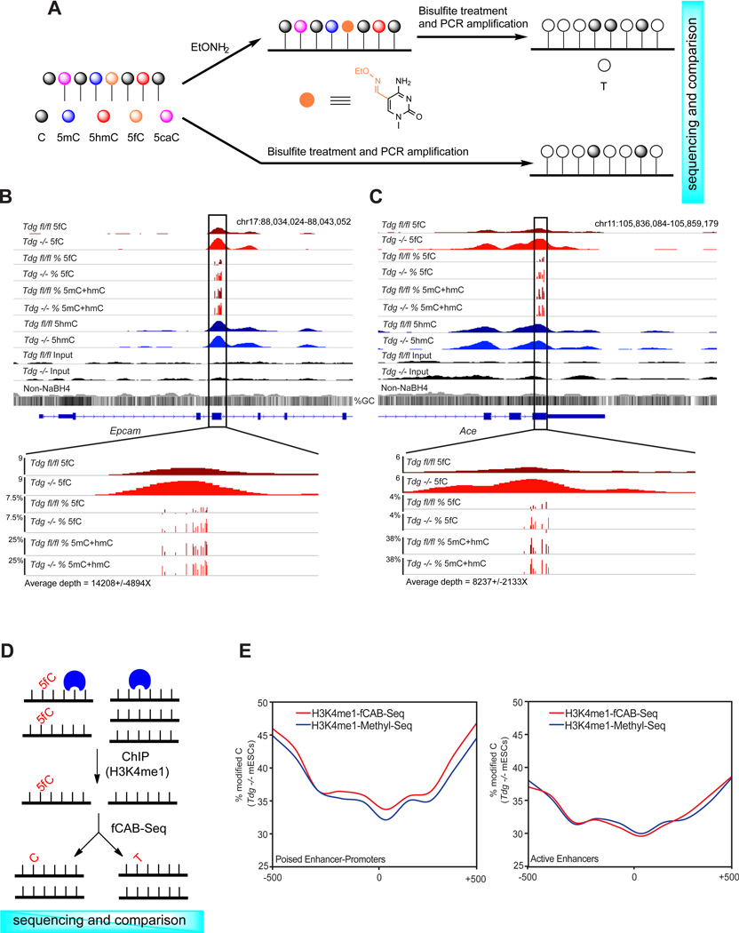
TET proteins oxidize 5-methylcytosine (5mC) to 5-hydroxymethylcytosine (5hmC), 5-formylcytosine (5fC), and 5-carboxylcytosine (5caC). 5fC and 5caC are excised by mammalian DNA glycosylase TDG, implicating 5mC oxidation in DNA demethylation. Here, we show that the genomic locations of 5fC can be determined by coupling chemical reduction with biotin tagging. Genome-wide mapping of 5fC in mouse embryonic stem cells (mESCs) reveals that 5fC preferentially occurs at poised enhancers among other gene regulatory elements. Application to Tdg null mESCs further suggests that 5fC production coordinates with p300 in remodeling epigenetic states of enhancers. This process, which is not influenced by 5hmC, appears to be associated with further oxidation of 5hmC and commitment to demethylation through 5fC. Finally, we resolved 5fC at base resolution by hydroxylamine-based protection from bisulfite-mediated deamination, thereby confirming sites of 5fC accumulation. Our results reveal roles of active 5mC/5hmC oxidation and TDG-mediated demethylation in epigenetic tuning at regulatory elements.


Figures

Figure 1
Figure 1. Selective labeling of 5fC in genomic DNA
(A) 5fC is selectively reduced to 5hmC. (B) General procedure for fC-Seal. Endogenous 5hmC is blocked by a regular glucose through βGT-catalyzed glucosylation. 5fC is then reduced to 5hmC by NaBH4. The newly generated 5hmC from 5fC can be specifically enriched for sequencing using the 5hmC-selective chemical labeling method (hMe-Seal). (C) MALDI-TOF characterization of 5fC-containing 9mer duplex DNA in βGT-catalyzed blocking with unmodified glucose, NaBH4-based reduction, and βGT-catalyzed azide-glucose labeling. Calculated MS shown in black, observed MS shown in Red. (D) Enrichment tests of a single pool of spike-in amplicons containing C, 5mC, 5hmC, 5fC, or 5caC, separately, using hMe-Seal and fC-Seal with NaBH4 or a control with methanol only. Values shown are fold-enrichment over input, normalized to 5mC-modified DNA (n = 3, mean ± s.e.m). See also Figure S1.
Figure 2
Figure 2. Annotation and comparison of 5hmC- and 5fC-containing regions in the wild-type (Tdgfl/fl) mESCs
(A) Genome browser view of the En2 locus in 5fC- and 5hmC-specific profiling, along with the input as well as the glucose-blocked, non-NaBH4-treated control. Below each track are regions defined as marked with each respective mark. The gold track at the bottom corresponds to known poised enhancers at En2 (Shen et al., 2012). (B) Quantification of %5mC+5hmC at 5fC- and 5hmC-marked regions in Tdgfl/fl mESCs. (C) Quantification of %5hmC at 5fC- and 5hmC-marked regions in Tdgfl/fl mESCs. (D) The relative enrichment of single-base 5hmC calls from (Yu et al., 2012) within fhMRs and hMRs Tdgfl/fl mESCs. mC random are randomly sampled 5mC bases defined by conventional bisulfite sequencing (Stadler et al., 2011) (10 iterations, mean ± s.d.). 5hmC base call counts are normalized in the genomic space covered by each set of enriched/random regions in megabases (MB) and divided by 103. Values above bars indicate the o/e ratios. (E) Percentage of fhMRs and hMRs overlapping a given genomic/epigenomic annotation compared to the average percent overlap of 10 randomized sets of equal number and length (mean ± s.d). Vertical values above bars indicate the o/e ratios with significant enrichment (p < 1e–15, Fisher’s exact). Genomic annotations are listed in the left panel and epigenomic annotations are listed on the right. (F) H3K4me1 and H3K27ac normalized read densities at fhMRs (red) and hMRs (blue). (G) %5hmC (top panel) and %5mC (bottom panel) at fhMRs (red) and hMRs (blue) associated with poised Enhancer-Promoters (Poised EP) and active enhancers (black). See also Figure S2.
Figure 3
Figure 3. Comparison of 5fC and 5hmC signals in Tdgfl/fl and Tdg−/− mESCs
(A–B) Mass spectrometry quantification of the genomic content of 5fC (A) and 5hmC (B) relative to cytosine in Tdgfl/fl and Tdg−/− mESCs in fC-Seal. Error bars indicate s.e.m. for n = 4 experiments. The red dotted lines indicate the detection limits under the assay conditions. (C–D) Scatter plot of input-normalized 5fC (C) and 5hmC (D) read counts (reads/million) in 10kb bins genome-wide in Tdgfl/fl and Tdg−/− mESCs. Read counts per 10kb bin are normalized to the total number of reads in millions and similarly normalized values from input control genomic DNA subtracted. R2 values are denoted in the upper left-hand corner and the black diagonal is provided for reference. (E) Venn diagram of the number of 5fC-marked regions (red) overlapping 5hmC-enriched regions (blue) in Tdgfl/fl (top) and Tdg−/− (bottom) mESCs. (F–G) The percentage of the genomic regions with 5fC also marked with 5hmC (F) and the percentage of 5hmC-enriched regions also containing 5fC (G). See also Figure S3.
Figure 4
Figure 4. TDG-dependent 5fC regulation at defined gene regulatory elements
(A–D) Log2 ratios of 5fC- and 5hmC-normalized read densities (reads/million/base, Tdg−/−: Tdgfl/fl) at genomic elements enriched for 5fC in Tdgfl/fl and Tdg−/− mESCs. (A) Tet1, (B) CTCF, (C) p300, and (D) H3K4me1+DHS. Each region of interest, denoted as the central portion of the x-axis, was divided into bins of 10 equal portions and reads were intersected to 10 bins within, upstream, and downstream of each region. (E) Heatmap representation of the Log2 ratios of 5fC-read densities (reads/million/base, Tdg−/−: Tdgfl/fl) at 22 distinct sets of transcription factor (TF)-binding sites. (F) Log2 ratio of the normalized 5fC and 5hmC read densities (reads/million/base, Tdg−/−: Tdgfl/fl) at FMRs, LMRs, and UMRs. Normalized read densities are plotted ± 3kb from the center of each segment as log2 fold-enrichment over input normalized read densities. (G) Heatmap representations of 5fC-normalized read densities (reads/million/base) at RefSeq TSSs/TESs (± 5kb). 5fC signals at genes that are ranked by RPKM in descending order. Heatmap scales correspond to normalized read densities. (H) Heatmap representations of 5hmC-normalized read densities (reads/million/base) at RefSeq TSSs/TESs (± 5kb). 5hmC signals at genes that ranked by RPKM in descending order. Heatmap scales correspond to normalized read densities. See also Figure S4.
Figure 5
Figure 5. TDG-dependent p300 binding in Tdgfl/fl and Tdg−/− mESCs
(A) Venn diagram summarizing the total number of p300 ChIP-Seq peaks identified in Tdgfl/fl (32,160) and Tdg−/− mESCs (42,202), the number of Tdg−/− p300 sites overlapping with Tdgfl/fl sites (25,699), and the number of p300 sites unique to Tdgfl/fl (6,683) and Tdg−/− mESCs (16,503). (B) p300 ChIP-Seq signals (reads/million/base) at the Tdg−/− specific p300-binding sites (16,503). (C) Log2-fold-change in 5fC and 5hmC signals at the Tdg−/− specific p300-binding sites (16,503). (D–E) p300 ChIP-Seq signals (reads/million/base, Tdg−/− mESCs) (D) and percent 5mC+5hmC (E) at p300 sites specific to Tdg−/− (16,503) and the 5fC negative p300-binding sites common to Tdgfl/fl and Tdg−/− (16,323). (F) The fraction of Tdg−/− specific p300-binding sites (16,503) and 5fC negative p300-binding sites common to Tdgfl/fl and Tdg−/−(16,323) that occur at active and poised enhancers. (G) Genome browser view of the Fgf4 locus at which multiple strong p300 sites lacking 5fC and 5hmC occur surrounding Fgf4 (gray), with a downstream poised enhancer (Shen et al., 2012) displaying a gain in 5fC and p300 in Tdg−/− (yellow). Shown below each track are the regions defined as marked for each respective mark. See also Figure S5.
Figure 6
Figure 6. fCAB-Seq for base-resolution detection of 5fC
(A) Schematic diagram of EtONH2-modified bisulfite sequencing for base-resolution detection of 5fC in genomic DNA (fCAB-Seq). (B) fCAB-Seq validation of TDG-dependent 5fC in genomic DNA. An example of 5fC detection by fCAB-Seq amplicon deep sequencing at a region of Epcam. Sequencing depth = 14,208 ± 4894. (C) fCAB-Seq validation of TDG-dependent 5fC in genomic DNA. An example of 5fC detection by fCAB-Seq amplicon deep sequencing at a region of Ace. Sequencing depth = 8,237 ± 2,133. For (B) and (C) 5fC track is equivalent to the signal from EtONH2 treatment minus (5mC+hmC) signal. All 5fC bases shown have p ≤ 0.005, Fisher’s exact. (D) Schematic diagram of ChIP-fCAB-Seq. DNA fragments associated with H3K4me1 are enriched in ChIP and then subjected to fCAB-Seq for the determination of 5fC at base resolution. (E) H3K4me1-ChIP-fCAB (Red) and H3K4m1-ChIP-Methyl-Seq (Blue) signals at 5fC-positive poised enhancers predicted as linked to promoters (left) and at all active enhancers (right) in Tdg− /− mESCs. Plotted are the weighted methylation signals in 100bp bins within the 1kb enhancer region. See also Figure S6.

Comment in

Similar articles

Cited by

References

    1. Bhutani N, Burns DM, Blau HM. DNA demethylation dynamics. Cell. 2011;146:866–872. - PMC - PubMed
    1. Booth MJ, Branco MR, Ficz G, Oxley D, Krueger F, Reik W, Balasubramanian S. Quantitative sequencing of 5-methylcytosine and 5-hydroxymethylcytosine at single-base resolution. Science. 2012;336:934–937. - PubMed
    1. Brinkman AB, Gu H, Bartels SJ, Zhang Y, Matarese F, Simmer F, Marks H, Bock C, Gnirke A, Meissner A, et al. Sequential ChIP-bisulfite sequencing enables direct genome-scale investigation of chromatin and DNA methylation cross-talk. Genome Res. 2012;22:1128–1138. - PMC - PubMed
    1. Chen Q, Chen Y, Bian C, Fujiki R, Yu X. TET2 promotes histone O-GlcNAcylation during gene transcription. Nature. 2012 - PMC - PubMed
    1. Cortazar D, Kunz C, Selfridge J, Lettieri T, Saito Y, MacDougall E, Wirz A, Schuermann D, Jacobs AL, Siegrist F, et al. Embryonic lethal phenotype reveals a function of TDG in maintaining epigenetic stability. Nature. 2011;470:419–423. - PubMed

Publication types

Associated data