. 2013 Jun 7;288(23):16937-16948.
doi: 10.1074/jbc.M113.463711. Epub 2013 Apr 18.

β-Amyloid (Aβ) oligomers impair brain-derived neurotrophic factor retrograde trafficking by down-regulating ubiquitin C-terminal hydrolase, UCH-L1

Affiliations

β-Amyloid (Aβ) oligomers impair brain-derived neurotrophic factor retrograde trafficking by down-regulating ubiquitin C-terminal hydrolase, UCH-L1

Wayne W Poon et al. J Biol Chem. .

Abstract

We previously found that BDNF-dependent retrograde trafficking is impaired in AD transgenic mouse neurons. Utilizing a novel microfluidic culture chamber, we demonstrate that Aβ oligomers compromise BDNF-mediated retrograde transport by impairing endosomal vesicle velocities, resulting in impaired downstream signaling driven by BDNF/TrkB, including ERK5 activation, and CREB-dependent gene regulation. Our data suggest that a key mechanism mediating the deficit involves ubiquitin C-terminal hydrolase L1 (UCH-L1), a deubiquitinating enzyme that functions to regulate cellular ubiquitin. Aβ-induced deficits in BDNF trafficking and signaling are mimicked by LDN (an inhibitor of UCH-L1) and can be reversed by increasing cellular UCH-L1 levels, demonstrated here using a transducible TAT-UCH-L1 strategy. Finally, our data reveal that UCH-L1 mRNA levels are decreased in the hippocampi of AD brains. Taken together, our data implicate that UCH-L1 is important for regulating neurotrophin receptor sorting to signaling endosomes and supporting retrograde transport. Further, our results support the idea that in AD, Aβ may down-regulate UCH-L1 in the AD brain, which in turn impairs BDNF/TrkB-mediated retrograde signaling, compromising synaptic plasticity and neuronal survival.

Keywords: Alzheimer Disease; Amyloid; BDNF; Microfluidic Device; Trafficking; Ubiquitin.


Figures
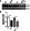
FIGURE 1.
FIGURE 1.
Aβ oligomers do not affect the internalization of BDNF receptors. A, cell surface biotinylation was employed to measure TrkB levels at the cell surface following BDNF treatment (50 ng/ml, 30 min) as described under “Experimental Procedures.” BDNF addition led to a decrease in cell surface levels of full-length TrkB (TrkB-FL) and truncated TrkB (TrkB.T1). Preincubation with Aβ oligomers (24 h) does not impair TrkB-FL or TrkB-T1 internalization. BDNF specifically caused internalization of BDNF receptors, but not the internalization of neural cell adhesion molecule (NCAM). B, cell surface TrkB-FL was quantitated using ImageJ (National Institutes of Health). The mean ± S.E. represents TrkB-FL levels normalized to vehicle-treated neurons (n = 3). Following BDNF treatment, we found that 38.3 ± 3.40% of TrkB-FL was internalized (black bar) when compared with vehicle (white bar) (**, p < 0.001). In the presence of Aβ, BDNF led to 41.5 ± 9.80% of TrkB-FL internalized (hatched bar) when compared with Aβ-only (gray bar) (*, p < 0.05). Veh, vehicle.
FIGURE 2.
FIGURE 2.
Aβ oligomers impair the trafficking of BDNF-GFP endosomes. A, in the presence of Aβ, the average velocity of BDNF-GFP-containing endosomes was 1.73 ± 0.378 μm/s. This represented a 38.4 ± 13.4% (*, p < 0.01) decrease when compared with the average velocity of endosomes in the absence of velocities of BDNF-GFP-positive endosomes determined as described previously (22). B, distribution plot of the vesicle velocities reveal that in the presence of Aβ oligomers, the percentage of vesicle velocities >2 mm/s was greatly reduced (gray bars), and the majority of the vesicle velocity was <1 μm/s. C, representative time lapse image of BDNF-GFP containing endosomes demonstrates the amount of BDNF-TrkB complex that undergoes retrograde transport of the (from right to left). Scale bar, 10 μm.
FIGURE 3.
FIGURE 3.
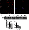
Aβ oligomers impair the trafficking of the signaling endosome complex including p-ERK5. Microfluidic devices were employed to measure the retrograde transport dependent ERK5 activation. A–D, representative images demonstrating p-ERK5 levels (red) in the somal compartment of microfluidic chamber of vehicle-treated neurons (A) and following BDNF treatment (B), in the presence of Aβ oligomers only (C), and after BDNF treatment (D). Scale bar, 200 μm. E–H, representative images of a neuron demonstrating the co-localization of BDNF-GFP (green) on the outer surface of the nucleus (blue). Also, p-ERK5 (red) co-localized with the nucleus, suggesting that the BDNF-mediated retrograde signal, i.e., the signaling endosome, undergoes retrograde transport from the axonal compartment to the soma and specifically the nucleus. Scale bar, 20 μm. I, Western blot analysis of p-ERK5 and T-ERK5 isolated from the somal compartment of microfluidics devices as described under “Experimental Procedures.” J, somal p-ERK5 was quantitated as described under “Experimental Procedures.” BDNF leads to a 68.1 ± 8.4% (*, p < 0.05) increase in p-ERK5. However, in neurons preincubated with Aβ oligomers, p-ERK5 levels are not increased following axonal BDNF treatment. K, quantification of p-ERK5 relative to total ERK5 levels in the somal compartment following BDNF treatment. BDNF leads to a 284% increase in p-ERK5. However, in the presence of Aβ oligomers, BDNF does not lead to increased p-ERK5. Also, a UCH-L1 inhibitor (LDN-57444) was used to inhibit deubiquitinating activity and revealed that it could mimic the effect of Aβ oligomers in impairing BDNF-mediated retrograde signaling. Veh, vehicle; LDN, LDN-57444.
FIGURE 4.
FIGURE 4.
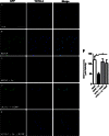
Aβ oligomers lead to decreased CREB-dependent gene expression. Microfluidic devices were used to assess CREB-mediated gene expression. Rat primary neurons (embryonic day 18) were transfected with a CRE-GFP reporter construct to assess CREB-mediated gene activation. At 7 DIV, the axonal compartment was treated with BDNF (50 ng/ml, 2 h), and the chambers were processed for immunochemical analysis as described under “Experimental Procedures” using polyclonal anti-GFP (Invitrogen) to measure CRE-GFP levels and were normalized to the neuronal marker, BIII-tubulin (red). A, representative image of CRE-GFP levels (green) within the somal compartment and co-imaged with the neuronal marker, BIII-tubulin (red) in vehicle-treated neurons, and in neurons treated with BDNF. B and C, in the presence of Aβ oligomers, base-line levels of CRE-GFP are not significantly reduced when compared with vehicle. D, in the presence of Aβ oligomers, the increase in the amount of CRE-GFP is greatly reduced. E, CRE-GFP levels were quantified, and the means ± S.E. represent n = 4 and demonstrate that BDNF treatment to the axonal compartment led to a 35.9 ± 4.73 (*, p < 0.01) increase in somal CRE-GFP immunoreactivity when compared with vehicle (white bar). However, in the presence of Aβ oligomers, CRE-GFP was only increased by 14.6 ± 5.23%, when compared with Aβ oligomer only cells, but this was not significant (p = 0.076). Therefore, in the presence of Aβ, axonal BDNF leads to reduced CRE-GFP immunoreactivity (hatched bar) when compared with vehicle treated neurons (black bar) (*, p < 0.01). Scale bar, 200 μm. Veh, vehicle.
FIGURE 5.
FIGURE 5.
The UCH-L1 inhibitor LDN mimics the effect of Aβ oligomers on BDNF-dependent retrograde signaling. To assess the effect of LDN on BDNF-dependent retrograde signaling, we measured p-ERK5 activation in the presence of LDN. A, representative images that demonstrate that BDNF led to an increase in somal p-ERK5, but the UCH-L1 inhibitor, LDN, led to decreased basal somal p-ERK5, and in the presence of BDNF, the nuclear translocation of p-ERK5 is not detected. p-ERK5 immunoreactivity was normalized to the nuclear counterstain, TOTO-3. B, quantification of somal p-ERK5 levels demonstrate that although BDNF treatment (black bar) leads to a 45.7 ± 10.2% (*, p < 0.05) increase in p-ERK5 when compared with vehicle (white bar), in neurons preincubated with LDN (gray bar), we do not observe an increase in somal p-ERK5 following BDNF (hatched bar). C, cell surface biotinylation assays were employed to determine the effect of LDN on TrkB internalization and demonstrate that LDN does not affect the internalization of full-length TrkB (TrkB-FL) or truncated TrkB (TrkB.T1). D, quantification of cell surface TrkB-FL demonstrates that in vehicle-treated neurons (white bar), the addition of BDNF (black bar) led to a 45.7 ± 10.2% (*, p < 0.05) decrease in cell surface TrkB levels, whereas in the presence of LDN (gray bar), BDNF led to a 44.3 ± 8.56 (*, p < 0.05) decrease in TrkB-FL. The mean ± S.E. represents n = 3. Scale bar, 20 μm. Veh, vehicle; LDN, LDN-57444.
FIGURE 6.
FIGURE 6.
Transduction of UCH-L1 rescued Aβ-mediated retrograde transport deficits. To assess whether increasing UCH-L1 could rescue Aβ-mediated transport deficits, we measured the extent of BDNF-GFP trafficking in neurons transduced with UCH-L1. A and B, representative images demonstrate that somal levels of BDNF-GFP are increased in neurons following BDNF-GFP treatment. GFP immunoreactivity was normalized to the nuclear marker, TOTO-3. C, pretreatment with Aβ led to decrease in BDNF-GFP when compared with vehicle-treated neurons. D, the addition of UCH-L1 alone led to increased somal BDNF-GFP immunoreactivity. E, UCH-L1 rescues the deficit in BDNF-GFP trafficking caused by Aβ. F, quantification of somal BDNF levels normalized to cell number (TOTO-3-positive nuclei) reveals that Aβ causes a 60.3 ± 7.1% (*, p = 0.003) decrease in the retrograde transport of BDNF-GFP back to soma compared with vehicle-treated neurons. In the presence of UCH-L1, BDNF-GFP levels were 92.6 ± 12.0% of vehicle plus BDNF. Importantly, UCH-L1 rescued the deficit in BDNF-GFP trafficking caused by Aβ. BDNF-GFP levels were 86.3 ± 14.1% of vehicle treated and revealed that UCH-L1 restored trafficking deficits caused by Aβ oligomers (*, p = 0.04). Scale bar, 200 μm. Veh, vehicle.
FIGURE 7.
FIGURE 7.
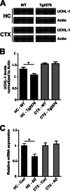
UCH-L1 is decreased in Tg2576 mice and in the Alzheimer disease brain. A, levels of UCH-L1 are decreased in APP-Tg2576 hippocampus. Hippocampal lysates were prepared as described under “Experimental Procedures.” Protein was separated on SDS-PAGE, and Western blot analysis was carried out to determine the amount of UCH-L1 protein in wild-type and Tg2576 mouse brain. B, UCH-L1 protein levels are decreased within the hippocampus but not the cortex of 15-month-old Tg2576 mice (*, p < 0.03). UCH-L1 levels were quantitated and normalized to actin protein as described under “Experimental Procedures.” C, UCHL-1 gene expression is lower in AD brain. Expression profiles were obtained from a microarray database consisting of brain tissue from AD cases (n = 26; range, 74–95 years; mean age, 85.7 ± 6.5 years) and age-matched controls (n = 33; range, 69–99 years; mean age, 84.2 ± 8.9 years) and were generated using Affymetrix HgU133 plus 2.0 arrays as described previously (44). Two probe sets corresponding to UCHL-1 (Unigene Hs.518731) were identified on the HgU133 plus 2.0 array, both of which had Present flags in all microarrays, indicating high expression reliability of the probes. Expression values were averaged across the probe sets to obtain an overall value for each case, followed by t test comparisons for each region and significance set at p < 0.05 (*).

Similar articles

Cited by

References

    1. Jack C. R., Jr., Knopman D. S., Jagust W. J., Shaw L. M., Aisen P. S., Weiner M. W., Petersen R. C., Trojanowski J. Q. (2010) Hypothetical model of dynamic biomarkers of the Alzheimer's pathological cascade. Lancet Neurol. 9, 119–128 - PMC - PubMed
    1. Näslund J., Haroutunian V., Mohs R., Davis K. L., Davies P., Greengard P., Buxbaum J. D. (2000) Correlation between elevated levels of amyloid β-peptide in the brain and cognitive decline. JAMA 283, 1571–1577 - PubMed
    1. Lue L. F., Kuo Y. M., Roher A. E., Brachova L., Shen Y., Sue L., Beach T., Kurth J. H., Rydel R. E., Rogers J. (1999) Soluble amyloid β peptide concentration as a predictor of synaptic change in Alzheimer's disease. Am. J. Pathol. 155, 853–862 - PMC - PubMed
    1. McLean C. A., Cherny R. A., Fraser F. W., Fuller S. J., Smith M. J., Beyreuther K., Bush A. I., Masters C. L. (1999) Soluble pool of Aβ amyloid as a determinant of severity of neurodegeneration in Alzheimer's disease. Ann. Neurol. 46, 860–866 - PubMed
    1. Walsh D. M., Klyubin I., Fadeeva J. V., Cullen W. K., Anwyl R., Wolfe M. S., Rowan M. J., Selkoe D. J. (2002) Naturally secreted oligomers of amyloid β protein potently inhibit hippocampal long-term potentiation in vivo. Nature 416, 535–539 - PubMed

Publication types

MeSH terms

LinkOut - more resources