. 2013 Jun 6;121(23):4729-39.
doi: 10.1182/blood-2012-12-471094. Epub 2013 Apr 18.

Nucleolin inhibits Fas ligand binding and suppresses Fas-mediated apoptosis in vivo via a surface nucleolin-Fas complex

Affiliations

Nucleolin inhibits Fas ligand binding and suppresses Fas-mediated apoptosis in vivo via a surface nucleolin-Fas complex

Jillian F Wise et al. Blood. .

Abstract

Resistance to Fas-mediated apoptosis is associated with poor cancer outcomes and chemoresistance. To elucidate potential mechanisms of defective Fas signaling, we screened primary lymphoma cell extracts for Fas-associated proteins that would have the potential to regulate Fas signaling. An activation-resistant Fas complex selectively included nucleolin. We confirmed the presence of nucleolin-Fas complexes in B-cell lymphoma cells and primary tissues, and the absence of such complexes in B-lymphocytes from healthy donors. RNA-binding domain 4 and the glycine/arginine-rich domain of nucleolin were essential for its association with Fas. Nucleolin colocalized with Fas on the surface of B-cell lymphoma cells. Nucleolin knockdown sensitized BJAB cells to Fas ligand (FasL)-induced and Fas agonistic antibody-induced apoptosis through enhanced binding, suggesting that nucleolin blocks the FasL-Fas interaction. Mice transfected with nucleolin were protected from the lethal effects of agonistic anti-mouse Fas antibody (Jo2) and had lower rates of hepatocyte apoptosis, compared with vector and a non-Fas-binding mutant of nucleolin. Our results show that cell surface nucleolin binds Fas, inhibits ligand binding, and thus prevents induction of Fas-mediated apoptosis in B-cell lymphomas and may serve as a new therapeutic target.


Figures
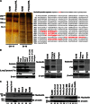
Figure 1
Figure 1
Nucleolin binds activation-resistant Fas and shows altered expression in B-cell lymphomas. (A) Silver-stained gel separating primary NHL and BJAB samples subjected to Fas activation and IP with agonistic antibody CH-11 (BJAB and primary NHL CH-11). The remaining lysates were subjected to a second Fas IP (B-10) of any remaining activation-resistant Fas (BJAB and primary NHL B-10). Specific activation-resistant Fas bands were excised, digested with trypsin, and analyzed by nanoflow-LC-MS/MS fragmentation and collision-induced dissociation spectra profiling. A 100 kDa band of interest (asterisk) reveals the protein that is the focus of the current study. The protein sequence of nucleolin is shown with peptides (red text) from the 100 kDa band identified by nanoflow-LC-MS/MS spectra. Peptides map to the RNA binding domains in the C-terminal region of nucleolin. (B) Whole-cell extracts from BJAB, Raji, Daudi, BC-3, and a histocytic lymphoma (U937) cell line and two B-lymphocyte isolations from healthy donors were subjected to IP with B-10 anti-Fas agarose, which recognizes an epitope within the cytoplasmic region of Fas, and analyzed by IB for the presence of nucleolin with MS-3 anti-nucleolin antibody. Representative data from 3 different experiments are shown. Shown is an analysis of 2 primary lymphoma samples (diffuse large B-cell lymphoma [DLBCL] and mantle cell lymphoma [MCL]) demonstrating nucleolin-Fas complex from a screen of 4 DLBCL and 6 MCL primary samples. Extracts were immunoprecipitated with B-10 and analyzed by IB for the presence of nucleolin in precipitated complexes. BJAB cells and isotype IP were used as positive and negative controls, respectively. β-actin was used as a loading control. (C) Whole-cell lysates of 5 lymphomas cell lines (BJAB, Raji, Daudi, BC-3, and U937) and healthy donor B-lymphocyte populations were analyzed by IB for nucleolin expression. β-actin was used as a loading control (bottom panel). Whole-cell lysates of 4 primary hematologic cancer tissues multiple myeloma (MM), MCL, chronic lymphocytic leukemia (CLL) and DLBCL were subjected to IB analysis of nucleolin. β-actin was used as a loading control. Ratio of nucleolin to β-actin levels determined by densitometry is shown below each sample lane. Representative data from 3 different experiments are shown.
Figure 2
Figure 2
R4 and GAR domains of nucleolin are necessary for its interaction with Fas. (A) Schematic of the nucleolin domains and its known modifications: N-terminal domain region (NDR), nuclear localization signal (NLS), RNA binding domains 1-4 (R1-4), and glycine/arginine-rich domain (GAR). Glycosylation sites are represented by Y, phosphorylation sites by yellow circles, protein-binding sites by stars, and 3 defined proteolytic cleavage sites (of a potential 18 putative sites) by a blue lightning bolt. (B) Domain deletions were created by using the Stratagene Quick Change II XL mutagenesis kit using C-terminal DDK/myc-tagged PCMV-ENTRY construct of full-length nucleolin (Origene) as a template. (C) 293T HEK cells were transfected with the indicated nucleolin domain deletion mutants and lysed for IP/IB analysis. Whole-cell lysates were subjected to Fas IP with agarose conjugated with B-10 anti-Fas antibody. Proteins were separated and immunoblotted for detection of coprecipitated domain mutants with an anti-DDK-HRP antibody. A mixture of all domain mutants was precipitated with mouse IgG and protein G agarose as a negative control. Whole-cell lysate samples prior to IP were immunoblotted with anti-DDK-HRP to reveal expression levels of the transfected constructs. Representative data from 3 different experiments are shown. (D) A chimeric Fc:Fas (Fc:extracellular domain of Fas) was incubated with varying concentrations of recombinant nucleolin-GST for 1.5 hour at 4°C. Fc:Fas was immunoprecipitated with protein A overnight and precipitates were separated by sodium dodecyl sulfate-polyacrylamide gel electrophoresis. IB revealed nucleolin-GST present in Fas-precipitated complexes in a dose-dependent manner. Mouse IgG-1 was used as a control for Fc fragment binding.
Figure 3
Figure 3
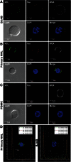
Nucleolin associates with Fas on the surface of primary B-cell lymphoma cells. Localization of nucleolin (NCL)-Fas complexes in B-cell lymphomas. BJAB cells (A) a primary NHL (B) and PBMCs (C) were incubated with an anti-NCL antibody (MS-3), anti-Fas antibody (Abcam), and respective secondary antibodies stepwise at 4°C. Subsequent incubation with wheat germ agglutinin alexa 555 was followed by mounting with prolonged gold anti-fade reagent containing 4,6 diamidino-2-phenylindole (DAPI) and examination by confocal microscopy. The images were captured by the Nikon A1R confocal laser microscope system (Nikon Instruments). All images were acquired at similar voltages for Channel 1 (488 nm) 605-620 V and Channel 3 (647 nm) 510-517 V. An aberration corrected objective (Paplon 1:40) and Nomarski prism for Brightfield DIC image was used for acquiring images. Original image size, 1024 × 1024 with clip size of 302 × 280. PowerPoint was used for further image processing and all panels were adjusted for brightness at correction 44. Top panels (left to right): NCL staining on the surface of BJAB cells (green). Fas staining on the surface of BJAB cells (red). Wheat germ agglutinin revealing surface sialic acid-modified proteins (white) was used as a control for cell membrane localization. Bottom panels (left to right): brightfield image revealing whole cell structure. DAPI nuclear stain (blue). Merged/overlaid images of NCL, Fas, and DAPI; note an almost a complete colocalization of Fas and NCL throughout the surface of BJAB cells (A) and the primary NHL (B) (yellow) and no colocalization on the surface of a healthy lymphocyte, largely because of a lack of surface Fas and low levels of NCL (C). For colocalization staining, we selected a healthy PBMC that had slight positive NCL surface staining as 2 of 15 B-cells scanned by flow cytometry for surface NCL revealed a small shift in staining intensity (MFI) (data not shown) (D) Intensity profile and Pearson’s coefficient analysis for colocalization, revealing positive colocalized staining of Fas and NCL in primary NHL and BJAB cells (merge image from A-B).
Figure 4
Figure 4
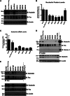
Downregulation of nucleolin removes surface nucleolin and eradicates the nucleolin-Fas complex. A nucleolin-specific (906), nontargeting (nonsilencing [NS]) control, and a glyceraldehyde-3-phosphate dehydrogenase (GAPDH) targeting short hairpin RNA miR30 constructs were used for transfection of BJAB cells to create a pooled NS control, GAPDH control, and nucleolin pKO (906P1) cell line. Four single-cell clones (906S1, 906S2, 906S4, 906S5) were derived from the original pooled cell line 906P1. (A) Whole-cell lysates were analyzed by IB for nucleolin and Fas protein expression. β-actin was used as a loading control. (B) Densitometry analysis revealed a minimum of 50% knockdown of nucleolin in the pKO cells compared with parental BJAB cells, nonsilencing controls, and GAPDH controls (906P1: P < .026; 906S1: P < .035; 906S2: P < .027; 906S4: P < .013; 906S5: P < .0114). Mean value and standard error of the mean of 3 independent experiments are shown. (C) Nucleolin pKO cells, parental BJABs, and nonsilencing controls were analyzed for nucleolin mRNA levels and normalized to GAPDH (906P1: P < .031, 906S1: P < .095, 906S2: P < .038, 906S4: P < .026, 906S5: P < .038). (D) Surface levels of nucleolin and Fas were analyzed in the parental BJAB, NS control, and nucleolin pKOs by biotinylation followed by strepavidin agarose IP. Histone 3 IB was used as a control for purity of the surface fraction. BJAB whole-cell extracts were used as a positive control for antibody specificity. Input levels of nucleolin and β-actin were used as a loading control. Representative data from 3 different experiments are shown. (E) Association of nucleolin and Fas was analyzed in parental BJAB cells, a NS control, a GAPDH control, and pKOs of nucleolin 906P1, 906S1, 906S2, 906S4, and 906S5 by IP with Fas (B-10) agarose beads. Nucleolin was detected by IB. Mouse isotype-matched IgG and protein G agarose were used as a negative control for nonspecific binding. BJAB whole-cell extracts were used as a positive protein control. Representative data from 3 different experiments are shown.
Figure 5
Figure 5
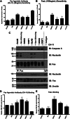
Loss of surface nucleolin and nucleolin-Fas complex sensitizes B-cell lymphomas to Fas-mediated apoptosis. (A) Indicated cells were challenged with the agonistic Fas antibody CH-11 (25 ng/mL) overnight and analyzed for apoptosis levels by Annexin V/7AAD staining and flow cytometry. The nucleolin pKO cells 906S2, 906S5, and 906P1 showed significant increases in sensitivity to agonistic antibody (906S2: P < .001, 906S5: P < .001, 906P1: P < .02). A Fas-sensitive T-cell line, Jurkat, was used as a positive control for Fas activation. Mean value and standard error of the mean (SEM) of 3 or more independent experiments each with 3 replicates are shown. (B) Cells were challenged with FasL (100 ng/mL) overnight and analyzed for apoptosis levels as in (A). The nucleolin pKO cells showed significant increases in FasL sensitivity compared with the nonsilencing control (906S2: P < .001, 906S5: P < .02, 906P1: P < .01). Mean value and SEM of 3 independent experiments each with 3 replicates are shown. (C) Parental BJAB, nonsilencing control, 906P1, and 906S2 cells were subjected to IP of Fas pre-challenge and 1 hour post-challenge with agonistic antibody CH-11 (25 ng/mL). The immunoprecipitated proteins were separated and Fas, caspase-8, and nucleolin were visualized by IB. Note a lack of nucleolin binding to Fas in 906P1 and 906S2 cells. IB of Fas was used as an IP control. Expression levels of nucleolin, Fas, and β-actin in whole-cell extracts as determined by IB analysis as input and loading controls. Representative data from 3 different experiments are shown. (D) Indicated cell lines were incubated with CH-11 (IgM subclass) for 20 minutes and the amount of bound antibody was analyzed by flow cytometry by measuring an anti-IgM–Allophycocyanin secondary antibody signal. The nucleolin pKO cells showed a significant increase in CH-11 signal (906S2: P < .001, 906S5: P < .03, 906P1: P < .04). Mean value and SEM of 3 independent experiments each with 3 replicates are shown. (E) Cells were incubated with FLAG-tagged FasL for 20 minutes and analyzed for the presence of ligand with an anti-FLAG-phycoerythrin secondary antibody by flow cytometry. The nucleolin pKO cells showed significantly increased FasL binding (906S2: P < .01, 906S5: P < .01, 906P1: P < .03). Mean value and SEM of 3 independent experiments each with 3 replicates are shown.
Figure 6
Figure 6
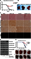
Overexpression of nucleolin protects mice from a lethal Fas activation through nucleolin-Fas complex. (A) Mice were hydrodynamically transfected with a vector control or a plasmid expressing DDK-tagged full-length nucleolin. The mice were challenged 24 hours later with a lethal dose of Jo2 agonistic anti-Fas antibody (2 µg/g weight) and monitored for survival for up to 8 hours post-challenge. The survival rate of nucleolin-transfected mice was significantly higher than mice transfected with vector alone (P < .006; log rank Mantel-Cox test). Representative data from 3 different experiments are shown. (B) Gross examination of vector and nontransfected livers challenged with Jo2 revealed massive hemorrhaging as indicated by darkening and swelling. Nucleolin-expressing livers challenged with Jo2 showed decreased hemorrhaging. (C) Livers were harvested, resected, and stained with hematoxylin and eosin (upper panel), or were analyzed using cleaved caspase-3 antibody, cleaved PARP antibody, or TUNEL assay to evaluate apoptosis. The images were captured by the Olympus BX41 (Olympus) UPlan FL N 40×/0.75 objective. Images were acquired with DP Controller (Olympus) with a −2 exposure adjustment for TUNEL staining with a fluorescein isothiocyanate filter (Olympus). Adobe Photoshop PS2 was used for further image enhancement of green fluorescent protein with a +30 brightness for all 4 panels equally. (D) Homogenized vector-tramsfected, nucleolin-transfected, a nontransfected Jo2-challenged and Jo2-unchallenged liver samples were subjected to lysis, sodium dodecyl sulfate-polyacrylamide gel electrophoresis and IB analysis of nucleolin, caspase-8, caspase-3, PARP, Bcl-2, and β-actin. (E) Mice were transfected with vector control, DDK-tagged full-length nucleolin, or DDK-tagged mutant lacking the Fas-nucleolin binding domain NR123 plasmids. Mice were challenged with a lethal dose of Jo2-agonistic anti-Fas antibody (.5 µg/g weight) and monitored for survival for up to 8 hours post-challenge. On gross examination, nontransfected, vector-transfected, and NR123-transfected livers challenged with Jo2 exhibited massive hemorrhaging as shown by darkening and swelling. Nucleolin livers challenged with Jo2 showed decreased hemorrhaging as shown by light spotting. We confirmed expression of nucleolin and NR123 by IB (data not shown). The survival rate was significantly higher for nucleolin-transfected mice than for mice transfected with vector alone or nonbinding mutant NR123 (P < .003; log rank Mantel-Cox test). Combined data from 2 independent experiments are shown.

Similar articles

Cited by

References

    1. Siegel R, Ward E, Brawley O, Jemal A. Cancer statistics, 2011: the impact of eliminating socioeconomic and racial disparities on premature cancer deaths. CA Cancer J Clin. 2011;61(4):212–236. - PubMed
    1. Kondo E, Yoshino T. Expression of apoptosis regulators in germinal centers and germinal center-derived B-cell lymphomas: insight into B-cell lymphomagenesis. Pathol Int. 2007;57(7):391–397. - PubMed
    1. Timmer T, de Vries EG, de Jong S. Fas receptor-mediated apoptosis: a clinical application? J Pathol. 2002;196(2):125–134. - PubMed
    1. Lynch DH, Watson ML, Alderson MR, et al. The mouse Fas-ligand gene is mutated in gld mice and is part of a TNF family gene cluster. Immunity. 1994;1(2):131–136. - PubMed
    1. Rieux-Laucat F, Le Deist F, Hivroz C, Roberts IA, Debatin KM, Fischer A, de Villartay JP. Mutations in Fas associated with human lymphoproliferative syndrome and autoimmunity. Science. 1995;268(5215):1347–1349. - PubMed

Publication types

MeSH terms