. 2013 Apr 15;201(2):233-47.
doi: 10.1083/jcb.201208172.

Microtubule-dependent endosomal sorting of clathrin-independent cargo by Hook1

Affiliations

Microtubule-dependent endosomal sorting of clathrin-independent cargo by Hook1

Lymarie Maldonado-Báez et al. J Cell Biol. .

Abstract

Many plasma membrane (PM) proteins enter cells nonselectively through clathrin-independent endocytosis (CIE). Here, we present evidence that cytoplasmic sequences in three CIE cargo proteins-CD44, CD98, and CD147-were responsible for the rapid sorting of these proteins into endosomal tubules away from endosomes associated with early endosomal antigen 1 (EEA1). We found that Hook1, a microtubule- and cargo-tethering protein, recognized the cytoplasmic tail of CD147 to help sort it and CD98 into Rab22a-dependent tubules associated with recycling. Depletion of Hook1 from cells altered trafficking of CD44, CD98, and CD147 toward EEA1 compartments and impaired the recycling of CD98 back to the PM. In contrast, another CIE cargo protein, major histocompatibility complex class I, which normally traffics to EEA1 compartments, was not affected by depletion of Hook1. Loss of Hook1 also led to an inhibition of cell spreading, implicating a role for Hook1 sorting of specific CIE cargo proteins away from bulk membrane and back to the PM.


Figures

Figure 1.
Figure 1.
CD44 and CD147 contain sorting sequences that direct their traffic to recycling tubular endosomes. (A) HeLa cells were incubated with primary antibodies against endogenous CD44, endogenous CD147, or Tac for 1 h at 37°C to allow internalization of the cargo proteins. After fixation, cells were immunolabeled with antibodies to EEA1, followed by secondary antibodies to detect EEA1 (red) and cargo proteins (green). Insets show enlarged views of the boxed regions. (B) Schematic representation of Tac, CD44, and CD147 chimeras used to identify potential sorting sequences in these CIE cargo proteins. Tac sequences are in orange, CD44 sequences in green, and CD147 sequences in purple. (C) HeLa cells transfected with either Tac or the different chimeric proteins were incubated with anti-CD44, anti–rat CD147, or anti-Tac antibodies for 1 h at 37°C to allow internalization of the antibody-bound cargo. Immunofluorescence analysis of all experiments was performed as described in Materials and methods. Arrows in A and C point to tubular endosomes. Bars: (A and C) 10 µm; (insets) 3 µm.
Figure 2.
Figure 2.
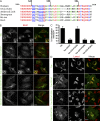
CD147 cytoplasmic sequence is sufficient for sorting directly into the recycling route. (A) Sequence alignment of the cytoplasmic tail (carboxyl terminal) of CD147, showing location of the truncated constructs at positions 235, 246, and 261. Identical sequences are highlighted in red, strongly similar in blue, and weakly similar in green. (B) Tac-CD147 carboxyl-terminal truncations were expressed in HeLa cells, and their locations were assessed after internalization for 1 h at 37°C using an anti-Tac antibody. Cells were fixed and processed for immunofluorescence as described in Materials and methods. Insets show enlarged views of the boxed regions. (C) The percentage of colocalization of internal Tac-CD147 truncated chimeras with EEA1 was quantified using MetaMorph software (see Materials and methods). The data presented is the mean of three independent experiments ± SD (error bars). (D) Cells expressing the different Tac-CD147 chimeras were incubated with anti-Tac for 1 h at 37°C. Then the cells were washed with media and chased in media containing 15 mM NH4Cl for 22 h. The arrow in the top row points to the cell surface. Other arrows point to tubular endosomes. The chimeric proteins were visualized with Alexa Fluor 488–conjugated goat anti–mouse, and late endosomes were visualized with antibodies to Lamp1. Bars: (B and D) 10 µm; (B, insets) 5 µm.
Figure 3.
Figure 3.
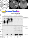
Hook1 interacts with the cytoplasmic tail of CD147 and CD98 through its carboxyl-terminal region. (A) Y2H analysis of the interaction between the cytoplasmic sequence of CD147 and the carboxyl-terminal sequence of Hook1. Yeast cells coexpressing CD147 carboxyl-terminal tail or the CD147 carboxyl-terminal acidic-cluster mutant (prey) and the carboxyl-terminal sequence of Hook1 or the pGBKT7 empty vector (bait) were grown on high-stringency plates. Growth on the –Leu/–TRP plate confirmed the expression of bait and prey plasmids (see Materials and methods for details about the Y2H screen). (B) Schematic representation of the domain organization of Hook1 (the amino terminus is shown in yellow; aa 1–168, coiled-coil region in blue; carboxyl terminus in pink, aa 659–728). Y2H clone encoding for amino acids 486–728 of Hook1. (C) BG-biotin–labeled SNAP-CD147 and CD98-SNAP were immunoprecipitated from lysates of cells coexpressing the Hook1 Y2H clone and separated on SDS–PAGE and immunoblotted as described in Materials and methods. The Hook1 Y2H clone was detected with rabbit anti-Hook1 and visualized with goat anti–rabbit 800 (bottom). The biotin-labeled SNAP-CD147 and CD98-SNAP were detected with NeutrAvidin DyLight-680 (top). For post-immunoprecipitation lysate (lanes 7–12), 1/10 of total protein was loaded.
Figure 4.
Figure 4.
Hook1 colocalizes with CD44, CD98, and CD147 on tubular endosomes. (A) HeLa cells were incubated with anti-CD147, anti-CD44, or anti-CD98 for 30 min at 37°C to allow the internalization of the cargo-bound antibodies. After internalization, cells were treated for 1 min with 10 µg/ml digitonin and then fixed. Endogenous Hook1 was localized using a rabbit anti-Hook1 antibody. Arrows point to tubular endosomes. (B) HeLa cells overexpressing HA-Hook1 were incubated with anti-CD147 antibody for 30 min at 37°C and processed for immunofluorescence. Enlargements of the two boxed regions are shown below. Arrows in the middle row indicate tubes, and arrows in the bottom row indicate swollen regions of the tubes. (C) After antibody internalization, endogenous CD147 (green; Alexa Fluor 488) and endogenous Hook1 (red; Alexa Fluor 568) were visualized using super-resolution fluorescence microscopy (SIM). This image is one projection of a 3D SIM image of a region of interest in the cell where CD147 colocalizes with Hook1 on tubular endosomal structures. Insets show enlarged views of the boxed regions. (D) Projection of a 3D image (from C, box 1) from 0° to 18° with 2° increments showing colocalization between CD147 (green) and Hook1 (red) at the end of a tubular endosome. (E) Montage of a 3D image (from C, box 2) from 0° to 10° with 2° increments showing CD147 (green) and Hook1 (red) together on an endosome (F). Endogenous Hook1 (red) and internalized CD98 (green) colocalized in tubular endosomes. Shown is a montage of a projection of a 3D image from 0° to 12° with 2° increments. Arrows in C–F point to Hook1 and cargo colocalization positions. Bars: (A–C) 10 µm; (A, insets) 3 µm; (B, insets) 5 µm; (C, inset) 3 µm; (D) 2 µm; (E and F) 1 µm.
Figure 5.
Figure 5.
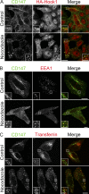
Endosomal sorting of CD147 requires microtubules. (A) HeLa cells overexpressing HA-Hook1 were untreated (Control) or pretreated with nocodazole for 2 h at 37°C before anti-CD147 antibody internalization for 30 min. (B) Untransfected HeLa cells were treated as described in A and then fixed and labeled with antibodies to EEA1. (C) Cells were incubated with CD147 antibody and Alexa Fluor 594–conjugated transferrin for 30 min at 37°C in control or nocodazole-containing media. Then the cells were fixed and stained with the respective secondary antibodies. Insets show enlarged views of the boxed panels. Bars: (A–C) 10 µm; (A and B, insets) 5 µm; (C, insets) 3 µm.
Figure 6.
Figure 6.
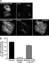
Expression of Hook1-Y2H results in loss of the recycling tubular endosomal compartment and the targeting of CIE cargo to EEA1-containing endosomes. (A) HeLa cells expressing endogenous levels of Hook1 (top), overexpressing Hook1-Y2H-HA (middle), or coexpressing Hook1-Y2H-HA and GFP-Rab22a (bottom) were incubated with anti-CD147 antibody for 1 h at 37°C to allow internalization of bound antibodies. Hook1-Y2H was localized using a rabbit anti-HA antibody. (B) The degree of restoration of the tubular endosomal network in Hook1-Y2H–expressing cells by GFP-Rab22a overexpression was determined by calculating the percentage of cells in each population exhibiting tubular endosomes. The data presented is the mean of three independent experiments ± SD (error bars). More than 100 cells were scored for each sample. Bars, 10 µm.
Figure 7.
Figure 7.
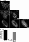
Hook1 and Rab22a regulate the sorting of CIE cargo proteins into the recycling tubular compartment. (A) HeLa cells expressing GFP-Rab22a S19N alone or coexpressing GFP-Rab22a S19N and HA-Hook1 were allowed to internalize anti-CD147 antibodies for 30 min at 37°C. HA-Hook1 was visualized using rabbit anti-HA and Alexa Fluor 594–conjugated anti–rabbit antibodies. Bars, 10 µm. (B) Quantitative analysis of the percentage of GFP-Rab22a S19N cells exhibiting tubular endosomes loaded with CD147 in the absence or presence of HA-Hook1. Data presented in the graph are the mean of three independent experiments ± SD (error bars).
Figure 8.
Figure 8.
Depletion of Rab22a affects CIE cargo tubular endosome biogenesis and sorting into EEA1-associated endosomes. (A) Rab22a protein levels in control or siRNA-treated cells were determined using Western blotting of cell lysates with an antibody against Rab22a and actin (loading control). (B and C) Control- or siRNA-treated cells were allowed to internalize antibody to CD147 and CD98 for 1 h at 37°C and processed for immunofluorescence and confocal microscopy. (D) Control- or siRNA-treated cells were scored for CIE cargo-containing tubules as described in Materials and methods. Results presented in the graph are the means of two independent experiments. (E) Quantitative analysis of colocalization between EEA1 and CIE cargo in control- and siRNA-treated cells. The graph shows the mean and standard deviation for the population of cells (20 cells per condition) from one representative experiment, repeated twice. Bars: (B and C) 10 µm; (B and C, insets) 5 µm.
Figure 9.
Figure 9.
Depletion of Hook1 affects tubular endosome formation and endosomal sorting of CIE cargo proteins. HeLa cells were treated with On-TARGET plus SMARTpool siRNA directed against Hook1 as described in Materials and methods. (A) Western blot showing the reduction of Hook1 protein levels in siRNA-treated versus control cells. Hook1 was detected using rabbit anti-Hook1 antibody and Alexa Fluor 680–conjugated anti–rabbit antibody. Actin was used as a loading control. (B and C) Control and Hook1 siRNA-depleted cells were incubated with CD147, CD98, and MHCI antibodies for 1 h at 37°C. After internalization, the cells were fixed and processed for immunofluorescence. Insets show enlarged views of the boxed regions. (D) Quantitative analysis of the percentage of cells exhibiting tubular endosomes loaded with CD147, CD98, and MHCI in control versus Hook1 siRNA-depleted cells. Data presented is the mean of three independent experiments ± SD (error bars). (E) The percentage of colocalization between CIE cargo and EEA1 after Hook1 depletion was determined using MetaMorph software (see Materials and methods). The graphs shows the mean and standard error for the population of cells (error bars; 20 cells per condition) from one experiment, repeated three times. Bars: (B and C) 10 µm; (B and C, insets) 5 µm.
Figure 10.
Figure 10.
Hook1 depletion inhibits recycling of CD98 and cell spreading in HeLa cells. (A) The amount of protein recycled back to the PM for CD98, MHCI, and the TfR in control and Hook1 siRNA-depleted cells was determined using the protocol described in Materials and methods. Cells were incubated with antibodies against CD98 or MCHI, or loaded with Alexa Fluor 594-Tf for 30 min at 37°C. After internalization, remaining surface-bound antibody was removed by a brief acid wash. Then the cells were incubated for 30 min at 37°C to allow recycling. The percentage of recycling for each cargo proteins was calculated, and shown here is the mean recycling from three independent experiments ± SD (error bars). To confirm the statistical significance of the results, the two-tailed Student’s t test was used. For CD98 recycling, the p-value for control versus Hook1-depleted cells was P < 0.05. For MHCI and TfR recycling, the values were not significantly different. (B) Control and Hook1 siRNA-treated cells were detached from the culture dish and replated onto coverslips for 6 h. The cells were fixed and stained with rhodamine phalloidin to facilitate the visualization and scoring of the phenotype. Bar, 10 µm. (C) The percentage of attached cells on the coverslips that were spread was determined by scoring >100 cells. The data shown are the means of “% cells spread” from three independent experiments ± SD (error bars).

Similar articles

Cited by

References

    1. Balasubramanian N., Scott D.W., Castle J.D., Casanova J.E., Schwartz M.A. 2007. Arf6 and microtubules in adhesion-dependent trafficking of lipid rafts. Nat. Cell Biol. 9:1381–1391 10.1038/ncb1657 - DOI - PMC - PubMed
    1. Balklava Z., Pant S., Fares H., Grant B.D. 2007. Genome-wide analysis identifies a general requirement for polarity proteins in endocytic traffic. Nat. Cell Biol. 9:1066–1073 10.1038/ncb1627 - DOI - PubMed
    1. Baron Gaillard C.L., Pallesi-Pocachard E., Massey-Harroche D., Richard F., Arsanto J.P., Chauvin J.P., Lecine P., Krämer H., Borg J.P., Le Bivic A. 2011. Hook2 is involved in the morphogenesis of the primary cilium. Mol. Biol. Cell. 22:4549–4562 10.1091/mbc.E11-05-0405 - DOI - PMC - PubMed
    1. Barral D.C., Cavallari M., McCormick P.J., Garg S., Magee A.I., Bonifacino J.S., De Libero G., Brenner M.B. 2008. CD1a and MHC class I follow a similar endocytic recycling pathway. Traffic. 9:1446–1457 10.1111/j.1600-0854.2008.00781.x - DOI - PMC - PubMed
    1. Cantor J.M., Ginsberg M.H. 2012. CD98 at the crossroads of adaptive immunity and cancer. J. Cell Sci. 125:1373–1382 10.1242/jcs.096040 - DOI - PMC - PubMed

Publication types

MeSH terms