. 2013 Apr 10;78(1):94-108.
doi: 10.1016/j.neuron.2013.02.003.

The CAMKK2-AMPK kinase pathway mediates the synaptotoxic effects of Aβ oligomers through Tau phosphorylation

Affiliations

The CAMKK2-AMPK kinase pathway mediates the synaptotoxic effects of Aβ oligomers through Tau phosphorylation

Georges Mairet-Coello et al. Neuron. .

Abstract

Amyloid-β 1-42 (Aβ42) oligomers are synaptotoxic for excitatory cortical and hippocampal neurons and might play a role in early stages of Alzheimer's disease (AD) progression. Recent results suggested that Aβ42 oligomers trigger activation of AMP-activated kinase (AMPK), and its activation is increased in the brain of patients with AD. We show that increased intracellular calcium [Ca²⁺](i) induced by NMDA receptor activation or membrane depolarization activates AMPK in a CAMKK2-dependent manner. CAMKK2 or AMPK overactivation is sufficient to induce dendritic spine loss. Conversely, inhibiting their activity protects hippocampal neurons against synaptotoxic effects of Aβ42 oligomers in vitro and against the loss of dendritic spines observed in the human APP(SWE,IND)-expressing transgenic mouse model in vivo. AMPK phosphorylates Tau on KxGS motif S262, and expression of Tau S262A inhibits the synaptotoxic effects of Aβ42 oligomers. Our results identify a CAMKK2-AMPK-Tau pathway as a critical mediator of the synaptotoxic effects of Aβ42 oligomers.


Figures

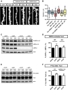
Figure 1
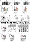
Figure 1. Aβ42-Dependent and Activity-Dependent Activation of the CAMKK2-AMPK Kinase Pathway in Cortical Neurons
(A–F) Primary cortical neurons isolated from E18.5 mouse embryos were cultured for 21 DIV and then treated with Aβ42 oligomers (1μM monomer equivalent for 24 hr) or INV42 (1 μM for 24 hr), KCl (20 mM for 15 min), and NMDA (50 μM for 15 min), in absence (CONT, control vehicle only) or presence of STO-609 (2.5 hr prior to treatment; 2.5 μM). Activation of AMPK was assessed by western blotting using the phospho-specific T172-AMPKα antibody and total AMPKα antibody (n = 6 experiments). Histograms in (B), (D), and (F) represent mean with SEM. (G–K′) Morphology of hippocampal neurons and their dendritic segments (secondary branches) treated at 21 DIV with Aβ42 oligomers (1 μM monomer equivalent for 24 hr) or INV42 (1 μM for 24 hr), or overexpressing CAMKK2, AMPKα1, or AMPKα2 (transfection at 15 DIV and visualization at 22 DIV). (L and M) Application of Aβ42 oligomers induced a significant reduction in dendritic spine density compared to INV42. Overexpression of CAMKK2 (L), AMPKα1, or AMPKα2 (M) resulted in dendritic spine loss compared to control neurons expressing GFP only. (N–Q) Hippocampal neurons treated at 21 DIV for 24 hr with compounds that activate AMPK such as AICAR, metformin, or A-769662 also induced rapid spine loss. Spine density was measured on 16–61 dendritic segments per condition. Box plots in (L), (M), (O), and (Q) represent data distribution (box 25th–75th percentiles, bars 10th–90th percentiles with central bar representing median). Statistical analysis was performed using ANOVA test followed by Dunnett posttest in (B), (D), (F), and (O) and Kruskal-Wallis with Dunn's posttest in (L), (M), and (Q). *p < 0.05; **p < 0.01; ***p < 0.001; ****p < 0.0001; ns, not significant p > 0.05. Scale bars, 5 μm (G–K) and 2 μm (G′, H′, I′, J′, K′, N, and P). See also Figures S1 and S2.
Figure 2
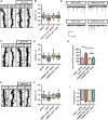
Figure 2. CAMKK2 and AMPKα1 Are Required for Aβ42 Oligomer-lnduced Spine Toxicity In Vitro
(A and C) Representative images of secondary dendritic segments of hippocampal neurons obtained from (A) control (CONT, CAMKK2+/+ or CAMKK2+/−) or CAMKK2 KO, and (C) WT (AMPKα1+/+) or AMPKα1 KO animals treated with Aβ42 oligomers or INV42 (1 μM for 24 hr). Hippocampal neurons were transfected with pCAG-EGFP plasmid at 11 DIV for spine visualization and treated at 20 DIV for 24 hr prior to fixation. (B and D) Neurons deficient for CAMKK2 (B) or AMPKα1 (D) were resistant to spine loss induced by Aβ42 oligomer exposure. Spine density was measured on 37–58 dendritic segments per condition. Box plots represent data distribution (box 25th–75th percentiles, bars 10th–90th percentiles with central bar representing median). Statistical analysis was performed using Kruskal-Wallis test followed by Dunn's posttest in (B) and (D). ***p < 0.001; ****p < 0.0001. Scale bars, 1 μm (A and C).
Figure 3
Figure 3. Inhibition of CAMKK2 or AMPK Activity Blocks the Synaptotoxic Effects of Aβ42 Oligomers in Hippocampal Neurons In Vitro
(A and B) Representative images of dendritic segments (A) and spine density quantification (B) of 21 DIV hippocampal neurons treated at 20 DIV with the CAMKK2 inhibitor STO-609 (2.5 μM) or DMSO (control) 2 hr prior to Aβ42 oligomer or INV42 application (1 μM for 24 hr). Neurons treated with STO-609 were resistant to spine loss induced by Aβ42 oligomers. (C and D) Representative images of dendritic segments (C) and quantification of spine density (D) of hippocampal neurons at 21 DIV expressing a KD version of CAMKK2 (K194A, transfected at 15 DIV), which significantly reduces the synaptotoxic effects of Aβ42 oligomers. (E and F) Representative images of dendritic segments (E) and quantification of spine density (F) of neurons that express a KD version of AMPKα2 (K45A, transfected at 15 DIV), which blocks the synaptotoxic effects of Aβ42 oligomers. Each condition is quantified from at least three independent experiments (19–151 dendritic segments quantified per condition). (G) Representative traces of mEPSCs recorded from 18 to 19 DIV hippocampal neurons expressing CAMKK2 KD or control vector (transfected at 11 DIV) and treated with Aβ42 oligomers or INV42 (1 μM for 24 hr). (H and I) Inter-mEPSC intervals (H) and mEPSC amplitudes (I) were quantified from 16 to 21 cells per condition (obtained from six independent experiments). Expression of CAMKK2 KD prevented the increase in inter-mEPSC interval induced by Aβ42 oligomers. There was no significant difference in mEPSC amplitude among the different conditions. Histograms represent mean with SEM. Box plots in (B), (D), and (F) represent data distribution (box 25 th–75th percentiles, bars 10 th–90th percentiles with central bar representing median). Statistical analysis was performed using ANOVA test followed by Dunnett's posttest in (B) and (F) and Kruskal-Wallis test followed by Dunn's posttest in (D), (H), and (I). *p < 0.05; **p < 0.01; ****p < 0.0001. Scale bars, 1 μm (A, C, and E).
Figure 4
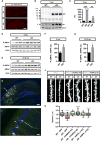
Figure 4. Inhibition of CAMKK2 or AMPK Activity Blocks the Synaptotoxic Effects of Aβ42 Oligomers in Hippocampal Neurons In Vivo
(A) Human APP and Aβ oligomers were detected by immunohistochemistry with the 6E10 antibody in the J20 transgenic mice (but not control littermates) as early as 3 months. (B and C) The hippocampus from 3-month-old WT and J20 mice was dissected, lysed, and the crude homogenate was processed to assess human APP and Aβ levels by western blotting with the 6E10 antibody. The J20 mice presented increased APP and Aβ levels compared to WT littermates (n = 3 animals per genotype). (D–G) The hippocampi of 4-month-old (D and F) and 8- to 12-month-old (E and G) WT and J20 mice were dissected and processed for cytosolic fractionation. AMPK activity for hippocampi isolated from individual mice was assessed by western blotting with pT172-AMPKα antibody, which was significantly increased in the cytosolic fraction of the J20 transgenic mice at both ages compared to littermate controls (n = 7–10 animals per genotype at both ages). (H and I) Inhibition of CAMKK2 or AMPKα activity in hippocampal pyramidal neurons of the CA3 region (arrow in H) was performed by in utero electroporation of CAMKK2 KD and AMPKα2 KD encoding plasmids in E15.5 WT and J20 mouse embryos. Dendritic spine density was quantified in the collateral processes (arrow in I) of basal dendrites (in stratum oriens) of CA3 pyramidal neurons in 3-month-old transfected animals. (J and K) Expression of CAMKK2 KD or AMPKα2 KD blocked the reduction of spine density observed in CA3 pyramidal neurons of 3-month-old J20 mice (33–53 dendritic segments from three to six animals per condition). Expression of CAMKK2 KD (but not AMPKα2 KD) induces a slight but significant increase in spine density in control mice. Box plots in (K) represent data distribution (box 25 th–75th percentiles, bars 10th–90th percentiles with central bar representing median). Histograms in (C), (F), and (G) represent mean ± SEM. Statistical analysis was performed using Mann-Whitney U test in (C), (F), and (G). In (K), Mann-Whitney U test was used to compare specific groups to WT-GFP (in blue) and to J20-GFP (in red). *p < 0.05; **p < 0.01; ****p < 0.0001. Scale bars, 2 μm (J), 50 μm (I), 100 μm (H), and 200 μm (A). See also Figure S3.
Figure 5
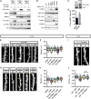
Figure 5. Phosphorylation of Tau on S262 Is Required for the Synaptotoxic Effects of Aβ Oligomers In Vitro and In Vivo
(A) Cotransfection of WT forms of AMPKα1 or α2 together with LKB1 is sufficient to trigger Tau phosphorylation on S262 residue in HeLa cells. Western blot on 25 μg of protein lysate with the indicated antibodies. (B) Upstream kinases LKB1, CAMKK1, and CAMKK2 display various abilities to phosphorylate AMPK upon expression in HEK293T cells. Western blot on 25 μg of protein lysate with the indicated antibodies. (C and D) AICAR treatment of cortical neurons at 21 DIV for 24 hr increased phosphorylation of Tau at epitope S262 compared to control (CONT, vehicle only). Histograms in (D) represent mean with SEM. (E–H) Hippocampal neurons were transfected at 11 DIV with constructs for different versions of Tau, treated with Aβ42 oligomers or INV42 at 20 DIV (1 μM for 24 hr), and spine density was assessed at 21 DIV. (E and F) Overexpression of WT Tau or its phospho-mimetic S262E version decreased spine density, whereas the nonphosphorylatable forms S262A and S356A had no effect. (G and H) Overexpression of mutant Tau S262A or S356A blocked oligomer-induced spine toxicity, whereas the WT form of Tau had no protective effects (n = 16–71 segments per condition). (I and J) Tau S262A was expressed in hippocampal pyramidal neurons of the CA3 region following in utero electroporation of E15.5 WT and J20 mouse embryos. Tau S262A expression induced a mild but significant decrease in spine density in WT animals but lessened the synaptotoxic effects of Aβ oligomers compared to J20 animals electroporated with control vector (19–53 dendritic segments from three to six animals per condition; compare figures with Figure 4J). Box plots in (F), (H), and (J) represent data distribution (box 25 th–75th percentiles, bars 10 th–90th percentiles with central bar representing median). Statistical analysis was performed using Mann-Whitney U test in (C) and ANOVA test followed by Dunnett's posttest in (F) and (H). In (J), Mann-Whitney U test was used to compare specific groups to WT-GFP (in blue) and to J20-GFP (in red). *p < 0.05; **p < 0.01; ***p < 0.001; ****p < 0.0001. Scale bars, 1 μM (E and G) and 2 μrn (I). See also Figure S4.
Figure 6
Figure 6. Phosphorylation of Tau on S262 by AMPKα1 Is Required for the Synaptotoxic Effects of Aβ42 Oligomers
(A and B) Hippocampal neurons were transfected at 11 DIV with control (GFP) or Tau S262A construct, treated with AMPK activator metformin or AICAR (100 μM) at 20 DIV for 24 hr, and spine density was assessed at 21 DIV. Overexpression of Tau S262A prevented the loss of spines induced by AMPKα overactivation (16–28 dendritic segments per condition). Box plots in (B) represent data distribution (box 25 th–75th percentiles, bars 10 th–90th percentiles with central bar representing median). (C-F) Cortical cells from WT or AMPKα1 KO animals were cultured for 20 DIV and then treated with Aβ42 oligomers or INV42 (1 μM for 24 hr). (C) AMPKα activation (phosphorylation of T172 of AMPKα1 and AMPKα2) and total AMPKα protein level were assessed by western blotting (n = 9–11 animals per genotype). (D) Aβ42 oligomers increased AMPK phosphorylation in WT cells, whereas oligomers had no effects in AMPKα1-deficient cells. (E and F) Phosphorylation of Tau on S262 and total Tau were assessed by western blotting (n = 9–10 animals per genotype). Aβ42 oligomers increased phosphorylation of Tau on S262 in WT cells, whereas no effect was detected in AMPKα1-deficient cells. Histograms in (D) and (F) represent mean with SEM. Statistical analysis was performed using ANOVA test followed by Dunnett's post hoc test in (B), and paired t test in (D) and (F). *p < 0.05; **p < 0.01. Scale bar, 1 μm (A).

Similar articles

Cited by

References

    1. Ageta-Ishihara N, Takemoto-Kimura S, Nonaka M, Adachi-Morishima A, Suzuki K, Kamijo S, Fujii H, Mano T, Blaeser F, Chatila TA, et al. Control of cortical axon elongation by a GABA-driven Ca2+/calmodulin-dependent protein kinase cascade. J Neurosci. 2009;29:13720–13729. - PMC - PubMed
    1. Alessi DR, Sakamoto K, Bayascas JR. LKB1-dependent signaling pathways. Annu Rev Biochem. 2006;75:137–163. - PubMed
    1. Anderson KA, Ribar TJ, Lin F, Noeldner PK, Green MF, Muehlbauer MJ, Witters LA, Kemp BE, Means AR. Hypothalamic CaMKK2 contributes to the regulation of energy balance. Cell Metab. 2008;7:377–388. - PubMed
    1. Barnes AP, Lilley BN, Pan YA, Plummer LJ, Powell AW, Raines AN, Sanes JR, Polleux F. LKB1 and SAD kinases define a pathway required for the polarization of cortical neurons. Cell. 2007;129:549–563. - PubMed
    1. Bezprozvanny I, Mattson MP. Neuronal calcium mishandling and the pathogenesis of Alzheimer's disease. Trends Neurosci. 2008;31:454–463. - PMC - PubMed

Publication types

MeSH terms