

Transcripts for combined synthetic microRNA and gene delivery
- PMID: 23579254
- PMCID: PMC3674133
- DOI: 10.1039/c3mb70043g
Transcripts for combined synthetic microRNA and gene delivery
Abstract
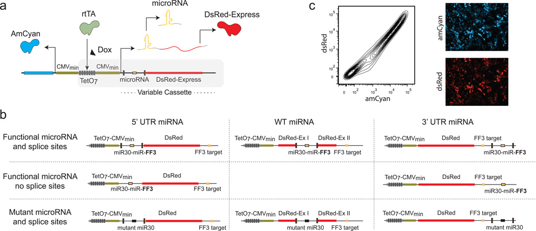
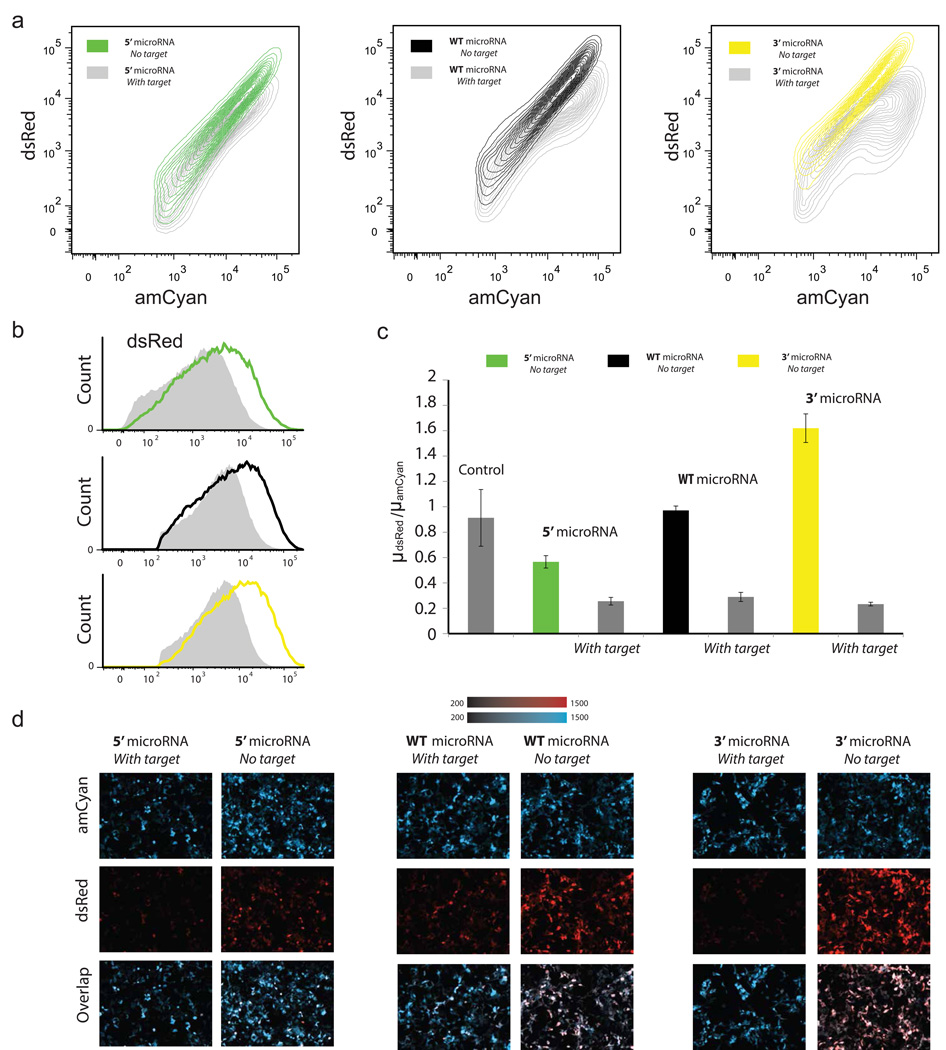
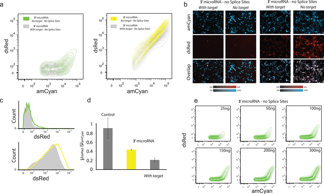
MicroRNAs (miRNAs) are a class of short noncoding RNAs which are endogenously expressed in many organisms and regulate gene expression by binding to messenger RNA (mRNA). MicroRNAs are either produced from their independent transcription units in intergenic regions or lie in intragenic regions. Intragenic miRNAs and their host mRNAs are produced from the same transcript by the microprocessor and the spliceosome protein complex respectively. The details and exact timing of the processing events have implications for downstream RNA interference (RNAi) efficiency and mRNA stability. Here we engineer and study in mammalian cells a range of synthetic intragenic miRNAs co-expressed with their host genes. Furthermore, we study transcripts which carry the target of the miRNA, thereby emulating a common regulation mechanism. We perform fluorescence microscopy and flow cytometry to characterize the engineered transcripts and investigate the properties of the underlying biological processes. Our results shed additional light on miRNA and pre-mRNA processing but importantly provide insight into engineering transcripts customized for combined delivery and use in synthetic gene circuits.
Figures

Similar articles
-
A multiplexed miRNA and transgene expression platform for simultaneous repression and expression of protein coding sequences.Mol Biosyst. 2016 Jan;12(1):295-312. doi: 10.1039/c5mb00506j. Mol Biosyst. 2016. PMID: 26617199
-
Gene Silencing In Vitro and In Vivo Using Intronic MicroRNAs.Methods Mol Biol. 2018;1733:107-126. doi: 10.1007/978-1-4939-7601-0_9. Methods Mol Biol. 2018. PMID: 29435927
-
miRNA and shRNA expression vectors based on mRNA and miRNA processing.Methods Mol Biol. 2013;936:195-207. doi: 10.1007/978-1-62703-083-0_16. Methods Mol Biol. 2013. PMID: 23007510
-
Gene silencing in vitro and in vivo using intronic microRNAs.Methods Mol Biol. 2006;342:295-312. doi: 10.1385/1-59745-123-1:295. Methods Mol Biol. 2006. PMID: 16957384 Review.
-
miRNA cassettes in viral vectors: problems and solutions.Biochim Biophys Acta. 2011 Nov-Dec;1809(11-12):732-45. doi: 10.1016/j.bbagrm.2011.05.014. Epub 2011 Jun 7. Biochim Biophys Acta. 2011. PMID: 21679781 Review.
Cited by
-
Model-guided quantitative analysis of microRNA-mediated regulation on competing endogenous RNAs using a synthetic gene circuit.Proc Natl Acad Sci U S A. 2015 Mar 10;112(10):3158-63. doi: 10.1073/pnas.1413896112. Epub 2015 Feb 23. Proc Natl Acad Sci U S A. 2015. PMID: 25713348 Free PMC article.
-
Mapping the operational landscape of microRNAs in synthetic gene circuits.NPJ Syst Biol Appl. 2018 Jan 11;4:6. doi: 10.1038/s41540-017-0043-y. eCollection 2018. NPJ Syst Biol Appl. 2018. PMID: 29354284 Free PMC article.
-
Functional intron-derived miRNAs and host-gene expression in plants.Plant Methods. 2018 Sep 24;14:83. doi: 10.1186/s13007-018-0351-2. eCollection 2018. Plant Methods. 2018. PMID: 30258486 Free PMC article.
-
Synthetic regulatory RNAs selectively suppress the progression of bladder cancer.J Exp Clin Cancer Res. 2017 Oct 30;36(1):151. doi: 10.1186/s13046-017-0626-x. J Exp Clin Cancer Res. 2017. PMID: 29084575 Free PMC article.
-
Robust Filtering and Noise Suppression in Intragenic miRNA-Mediated Host Regulation.iScience. 2020 Sep 21;23(10):101595. doi: 10.1016/j.isci.2020.101595. eCollection 2020 Oct 23. iScience. 2020. PMID: 33083753 Free PMC article.
References
-
- Bartel DP. MicroRNAs: Genomics, biogenesis, mechanism, and function. Cell. 2004;116(2):281–297. - PubMed
-
- Stark A, Brennecke J, Bushati N, Russell RB, Cohen SM. Animal microRNAs confer robustness to gene expression and have a significant impact on 3′ UTR evolution. Cell. 2005;123(6):1133–1146. - PubMed
-
- Pillai RS, Bhattacharyya SN, Filipowicz W. Repression of protein synthesis by miRNAs: How many mechanisms? Trends Cell Biol. 2007;17(3):118–126. - PubMed
Publication types
MeSH terms
Substances
Grants and funding
LinkOut - more resources
Full Text Sources
Other Literature Sources