. 2013 Jun 15;452(3):509-18.
doi: 10.1042/BJ20121908.

A covalent protein-DNA 5'-product adduct is generated following AP lyase activity of human ALKBH1 (AlkB homologue 1)

Affiliations

A covalent protein-DNA 5'-product adduct is generated following AP lyase activity of human ALKBH1 (AlkB homologue 1)

Tina A Müller et al. Biochem J. .

Abstract

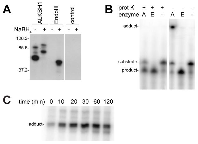
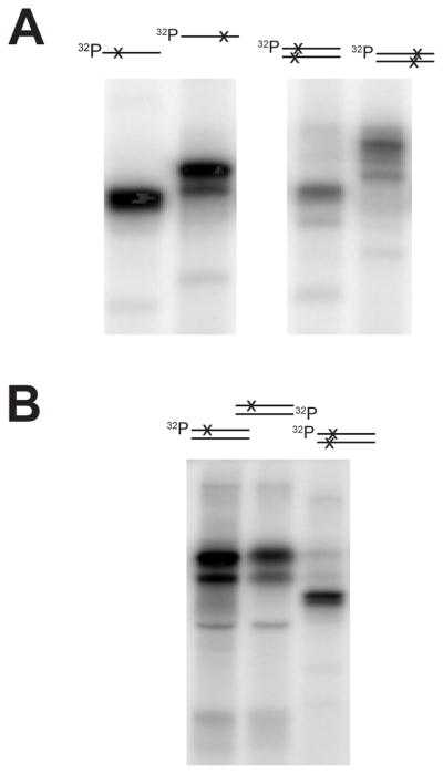
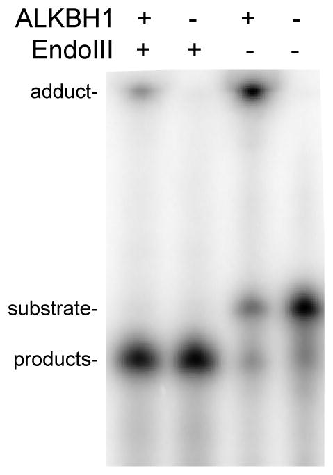
ALKBH1 (AlkB homologue 1) is a mammalian AlkB (2-oxoglutarate-dependent dioxygenase) homologue that possesses AP (abasic or apurinic/apyrimidinic) lyase activity. The AP lyase reaction is catalysed by imine formation with an active site lysine residue, and a covalent intermediate can be trapped in the presence of NaBH4. Surprisingly, ALKBH1 also forms a stable protein-DNA adduct in the absence of a reducing agent. Experiments with different substrates demonstrated that the protein covalently binds to the 5' DNA product, i.e. the fragment containing an α,β-unsaturated aldehyde. The N-terminal domain of ALKBH1 was identified as the main site of linkage with DNA. By contrast, mutagenesis studies suggest that the primary catalytic residue forming the imine linkage is Lys133, with Lys154 and other lysine residues in this region serving in opportunistic roles. These findings confirm the classification of ALKBH1 as an AP lyase, identify the primary and a secondary lysine residues involved in the lyase reaction, and demonstrate that the protein forms a covalent adduct with the 5' DNA product. We propose two plausible chemical mechanisms to account for the covalent attachment.


Figures

Figure 1
Figure 1. Comparison of AP hydrolase and AP lyase reactions
AP sites can be cleaved by an AP hydrolase reaction (top) or by an AP lyase reaction acting on the open-ring aldehyde form of the deoxyribose (bottom).
Figure 2
Figure 2. ALKBH1 covalently binds AP-containing DNA in the absence of NaBH4 reducing agent
(A) Formation of an ALKBH1-DNA adduct. N-terminal His6-tagged ALKBH1 (2.5 μM), and EndoIII (10 U) were incubated with the radioactively labeled UDG-treated substrate oligo1+2 (1 μM) for 1 h at 37 °C in the absence and presence of the reducing agent NaBH4 (10 mM). Control lanes lack either lyase. The samples were denatured in a boiling water bath for 3 min prior to resolving by 12% SDS-PAGE and visualizing by autoradiography. The Schiff base intermediate formed with ALKBH1 and EndoIII was trapped with NaBH4 as expected, but ALKBH1 also formed an adduct with the DNA in the absence of the reducing agent. The positions of three protein molecular mass markers (in kDa) are indicated to the left of the gel. (B) AP lyase activity assays with and without proteinase K treatment. His6-tagged ALKBH1 (labeled A, 5 μM) and EndoIII (labeled E, 1 U/assay) were incubated with radioactively labeled UDG-treated oligo1+2 (1 μM) for 1 h at 37 °C. Half of the sample was subjected to proteinase K (prot K, 1 U) digestion at 65 °C for 30 min, whereas the other half was kept on ice. Control lanes did not contain either lyase. All samples were analyzed by denaturing 7 M urea PAGE and the 5′ radioactively labeled DNA species were detected by autoradiography. (C) Time course studies. ALKBH1 was incubated with UDG-treated oligo1+2 for the times indicated and examined directly by 12% SDS-PAGE to assess adduct formation.
Figure 3
Figure 3. Demonstration of an adduct between ALKBH1 and the 5′-DNA product
(A) Comparison of the DNA-ALKBH1 adducts with single and double stranded substrates. N-terminal His6-tagged ALKBH1 (5 μM) was incubated with DNA substrates (1 μM) for 1 h at 37 °C. UDG-treated oligo3 or oligo4 was used directly as single stranded species (left) or formed into double stranded oligo3+4 species, as schematically indicated above the autoradiographs (the AP sites are indicated by x and the location of the radiolabel is shown). After adduct formation, the DNA-protein adduct was analyzed by 12% SDS-PAGE and visualized by autoradiography. (B) Comparison of the DNA-ALKBH1 adducts with double stranded DNA containing one or two AP sites. His6-tagged ALKBH1 (5 μM) was incubated with 1 μM substrate containing one AP site, the UDG-treated oligo3+5 radioactively labeled on either oligo3 (lane 1) or oligo5 (lane 2), or substrate containing two AP sites in close proximity on opposing strands (UDG-treated oligo3+4, lane 3) for 1 h at 37 °C. The adducts were then analyzed as described in panel A.
Figure 4
Figure 4. The amino-terminal region of ALKBH1 forms a covalent bond to the 5′-DNA product of AP-containing DNA
(A) BNPS-skatole cleavage of ALKBH1. N-terminal His6-tagged and thrombin-treated (untagged) ALKBH1 samples (5 μM) were incubated in the absence of substrate or with AP oligo4 (7.5 μM) for 1 h at 37 °C. Samples to be cleaved with BNPS-skatole were subjected to the reagent (1 mM) in 70% acetic acid containing 0.1% phenol for 72 h, whereas the control proteins were incubated in the same conditions without the reagent. The samples were analyzed by using a 10–16% Tris-Tricine gel and visualized by Coomassie staining. The migration positions of molecular mass markers are shown to the left. The numbers next to the bands indicate peptides involved in protein-DNA adduct formation. (B) LysC digest of selected Lys variants of ALKBH1. Selected Lys variants of N-terminal His6-tagged ALKBH1 were incubated with 32P-labelled UDG-treated oligo4 for 1 h at 37 °C, denatured at 95 °C, and subjected to LysC digestion (0.2 U) overnight. The samples were analyzed by 15% SDS-PAGE and the DNA-peptide adducts were visualized by autoradiography. A band of interest (see text) is marked *. (C) The His-tag is not required for adduct formation. His6-tagged and untagged ALKBH1 samples were incubated with UDG-treated oligo1+2. Aliquots were removed at the indicated time intervals, frozen, and analyzed by 6% PAGE. (D) C-terminal His6-tagged ALKBH1 has AP-lyase activity and forms the enzyme-product adduct. N- and C-terminal His6-tagged ALKBH1 as well as N-terminal His6-tagged ALKBH1 treated with thrombin were incubated with AP-oligo1+2 for 1 h at 37 °C. One series of samples was treated with proteinase K (1 U) for 30 min at 65 °C, whereas the other half was kept on ice. All samples were analyzed by 18% native PAGE and the DNA visualized by ethidium bromide staining.
Figure 5
Figure 5. ALKBH1’s AP lyase activity yields a 5′-phosphorylated 3′ product
N-terminal His6-tagged (ALKBH1His) or untagged ALKBH1 (5 μM), Ape1 (A, 10 U), and EndoIII (E, 10U) were incubated with unlabeled, single stranded AP-containing oligo4 (1 μM) for 1 h at 37 °C and either used directly or treated with proteinase K (2 U). One aliquot of each sample was incubated with Antarctic alkaline phosphatase (5 U) for 30 min at 37 °C followed by an inactivation step at 65 °C for 30 min, whereas a second aliquot was left on ice. All samples were incubated with 10 U of PNK plus [γ-32P] ATP and examined by denaturing 20% PAGE gel. The signals were detected by exposing the dried gel to a phosphorimager.
Figure 6
Figure 6. ALKBH1 forms a covalent bond with the α,β-unsaturated aldehyde
ALKBH1 forms an adduct with the product of the AP lyase activity of EndoIII. Radioactively labeled UDG-treated oligo1 (1 μM) was incubated with EndoIII (1 U) for 1 h at 37 °C. This sample was directly examined or His6-tagged ALKBH1 (5 μM) was added to the pre-cleaved products and allowed to form the adduct for 1 h at 37 °C. Adduct formation of ALKBH with radioactively labeled UDG-treated oligo1 and a lane lacking either enzyme are shown as additional controls. All protein-DNA adducts were analyzed by denaturing 7 M urea PAGE (20%).
Figure 7
Figure 7. Characterization of ALKBH1 Lys variants and elucidation of the AP lyase active site Lys residue
(A) AP lyase activity of single Lys variants of ALKBH1. All 22 Lys residues in N-terminal His6-tagged ALKBH1 were individually substituted by Ala using site-directed mutagenesis and the purified proteins were analyzed for AP lyase activity (5 μM variant protein and 1 μM double stranded UDG-treated oligo1+2 for 1 h at 37 °C, proteinase K (0.2 U) digested for 30 min at 65 °C, and the products visualized by ethidium bromide treatment after 18% PAGE). The 100% activity of WT ALKBH1 corresponds to cleavage of 81% of the substrate. (B) AP lyase activity of ALKBH1 double variants. Selected variants of K133A ALKBH1 were constructed, purified, and analyzed as above. The activity of K133A ALKBH1 was set to 100%.
Figure 8
Figure 8. Hypothetical mechanisms for the covalent attachment of ALKBH1 to the AP DNA product
(A) Overview of interaction between DNA and ALKBH1, with its separate Fe-containing demethylase and Lys-dependent AP lyase active sites. The line connecting protein and DNA indicates a covalent linkage between ALKBH1 and the 5′ DNA product. (B) One plausible mechanism for generating the covalent linkage uses a nucleophile to attacks the C3′ atom of the α,β-unsaturated aldehyde. The resulting adduct then forms a more stable cyclic hemiacetal which may be further stabilized by dehydration. (C) An alternative hypothesis has the nucleophile being involved in a phosphotransfer reaction with the phosphate preceding the α,β-unsaturated aldehyde with release of the five-carbon unit.

Similar articles

Cited by

References

    1. Christmann M, Tomicic MT, Roos WP, Kaina B. Mechanisms of human DNA repair: an update. Toxicology. 2003;193:3–34. - PubMed
    1. Lhomme J, Constant JF, Demeunynck M. Abasic DNA structure, reactivity, and recognition. Biopolymers. 1999;52:65–83. - PubMed
    1. Lindahl T. Instability and decay of the primary structure of DNA. Nature. 1993;362:709–715. - PubMed
    1. Dianov GL, Sleeth KM, Dianova II, Allinson SL. Repair of abasic sites in DNA. Mutat Res. 2003;531:157–163. - PubMed
    1. Hegde ML, Hazra TK, Mitra S. Early steps in the DNA base excision/single-strand interruption repair pathway in mammalian cells. Cell Res. 2008;18:27–47. - PMC - PubMed

Publication types

MeSH terms

LinkOut - more resources