Functional impairment of microglia coincides with Beta-amyloid deposition in mice with Alzheimer-like pathology
- PMID: 23577177
- PMCID: PMC3620049
- DOI: 10.1371/journal.pone.0060921
Functional impairment of microglia coincides with Beta-amyloid deposition in mice with Alzheimer-like pathology
Abstract
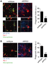
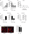
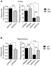
Microglial cells closely interact with senile plaques in Alzheimer's disease and acquire the morphological appearance of an activated phenotype. The significance of this microglial phenotype and the impact of microglia for disease progression have remained controversial. To uncover and characterize putative changes in the functionality of microglia during Alzheimer's disease, we directly assessed microglial behavior in two mouse models of Alzheimer's disease. Using in vivo two-photon microscopy and acute brain slice preparations, we found that important microglial functions - directed process motility and phagocytic activity - were strongly impaired in mice with Alzheimer's disease-like pathology compared to age-matched non-transgenic animals. Notably, impairment of microglial function temporally and spatially correlated with Aβ plaque deposition, and phagocytic capacity of microglia could be restored by interventionally decreasing amyloid burden by Aβ vaccination. These data suggest that major microglial functions progressively decline in Alzheimer's disease with the appearance of Aβ plaques, and that this functional impairment is reversible by lowering Aβ burden, e.g. by means of Aβ vaccination.
Conflict of interest statement
Figures


Similar articles
-
Fibrillar Aβ triggers microglial proteome alterations and dysfunction in Alzheimer mouse models.Elife. 2020 Jun 8;9:e54083. doi: 10.7554/eLife.54083. Elife. 2020. PMID: 32510331 Free PMC article.
-
Microglia contributes to plaque growth by cell death due to uptake of amyloid β in the brain of Alzheimer's disease mouse model.Glia. 2016 Dec;64(12):2274-2290. doi: 10.1002/glia.23074. Epub 2016 Sep 23. Glia. 2016. PMID: 27658617
-
Accelerated microglial pathology is associated with Aβ plaques in mouse models of Alzheimer's disease.Aging Cell. 2014 Aug;13(4):584-95. doi: 10.1111/acel.12210. Epub 2014 Mar 18. Aging Cell. 2014. PMID: 24641683 Free PMC article.
-
Review: experimental manipulations of microglia in mouse models of Alzheimer's pathology: activation reduces amyloid but hastens tau pathology.Neuropathol Appl Neurobiol. 2013 Feb;39(1):69-85. doi: 10.1111/nan.12002. Neuropathol Appl Neurobiol. 2013. PMID: 23171029 Free PMC article. Review.
-
Effects of CX3CR1 and Fractalkine Chemokines in Amyloid Beta Clearance and p-Tau Accumulation in Alzheimer's Disease (AD) Rodent Models: Is Fractalkine a Systemic Biomarker for AD?Curr Alzheimer Res. 2016;13(4):403-12. doi: 10.2174/1567205013666151116125714. Curr Alzheimer Res. 2016. PMID: 26567742 Review.
Cited by
-
Glial Purinergic Signaling in Neurodegeneration.Front Neurol. 2021 May 14;12:654850. doi: 10.3389/fneur.2021.654850. eCollection 2021. Front Neurol. 2021. PMID: 34054698 Free PMC article. Review.
-
The dynamics of monocytes and microglia in Alzheimer's disease.Alzheimers Res Ther. 2015 Apr 15;7(1):41. doi: 10.1186/s13195-015-0125-2. eCollection 2015. Alzheimers Res Ther. 2015. PMID: 25878730 Free PMC article.
-
The Emerging Role of the Interplay Among Astrocytes, Microglia, and Neurons in the Hippocampus in Health and Disease.Front Aging Neurosci. 2021 Apr 6;13:651973. doi: 10.3389/fnagi.2021.651973. eCollection 2021. Front Aging Neurosci. 2021. PMID: 33889084 Free PMC article. Review.
-
Astrocytic phagocytosis is a compensatory mechanism for microglial dysfunction.EMBO J. 2020 Nov 16;39(22):e104464. doi: 10.15252/embj.2020104464. Epub 2020 Sep 22. EMBO J. 2020. PMID: 32959911 Free PMC article.
-
Marked Response of Rat Ileal and Colonic Microbiota After the Establishment of Alzheimer's Disease Model With Bilateral Intraventricular Injection of Aβ (1-42).Front Microbiol. 2022 Feb 11;13:819523. doi: 10.3389/fmicb.2022.819523. eCollection 2022. Front Microbiol. 2022. PMID: 35222337 Free PMC article.
References
-
- Nimmerjahn A, Kirchhoff F, Helmchen F (2005) Resting microglial cells are highly dynamic surveillants of brain parenchyma in vivo. Science 308: 1314–1318. - PubMed
-
- Davalos D, Grutzendler J, Yang G, Kim JV, Zuo Y, et al. (2005) ATP mediates rapid microglial response to local brain injury in vivo. Nat Neurosci 8: 752–758. - PubMed
-
- Kettenmann H, Kirchhoff F, Verkhratsky A (2013) Microglia: new roles for the synaptic stripper. Neuron 77: 10–18. - PubMed
Publication types
MeSH terms
Substances
Grants and funding
LinkOut - more resources
Full Text Sources
Other Literature Sources
Medical
Molecular Biology Databases

