. 2013 Jun;87(12):6804-18.
doi: 10.1128/JVI.00243-13. Epub 2013 Apr 10.

Novel cis-acting element within the capsid-coding region enhances flavivirus viral-RNA replication by regulating genome cyclization

Affiliations

Novel cis-acting element within the capsid-coding region enhances flavivirus viral-RNA replication by regulating genome cyclization

Zhong-Yu Liu et al. J Virol. 2013 Jun.

Abstract

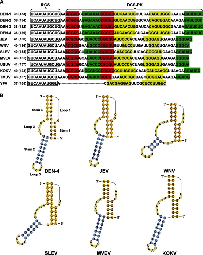
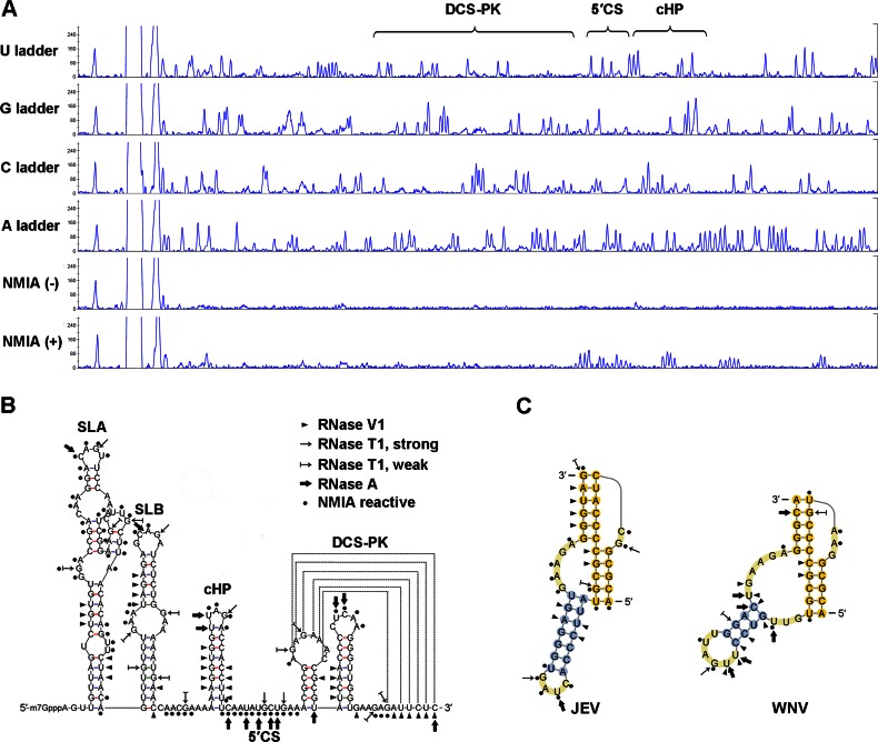
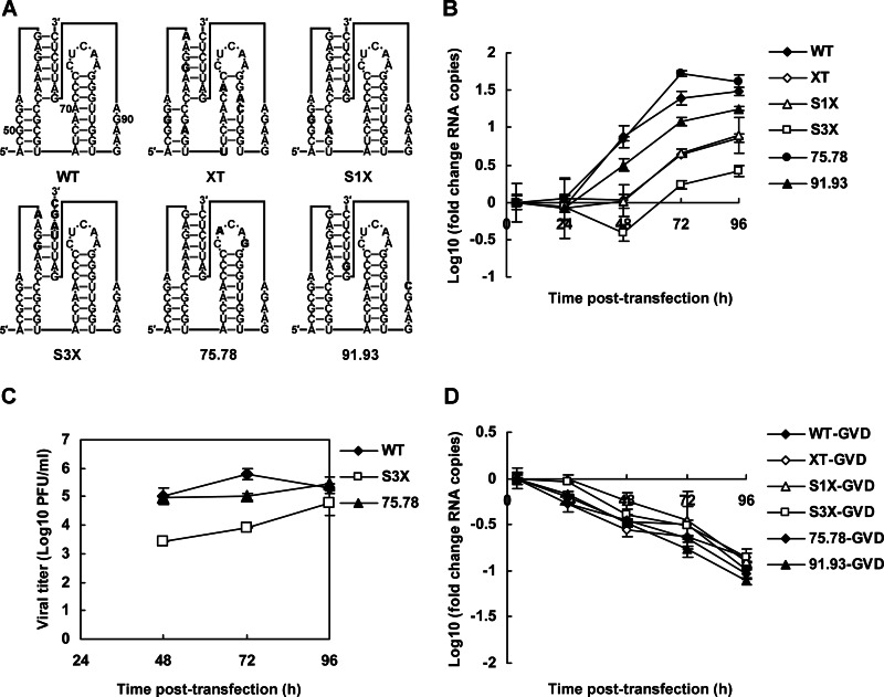
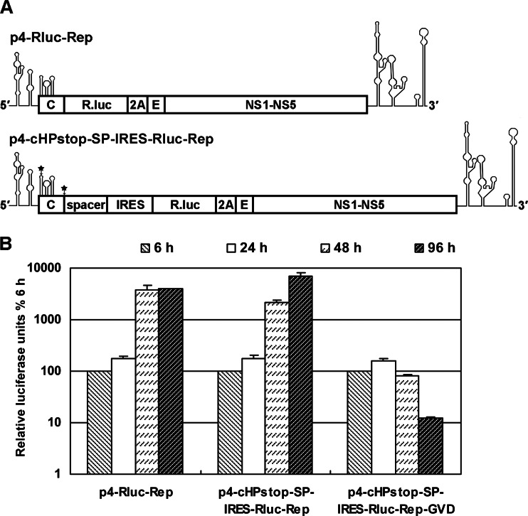
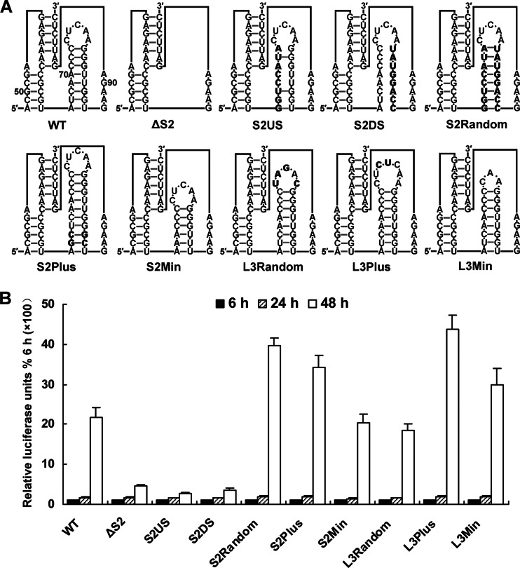
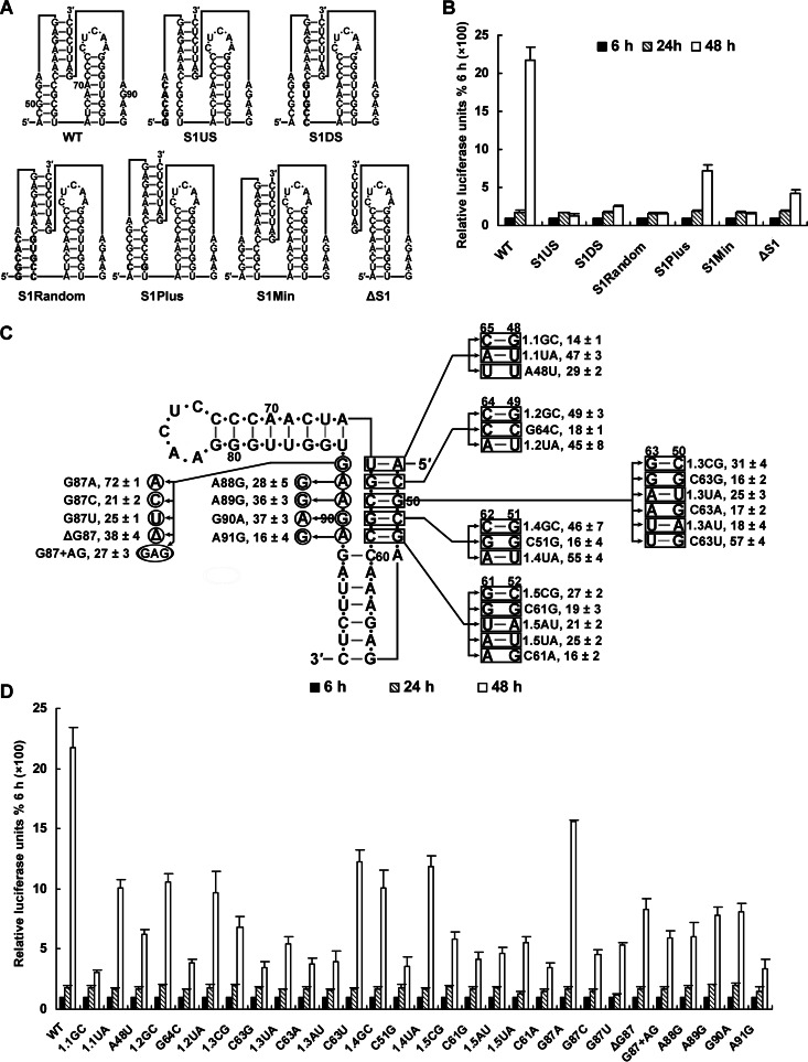
cis-Acting elements in the viral genome RNA (vRNA) are essential for the translation, replication, and/or encapsidation of RNA viruses. In this study, a novel conserved cis-acting element was identified in the capsid-coding region of mosquito-borne flavivirus. The downstream of 5' cyclization sequence (5'CS) pseudoknot (DCS-PK) element has a three-stem pseudoknot structure, as demonstrated by structure prediction and biochemical analysis. Using dengue virus as a model, we show that DCS-PK enhances vRNA replication and that its function depends on its secondary structure and specific primary sequence. Mutagenesis revealed that the highly conserved stem 1 and loop 2, which are involved in potential loop-helix interactions, are crucial for DCS-PK function. A predicted loop 1-stem 3 base triple interaction is important for the structural stability and function of DCS-PK. Moreover, the function of DCS-PK depends on its position relative to the 5'CS, and the presence of DCS-PK facilitates the formation of 5'-3' RNA complexes. Taken together, our results reveal that the cis-acting element DCS-PK enhances vRNA replication by regulating genome cyclization, and DCS-PK might interplay with other cis-acting elements to form a functional vRNA cyclization domain, thus playing critical roles during the flavivirus life cycle and evolution.


Figures

Fig 1
Fig 1
Structure prediction of DCS-PK among different mosquito-borne flaviviruses. (A) Nucleotide sequences of the downstream of 5′CS region of mosquito-borne flaviviruses. The 5′CSs are boxed, and predicted stem regions of DCS-PK are shown in different colors: red for stem 1, yellow for stem 2, and green for stem 3. The numbers at the beginning of the sequences represent the position of the first shown nucleotide in the capsid ORF, and its position in the viral genome (the latter are shown in parentheses). Abbreviations: DEN-1 to DEN-4, DENV serotypes 1 to 4; SLEV, Saint Louis encephalitis virus; MVEV, Murray Valley encephalitis virus; USUV, Usutu virus; KOKV, Kokobera virus; TMUV, Tembusu virus. The GenBank accession numbers of representative sequences are as follows: DEN-1, EU848545; DEN-2, AY702035; DEN-3, EU081188; DEN-4, AF326573; JEV, M55506; WNV, AY490240; SLEV, EU566860; MVEV, AF161266; USUV, EF206350; KOKV, AY632541; TMUV, JQ289550; YFV, AY640589. (B) DCS-PK structures predicted by pknotRG and drawn with PseudoViewer3. Note that constraints were applied to the stem 1-stem 3 interfaces of several viruses based on sequence comparison shown in panel A and structure probing results shown in Fig. 2.
Fig 2
Fig 2
Biochemical structure analysis of DCS-PK elements from different flaviviruses. (A) A representative SHAPE experiment for the analysis of the structure of DEN-4 5′ end RNA. Nucleotide ladders were generated by dideoxy-sequencing. The regions corresponding to DCS-PK and several 5′ end RNA elements are indicated. (B) Summary of the RNase probing and NMIA modification results of the DEN-4 5′ RNA containing the DCS-PK element. The major RNA elements of the 5′ end are indicated. The symbols used are explained in the right panel. (C) RNase probing and NMIA modification results of the DCS-PK elements from JEV and WNV. The symbols used are the same in panel B.
Fig 3
Fig 3
Characterization of DEN-4 mutants containing silent mutations in DCS-PK. (A) Demonstration of mutants containing silent mutations in the DCS-PK element. Mutations are indicated in the predicted pseudoknot structure of DCS-PK in boldface. The positions of several nucleotides of DCS-PK in the capsid ORF are indicated in the WT structure. Note that the secondary structures of DCS-PK mutants are only shown to provide a clear illustration and do not represent the actual folding of these mutants. (B) Replication of vRNA mutants in transfected BHK-21 cells. vRNA levels are expressed as fold changes relative to the RNA copies 4 h posttransfection. (C) Growth curves of viruses in the supernatants of vRNA-transfected BHK-21 cells. Virus titers were normalized to transfection efficiency as determined by qRT-PCR at 4 h after transfection. (D) Decay kinetics of various vRNA mutants, which all contained GDD-to-GVD mutations in NS5RdRp. The error bars indicate the standard deviations in all figures.
Fig 4
Fig 4
Effects of secondary interactions in DCS-PK on vRNA replication. (A) Mutations targeting various stem regions of DCS-PK. The mutations introduced are indicated in boldface. The nucleotide positions of DCS-PK in the capsid ORF are indicated in the S1POS structure. Note that the secondary structures of DCS-PK mutants are only shown to provide a clear illustration and do not represent the actual folding of these mutants. (B to D) Replication of vRNA mutants containing the disrupted or restored DCS-PK stem 1 (B), stem 2 (C), and stem 3 (D), which were determined by qRT-PCR. Metabolism curves of corresponding GVD-containing RNAs are shown in parallel. The data are expressed as fold changes relative to the RNA level 4 h posttransfection.
Fig 5
Fig 5
Design of replicons uncoupling the coding role of DCS-PK from its function in vRNA replication. (A) Organization of replicons used in the present study. In p4-Rluc-Rep, the Rluc ORF was fused in frame with the capsid ORF, whereas in p4-cHPstop-SP-IRES-Rluc-Rep, the EMCV IRES sequence was located upstream of the Rluc ORF and directed the translation of the Rluc-DENV polypeptide, and a spacer sequence separated the DENV 5′ end and IRES domain to ensure the correct folding of both. The foot-and-mouth disease virus 2A sequence was placed after the Rluc ORF in both replicons to cleave the luciferase peptide with DENV nonstructural proteins. The C terminus of the envelope protein was retained for correct anchoring of DENV polypeptide. The black stars indicate the positions where artificial stop codons were introduced. (B) Replication kinetics of different replicons. Relative luciferase units are expressed as the ratio of light units measured at different time points after transfection to the value at 6 h, which was set to 100%. A replicon containing a mutation in the conserved GDD motif of NS5RdRp (p4-cHPstop-SP-IRES-Rluc-Rep-GVD) is shown as control.
Fig 6
Fig 6
Mutagenesis of the stem 2-loop 3 hairpin of DCS-PK. (A) Mutations targeting stem 2-loop 3 hairpin are shown. The mutation sites are indicated in bold. The stem 2-loop 3 hairpin was deleted in ΔS2. The S2Random mutant replaced stem 2 with a randomized stem sequence. The S2US and S2DS mutants contained the 5′ and 3′ halves of the randomized stem, respectively. Stem 2 was lengthened in S2Plus, and it was shortened in S2Min. The L3Random, L3Plus, and L3Min included a randomized, lengthened, and shortened loop 3, respectively. This illustration follows the rules outlined in Fig. 3A. (B) Replication of different replicon mutants. Relative luciferase units are expressed as the ratio of light units measured at different time points after transfection to the value at 6 h, which was set to 100%.
Fig 7
Fig 7
Mutagenesis of the stem 1-loop 2 region of DCS-PK. (A) Mutations targeting stem 1 are shown in the pseudoknot structure of DCS-PK. The S1Random mutant replaced stem 1 with a randomized stem sequence. The S1US and S1DS mutants contained the 5′ and 3′ halves of the randomized stem, respectively. Stem 1 was lengthened in S1Plus, and it was shortened in S1Min. The ΔS1 mutant contained deletion of the A48 to U65 region in capsid ORF. This illustration follows the rules outlined in Fig. 3A. (B) Replication of replicon mutants shown in panel A. Relative luciferase units are expressed as the percentage of the value 6 h posttransfection, which was set to 100%. (C) Single-base-pair and -nucleotide mutations that were introduced into DCS-PK stem 1-loop 2 region, and their replication efficiency. Nucleotides in rectangles or circles were substituted with the nucleotides indicated by arrows. The “Δ” symbol means deletion. Numbers near the main DCS-PK structure indicate the positions of the corresponding nucleotides in the capsid ORF. The 1.1GC mutant contained the A48-U65 bp substituted with G48-C65, and 1.1UA had a U48-A65 substitution, whereas the A48U mutant, which indicates that A48 was mutated to U, disrupted this base pair. For each base pair, at least three mutants were constructed, and the nomenclature followed the example of the A48-U65 bp. The replication efficiency (mean ± the standard deviation) is listed after the name of the corresponding mutant and is expressed as the ratio of a mutant's relative luciferase units at 48 h after transfection to the value of the WT replicon, which was set to 100. (D) Replication of mutants shown in panel C. Relative luciferase units are expressed as the percentage of the value 6 h posttransfection, which was set to 100%.
Fig 8
Fig 8
Mutagenesis of the loop 1 region of DCS-PK. (A) Mutations introduced into DCS-PK loop 1 region. A53G and A53U replaced A53 with G and U, respectively. The A53+GA mutant increased the length of loop 1 by inserting GA after A53. A53 was deleted in ΔA53. In the L1J mutant, A53 was mutated to GC, which corresponded to the loop 1 sequence of JEV. In S3J, stem 3 was replaced by the homologous stem region from JEV. In S3L1J, the loop 1 and stem 3 sequences were from JEV. (B) The luciferase activity of the replicon mutants shown in panel A 6 to 48 h after transfection. Relative luciferase units are expressed as the percentage of the value 6 h posttransfection, which was set to 100%. (C) Capillary electrophoresis diagrams of a SHAPE experiment with A53U and A53G mutants are shown. ddG dideoxy sequencing is shown as a ladder. The regions corresponding to the DCS-PK and 5′CS are indicated by braces. (D) Comparison of the SHAPE activities of WT, A53G, and A53U DCS-PK. Nucleosides reactive with NMIA are indicated by a black square.
Fig 9
Fig 9
Mutagenesis of the stem 3 region of DCS-PK. (A) Demonstration of mutations targeting the entire stem 3 region of the DCS-PK element. The mutant S3Random had the 5′ helical region of stem 3 replaced by a random double-stranded region, whereas S3US and S3DS served as controls similar to the design of the stem 1 and stem 2 mutants. S3Plus had a lengthened stem 3, and S3Min had a stem 3 that was shortened by 1 bp. The 5′ helical region of stem 3 was replaced by the corresponding region from JEV in the S3.1.5-J mutant. (B) Replication kinetics of the replicon mutants shown in panel A. Relative luciferase units are expressed as the percentage of the value 6 h posttransfection, which was set to 100%. (C) Single-base-pair and -nucleotide mutations that were introduced into DCS-PK stem 3 region. The A59U mutation rendered stem 3 a continuous helix. The ΔA59, ΔA93, and ΔA59.A93 mutants deleted A59, A93, and both A59 and A93, respectively. The A59G and A93G mutants contained the corresponding A substituted with G. The mutant nomenclature and data representation follow the rules described in Fig. 7C. (D) Luciferase activity of the mutant replicon RNAs shown in panel C 6 to 48 h after transfection. Relative luciferase units are expressed as the percentage of the value 6 h after transfection, which was set to 100%. (E) Modeling of the cWW_tHS GCA base triple between loop 1 and stem 3. This illustration follows the Leontis-Westhof nomenclature. The top nucleotides in the shown triples are from loop 1 and the bottom base-pairing nucleotides are from stem 3. Structural files are from the RNA base triples database. Steric clashes are indicated by a cross symbol.
Fig 10
Fig 10
Interplay of DCS-PK with 5′CS. (A) Illustration of mutations introduced into the spacer region between the 5′CS and DCS-PK. The conserved AA was changed to GG and UU in the 46.47GG and 46.47UU mutants, respectively. Seven additional adenosines were inserted into the 46.47pA spacer sequence. pAGG and GGpA contained AAAAAAAGG and GGAAAAAAA spacer sequences, respectively. (B) The organization of replicon mutants, which contained DCS-PK duplications, and mutants, which contained substitutions of DCS-PK with other RNA elements. DCS-PKX contained a mutated DCS-PK. DCS-PKX-PK contained a mutated DCS-PK followed by a WT DCS-PK, and DCS-PKX-PKX contained two tandem-repeat mutated DCS-PKs. In DCS-SL6, DCS-PK was replaced by SL6 of TBEV. DCS-PK was substituted with pseudoknots from the bacteriophage T2 gene 32 mRNA (PDB 2TPK) in the DCS-2TPK mutant. In DCS-JEV, the DCS-PK of DEN-4 was replaced by the DCS-PK of JEV. (C) Replication of the corresponding replicon mutants. The data are presented as described in Fig. 5 to 9.
Fig 11
Fig 11
DCS-PK regulates vRNA cyclization. (A) RNA binding assay of 5′ RNA containing WT, S1X, or S3X DCS-PK with 3′ RNA. 5′ RNA and 3′ RNA alone were run in parallel as controls. The 5′ RNA concentration was increased as indicated from 1× to 10×. (B) RNA binding assay of 5′ RNA containing S2POS, S2NEG, or 75.78 DCS-PK with 3′ RNA. The sequences of the DCS-PK mutants are the same as those shown in Fig. 3 and 4.
Fig 12
Fig 12
Model of flavivirus genome cyclization. The terminal structures are shown in linear (top) and circular (bottom) conformations. The largest region (shown in a dashed-lined box) indicates the proposed cyclization domain. The inner-left boxed region demonstrates the cyclization core, which is composed of the 5′UAR, 5′DAR, cHP, and 5′CS. The inner-right boxed region shows the DCS-PK element. The double-headed arrow indicates the potential tertiary interactions between the enhancer and the cyclization core.

Similar articles

Cited by

References

    1. Li H, Clum S, You S, Ebner KE, Padmanabhan R. 1999. The serine protease and RNA-stimulated nucleoside triphosphatase and RNA helicase functional domains of dengue virus type 2 NS3 converge within a region of 20 amino acids. J. Virol. 73:3108–3116 - PMC - PubMed
    1. Bartelma G, Padmanabhan R. 2002. Expression, purification, and characterization of the RNA 5′-triphosphatase activity of dengue virus type 2 nonstructural protein 3. Virology 299:122–132 - PubMed
    1. Benarroch D, Selisko B, Locatelli GA, Maga G, Romette JL, Canard B. 2004. The RNA helicase, nucleotide 5′-triphosphatase, and RNA 5′-triphosphatase activities of dengue virus protein NS3 are Mg2+-dependent and require a functional Walker B motif in the helicase catalytic core. Virology 328:208–218 - PubMed
    1. Issur M, Geiss BJ, Bougie I, Picard-Jean F, Despins S, Mayette J, Hobdey SE, Bisaillon M. 2009. The flavivirus NS5 protein is a true RNA guanylyltransferase that catalyzes a two-step reaction to form the RNA cap structure. RNA 15:2340–2350 - PMC - PubMed
    1. Egloff MP, Benarroch D, Selisko B, Romette JL, Canard B. 2002. An RNA cap (nucleoside-2′-O)-methyltransferase in the flavivirus RNA polymerase NS5: crystal structure and functional characterization. EMBO J. 21:2757–2768 - PMC - PubMed

Publication types

MeSH terms

LinkOut - more resources