. 2013 May;172(2):333-48.
doi: 10.1111/cei.12056.

Human mesenchymal stem cells suppress donor CD4(+) T cell proliferation and reduce pathology in a humanized mouse model of acute graft-versus-host disease

Affiliations

Human mesenchymal stem cells suppress donor CD4(+) T cell proliferation and reduce pathology in a humanized mouse model of acute graft-versus-host disease

L M Tobin et al. Clin Exp Immunol. 2013 May.

Abstract

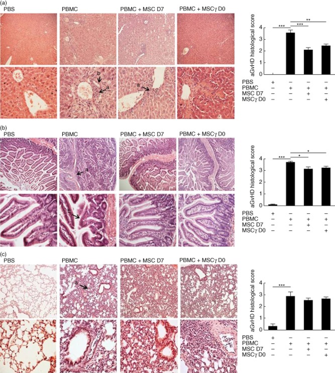
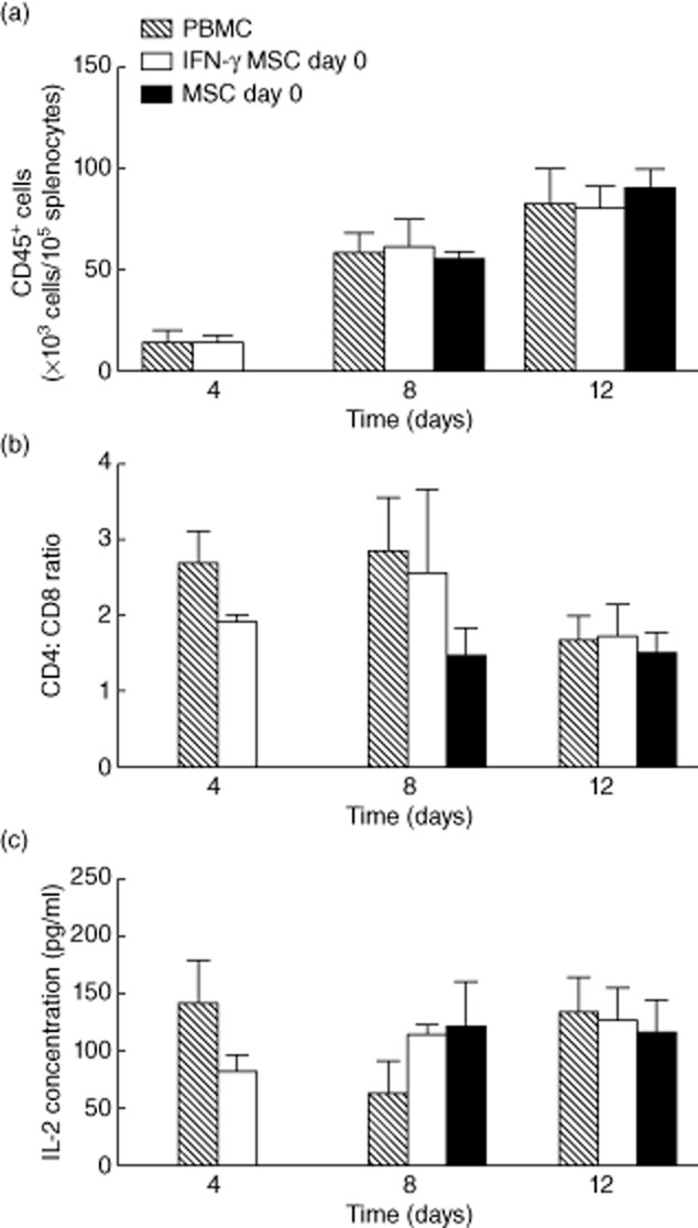
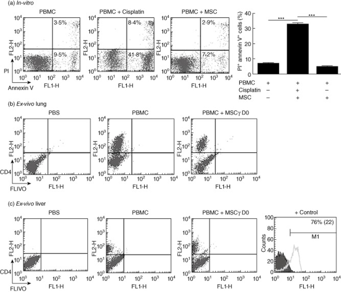
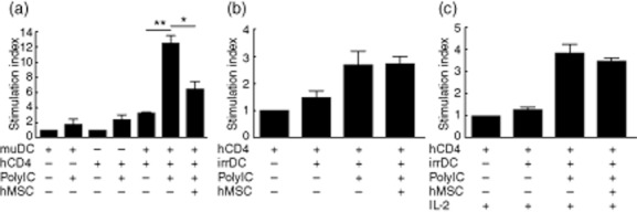
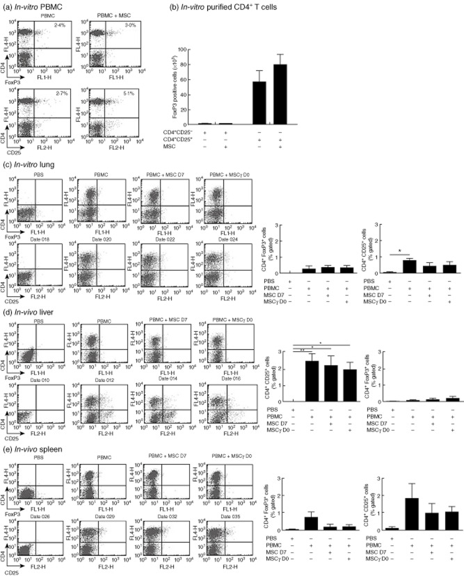
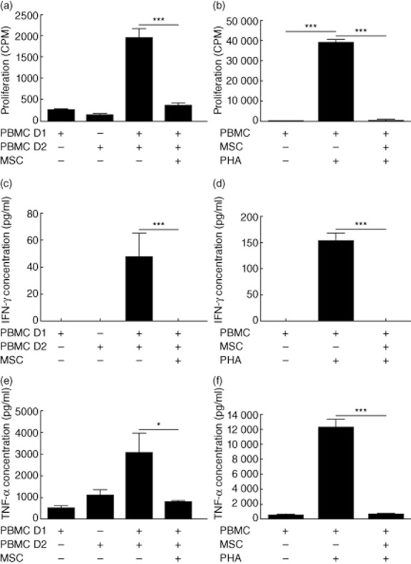
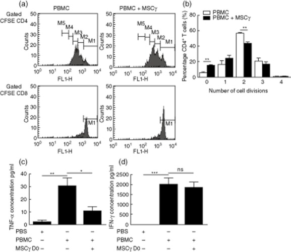
Acute graft-versus-host disease (aGVHD) is a life-threatening complication following allogeneic haematopoietic stem cell transplantation (HSCT), occurring in up to 30-50% of patients who receive human leucocyte antigen (HLA)-matched sibling transplants. Current therapies for steroid refractory aGVHD are limited, with the prognosis of patients suboptimal. Mesenchymal stem or stromal cells (MSC), a heterogeneous cell population present in many tissues, display potent immunomodulatory abilities. Autologous and allogeneic ex-vivo expanded human MSC have been utilized to treat aGVHD with promising results, but the mechanisms of therapeutic action remain unclear. Here a robust humanized mouse model of aGVHD based on delivery of human peripheral blood mononuclear cells (PBMC) to non-obese diabetic (NOD)-severe combined immunodeficient (SCID) interleukin (IL)-2rγ(null) (NSG) mice was developed that allowed the exploration of the role of MSC in cell therapy. MSC therapy resulted in the reduction of liver and gut pathology and significantly increased survival. Protection was dependent upon the timing of MSC therapy, with conventional MSC proving effective only after delayed administration. In contrast, interferon (IFN)-γ-stimulated MSC were effective when delivered with PBMC. The beneficial effect of MSC therapy in this model was not due to the inhibition of donor PBMC chimerism, as CD45(+) and T cells engrafted successfully in this model. MSC therapy did not induce donor T cell anergy, FoxP3(+) T regulatory cells or cause PBMC apoptosis in this model; however, it was associated with the direct inhibition of donor CD4(+) T cell proliferation and reduction of human tumour necrosis factor-α in serum.


Figures

Fig. 1
Fig. 1
Mesenchymal stem or stromal cell (MSC) therapy significantly prolongs the survival of non-obese diabetic (NOD) severe combined immunodeficient (SCID) interleukin (IL)-2rγnull (NSG) mice with acute graft-versus-host disease (aGVHD). NSG mice received 2·4 Gy low-dose irradiation; 4 h later peripheral blood mononuclear cells (PBMC) (6·3 × 105 g−1) were administered intravenously via the tail vein. Non-stimulated MSC were administered on day 7 post-PBMC transfusion or interferon (IFN)-γ-prestimulated MSC (MSCγ) (4·4 × 104 g1) were delivered on day 0, concurrent with PBMC. (a,d) Weight loss, (b,e) aGVHD score and (c,f) survival were recorded daily. Both non-stimulated and IFN-γ-stimulated MSC significantly prolonged the survival of NSG mice with aGVHD (P < 0·0001 and P < 0·0015, respectively), limiting weight loss and reducing the clinical signs of aGVHD development (n = 7 mice per group). Data shown are representative of at least two experiments.
Fig. 2
Fig. 2
Mesenchymal stem or stromal cells (MSC) cell therapy significantly reduces pathology in the liver and gut of non-obese diabetic (NOD) severe combined immunodeficient (SCID) interleukin (IL)-2rγnull (NSG) mice with acute graft-versus-host disease (aGVHD). aGVHD was established in NSG mice (as Fig. 1) and treated with non-stimulated MSC (day 7) or interferon (IFN-γ)-stimulated MSC (day 0). The livers, small intestine and lungs were harvested on day 12. (a) The livers of NSG mice displayed a significant increase in mononuclear cell infiltration (denoted by arrow and letter a) and increased endothelialitis, especially around hepatic ducts (denoted by arrow and letter b). Both MSC and interferon (IFN)-γ-prestimulated MSC (MSCγ) treatment significantly reduced this pathology. (b) aGVHD of the small intestine resulted in the accumulation of infiltrating cells into the lamina propria (denoted by arrow and letter a) and increased blunting of the villi (denoted by arrow and letter b). Similar to the liver, MSC or MSCγ cell therapy resulted in a significant decrease of cell infiltration and villous blunting. (c) aGVHD development in the lungs manifested by a significant increase of mononuclear cells into alveolar air spaces (denoted by arrow and letter a). Unlike the liver and small intestine, treatment of aGVHD NSG mice with MSC or MSCγ did not alleviate the symptoms of airway aGVHD. Bar charts in the right-hand panel summarize histological scores. Data are representative of a minimum five mice per treatment group and at least two independent experiments.
Fig. 3
Fig. 3
Mesenchymal stem or stromal cells (MSC) cell therapy did not interfere with donor peripheral blood mononuclear cells (PBMC) engraftment. Interferon (IFN)-γ-stimulated (day 0) or non-stimulated MSC (day 7) (4·4 × 104 g−1) were administered to irradiated non-obese diabetic (NOD) severe combined immunodeficient (SCID) interleukin (IL)-2rγnull NSG mice that had received PBMC (6·3 × 105 g−1) on day 0 (n = 5 per group). The spleens of the NSG mice from each treatment group were harvested on days 4, 8 and 12 and analysed for the expression of (a) human CD45+ and (b) the ratio of human CD4 to CD8 T cells by flow cytometry. Human PBMC engrafted readily, with a significant increase in CD45+ cells from days 4 to 8 (P < 0·05) and 12 (P < 0·002) compared to controls. MSC therapy had no significant effect on the engraftment of human PBMC. The number of CD45+ cells per 105 total splenocytes was enumerated using counting beads. (c) The level of human IL-2 present in the sera of mice from each treatment group was analysed by cytokine bead array. MSC therapy had no effect on the amount of human IL-2 detectable in sera.
Fig. 4
Fig. 4
Mesenchymal stem or stromal cells (MSC) did not induce T cell apoptosis in vitro or in vivo. (a) Peripheral blood mononuclear cells (PBMC) were cultured in the presence of cisplatin or MSC for 24 h. PBMC cultured alone or with cisplatin at 250 μg/ml were used as controls. The presence of MSC did not induce apoptosis of PBMC in vitro (P < 0·0002). The percentages of positive cells within the lower region are represented in the bar chart. Data are representative of two studies. Irradiated non-obese diabetic (NOD) severe combined immunodeficient (SCID) interleukin (IL)-2rγnull NSG mice received phosphate-buffered saline (PBS) or PBMC (6·3 × 105 g−1) on day 0. Interferon (IFN)-γ-prestimulated MSC (MSCγ) (4·4 × 104 g−1) were given on day 0, concurrent with PBMC. On day 12, FLIVO green dye (8 μg/mouse) was injected intravenously and left to circulate for 1 h. (b) The lungs and (c) livers were harvested from each mouse and analysed for FLIVO staining and human CD4 phycoerythrin (PE) labelling by flow cytometry. MSC cell therapy did not induce CD4 T cell apoptosis in vivo. Liver cells from lethally irradiated BALB/c were used as a positive control for FLIVO detection in vivo.
Fig. 5
Fig. 5
Mesenchymal stem or stromal cells (MSC) did not induce T cell anergy in vitro. (a) Human CD4+ T cells (1 × 10⁶/ml) were co-cultured with or without BALB/c bone marrow-derived dendritic cells (muDC) (1 × 10⁵/ml) matured using polyinosinic-polycytidylic acid (polyIC) (20 μg/ml) in the presence or absence of human MSC (hMSC) (1 × 10⁵/ml) for 5 days. [3H]-thymidine was then added to cultures for the final 6 h and proliferation was measured. PolyIC matured murine dendritic cells (DC) induced significant human CD4+ T cell proliferation (P < 0·0001). In the presence of hMSC, the proliferation of CD4+ T cell proliferation was significantly reduced (P < 0·05). Following co-culture, CD4+ T cells were repurified and co-cultured in a second-stage experiment with irradiated mature DC (irrDC/polyIC) for 72 h in the (b) absence or (c) presence of recombinant human interleukin (rhIL)-2 to assess anergy. T cell proliferation was analysed by [3H]-thymidine incorporation.
Fig. 6
Fig. 6
Human mesenchymal stem or stromal cells (hMSC) supported the expansion of CD4+ CD25+ forkhead box protein 3 (FoxP3)+ T cells in vitro but not in vivo. Expression of FoxP3 was measured in vitro (a,b) and in vivo (c–e). In vitro, (a) peripheral blood mononuclear cells (PBMC) or (b) sorted human CD4+ CD25+ or CD4+ CD25 T cells (0·5 × 106/ml) were co-cultured with or without MSC (1·5 × 105/ml) for 72 h. Cells were recovered and examined for the expression of CD4, CD25 (PBMC population) and FoxP3 by intracellular flow cytometry. (a) The percentage of double-positive PBMC expressing CD4/CD25 or CD4/FoxP3 is given in the upper quadrant. (b) The number of FoxP3-expressing CD4 cells sorted from whole PBMC is represented in the bar chart. MSC did not induce a population of FoxP3+ regulatory T cells (Treg) cells, but rather expanded an already existing population in vitro (data are representative of three independent experiments). In-vivo non-obese diabetic (NOD) severe combined immunodeficient (SCID) interleukin (IL)-2rγnull NSG mice received phosphate-buffered saline (PBS) or PBMC (6·3 × 105 g−1) on day 0 with or without non-stimulated MSC on day 7 or interferon (IFN)-γ-prestimulated MSC (MSCγ) on day 0 (4·4 × 104 g−1). On day 12, the (c) lungs, (d) livers and (e) spleens were harvested and analysed for the expression of human CD4, CD25 and FoxP3 by flow cytometry. Data are summarized in bar charts in the right-hand panels. CD4+ FoxP3+ Treg-like cells were not detected in vivo following MSC therapy. In this instance data are representative of six mice per group (n = 6).
Fig. 7
Fig. 7
Mesenchymal stem or stromal cells (MSC) inhibit peripheral blood mononuclear cell (PBMC) proliferation and suppress interferon (IFN)-γ and tumour necrosis factor (TNF)-α production in vitro. (a) PBMC (1 × 10⁶/ml) from two major histocompatibility complex (MHC) mismatched donors (D1 or D2) were cultured in the presence or absence of MSC (1 × 10⁵/ml) in a mixed lymphocyte reaction (MLR). MSC inhibited alloantigen-driven proliferation significantly (P < 0·0001). (b) Human MSC also suppressed mitogen [phytohaemagglutinin(PHA)]-driven proliferation of allogeneic human PBMC (P < 0·0001) in vitro. The inhibition of proliferation correlated with a significant decrease in the production of (c,d) IFN-γ (P < 0·0001) and (e,f) TNF-α (P < 0·0201, P < 0·0001, respectively), as measured by enzyme-linked immunosorbent assay. Data are representative of three experiments, each performed in triplicate.
Fig. 8
Fig. 8
Mesenchymal stem or stromal cells (MSC) reduced the proliferation of CD4+ T cells and suppressed tumour necrosis factor (TNF)-α production in vivo. Peripheral blood mononuclear cells (PBMC) labelled with 10 μM carboxyfluorescein succinimidyl ester (CFSE) were administered to conditioned non-obese diabetic (NOD) severe immunodeficient interleukin (IL)-2rγnull NSG mice with or without interferon (IFN)-γ-prestimulated MSC (MSCγ) on day 0. After 5 days, the lungs, livers and spleen were harvested. (a) The level of CFSE in CD4+ T cells was analysed by flow cytometry. MSC reduced the proliferation of CD4+ T cells in the lung at 5 days. Sufficient CFSE-stained CD4+ T cells were not recoverable from the livers or spleen after 5 days. (b) The percentage of CD4+ cells present in the lung at each division in vivo. Serum was taken from NSG mice on day 12 and analysed for the presence of (c) human TNF-α and (d) human IFN-γ by bead array. Prestimulated MSCγ reduced significantly the level of (c) human TNF-α (P < 0·0267) in the sera of NSG mice with acute graft-versus-host disease (aGVHD). Human MSC (hMSC) therapy had no significant effect on (d) human IFN-γ production in sera. Data are representative of five mice per group (n = 5).

Similar articles

Cited by

References

    1. Reddy P, Ferrara JL. Immunobiology of acute graft-versus-host disease. Blood Rev. 2003;17:187–194. - PubMed
    1. Ferrara JL, Levine JE, Reddy P, Holler E. Graft-versus-host disease. Lancet. 2009;373:1550–1561. - PMC - PubMed
    1. Billingham RE. The biology of graft-versus-host reactions. Harvey Lect. 1966;62:21–78. - PubMed
    1. Messina C, Faraci M, de Fazio V, Dini G, Calo MP, Calore E. Prevention and treatment of acute GvHD. Bone Marrow Transplant. 2008;41(Suppl. 2):S65–70. - PubMed
    1. Van Lint MT, Uderzo C, Locasciulli A, et al. Early treatment of acute graft-versus-host disease with high- or low-dose 6-methylprednisolone: a multicenter randomized trial from the Italian Group for Bone Marrow Transplantation. Blood. 1998;92:2288–2293. - PubMed

Publication types

MeSH terms