. 2013 May;14(5):514-22.
doi: 10.1038/ni.2569. Epub 2013 Apr 7.

Trypanosoma cruzi trans-sialidase initiates a program independent of the transcription factors RORγt and Ahr that leads to IL-17 production by activated B cells

Affiliations

Trypanosoma cruzi trans-sialidase initiates a program independent of the transcription factors RORγt and Ahr that leads to IL-17 production by activated B cells

Daniela A Bermejo et al. Nat Immunol. 2013 May.

Abstract

Here we identified B cells as a major source of rapid, innate-like production of interleukin 17 (IL-17) in vivo in response to infection with Trypanosoma cruzi. IL-17(+) B cells had a plasmablast phenotype, outnumbered cells of the TH17 subset of helper T cells and were required for an optimal response to this pathogen. With both mouse and human primary B cells, we found that exposure to parasite-derived trans-sialidase in vitro was sufficient to trigger modification of the cell-surface mucin CD45, which led to signaling dependent on the kinase Btk and production of IL-17A or IL-17F via a transcriptional program independent of the transcription factors RORγt and Ahr. Our combined data suggest that the generation of IL-17(+) B cells may be a previously unappreciated feature of innate immune responses required for pathogen control or IL-17-mediated autoimmunity.


Figures
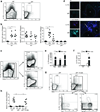
Figure 1
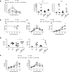
Figure 1. B cells from T. cruzi infected mice produce IL-17
(a) Representative flow cytometry plots showing IL-17A expression in B220+ cells in the spleen of wild-type (WT) and µMT mice infected with T. cruzi at 10 days (d) post-infection. (b) Number of IL-17A-expressing splenic CD4+, CD8+ and B220+ cells in uninfected (UI), or 10 and 19 d T. cruzi-infected mice (WT or µMT). Each symbol denotes a single animal and the line represents the mean value in each group (6–9 animals per group). Data are representative of 4 independent experiments. (c) IL-17A expression (at 10 d) in total splenocytes from T. cruzi-infected WT mice (left) and in gated CD19+ cells expressing the plasma cell marker, CD138, or the GC B cell marker, GL7 (right). Data are representative of 3 experiments. (d) Immunofluorescence staining of splenic sections from UI and T. cruzi infected (10 d) mice showing IL-17A and IgM expression (magenta and cyan, respectively, left; and merged images, right). Arrow indicates IL-17+IgM+ cells. Dashed lines surround less strongly staining (e.g. IgMlo) B cell follicles (*). Data are representative of 3 experiments. (e) IL-17A mRNA expression in total, sorted B220+ and B220 splenocytes from infected mice cultured in media alone or with PMA and ionomycin (PMA+Iono). HPRT was used for normalization (10 d, 3 replicates per condition). (f) IL-17A production by B220+ splenocytes from UI or infected (Inf) mice cultured with medium or PMA+Iono for 72 h (10 d, 3 replicates per condition). Data (e–f) are representative of 2 experiments. (g) IL-17A+ B220+ cells in T. cruzi-infected µMT or B-cell reconstituted µMT (µMT + B cells) mice (10 d). Lower panel shows IL-17A expression predominantly within the B220+CD138+ gate. Data are representative of 3 experiments. (h) Number of IL-17+ B220+ cells within the spleen of UI or Inf WT, µMT or B cell reconstituted µMT mice (10 d; 6–7 mice per condition). Data are representative of 3 experiments. All error bars denote s.e.m. *, P ≤ 0.05 **, P ≤ 0.005 (calculated by Mann-Whitney U-test).
Figure 2
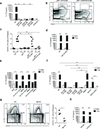
Figure 2. B cell IL-17A production is required for T. cruzi infection control
Control WT, µMT, or µMT mice reconstituted with WT or Il17a−/− B cells, were infected with T. cruzi and evaluated for: (a) Parasitemia (left panel: kinetic; right panels: day 10 and 15); (b) Survival based on Kaplan-Meier survival curve; (c) Serum IFNγ and TNF concentrations; and, (e) plasma aspartate aminotransferase (AST) and alanine aminotransferase (ALT) activity. Surviving animals were sacrificed at 20 d and (d) IFN-γ and TNF production by unstimulated splenocytes was quantified (72 hour in vitro culture). Each symbol denotes a single animal and the line represents the mean value in each group. All error bars denote s.e.m. *, P ≤ 0.05 **, P ≤ 0.005 (calculated by Mann-Whitney U-test). Results shown are from 2 independent experiments with 3–4 animals per group in each experiment.
Figure 3
Figure 3. Exposure to T. cruzi trypomastigotes or purified trans-sialidase is sufficient to trigger B cell IL-17 production
(a,d–g) IL-17A secretion by B220+CD19+ cells or (b–c) IL-17A and IL-17F production by follicular (FM), marginal zone (MZ) and transitional 1 (T1) B cell subsets purified from spleens of UI WT mice and stimulated for 72 h with: (a) various doses of live T. cruzi trypomastigotes (Tryp) or T. cruzi antigen (Ag); or (b–g) 1 × 10⁶ Tryp or different doses of active vs. inactive trans-sialidase (Tsial) with or without a Tsial-neutralizing mAb (α-Tsial) or IgG2a isotype control. (f) B220+CD19+ cells from UI WT mice were cultured with Tryp, Tsial or different doses of bacterial V. cholerae and C. perfringens neuramidases (NA). (g) B220+CD19+ cells from WT mice were cultured with Tryp or Tsial for 72 h in medium containing FBS or 2% BSA with alpha(2,3)-sialyllactose (α-2,3-SL) and alpha(2,6)-sialyllactose (α-2,6-SL) or in identical media lacking acceptor lactose. All error bars denote s.e.m. *, P ≤ 0.05 **, P ≤ 0.005 (calculated by Mann-Whitney U-test). Results are representative of 3 independent experiments with 2 replicates per condition. ND: None detectable.
Figure 4
Figure 4. CD45 and Btk are required for B cell IL-17 production in response to T. cruzi trypomastigotes or purified trans-sialidase
(a) IL-17A production in follicular B cells from WT mice in response to T. cruzi Tryp and treated with or without various doses of CD45 inhibitor. Similar analysis was performed using B cells isolated from Cd45−/− mice. (b) IL-17A expression in CD19+ splenic cells from 10 d infected µMT mice that were recipients of adoptively transferred WT (left) or Cd45−/− B cells (right). (c) Number of CD19+IL-17A+ cells in spleen of UI vs. 10 d infected animals including: WT or µMT mice, or µMT recipients of WT vs. Cd45−/− B cells. Each symbol denotes a mouse and the line represents the median value in each group. (d) In vitro IL-17A secretion by B220+CD19+ cells derived from WT vs. Gal-1−/−Gal-3−/− mice in response to Tryp-stimulation. (e) IL-17A production by WT FM B cells stimulated with Tryp and treated with different concentration of Src inhibitor (PP2) vs. DMSO control. (f) IL-17A production by WT FM B cells stimulated with Tryp and treated with increasing doses of Btk-specific inhibitors (AVL-292 or CNX-652) vs. DMSO control. IL-17A production by Tryp stimulated FO B cells derived from Btk−/− and Btk−/−Tec−/− mice is also shown. (g) Representative FACS plots (left panels) and total number of splenic CD19+IL-17A+ cells (right panel) in T. cruzi infected (10 d) WT or Btk−/− mice. (h) IL17A production in Tryp stimulated purified B cells derived from BALB/c (control) vs. Pkcβ−/− mice. All in vitro analyses were performed at 72 h and results are representative of three (a–c) or two (d–g) independent experiments for each strain or condition. Data are shown as means ± s.e.m. *, P ≤ 0.05 **, P ≤ 0.005 (calculated by one-way ANOVA with Bonferroni correction (a–f, h) and Mann-Whitney U-test (g)).
Figure 5
Figure 5. RORγt and AHR are not required for B cell IL-17 production in response to T. cruzi
(a) IL-17 secretion by WT and Rorγt−/− splenic B cells cultured in vitro for 72 hr with T. cruzi Tryp, with or without Tsial-neutralizing mAb (α-Tsial) (4 replicates per condition). (b) IL-17A production in non-stimulated and Tryp-stimulated, WT B cells (in presence or absence of the RORγt and RORα inhibitor SR1001, 25µM) and Ahr−/− B cells. Data are shown as means ± s.e.m. *, P ≤ 0.05 **, P ≤ 0.005 (calculated by one-way ANOVA with Bonferroni correction). (c) Number of splenic B220+IL-17A+ cells in T. cruzi infected (10 d) WT, Rorγt−/− and Ahr−/− mice. Each symbol denotes a mouse and the line represents the median value in each group. (d) Rorγt locus activation cannot be detected in vivo in B cells following T. cruzi infection. RORγt-cre × ROSA-RFP reporter mice were infected with T. cruzi and spleens collected at 10 d post-infection. ROSA-RFP (magenta) and IgM (cyan) expression in spleen sections from UI (upper panels) vs. 10 d-infected (lower panels) animals. Results are representative of three (a), two (b, c), or one (d) experiment for each strain or condition.
Figure 6
Figure 6. Primary human B cells stimulated with T. cruzi produce IL-17
Purified human CD19+ tonsillar B cells were cultured for 72 hr in presence of recombinant IL-10 or BAFF and incubated with T. cruzi Tryp with or without α-Tsial mAb. B cells were also treated with Btk (AVL-292) or CD45 inhibitors or DMSO control. Production of (a) IL-17A or (b) IL-17F determined by ELISA. Data (3 replicates per condition) are shown as means ± s.e.m. *, P ≤ 0.05 **, P ≤ 0.005 (calculated by one-way ANOVA with Bonferroni correction). Results are representative of 3 independent experiments.

Comment in

Similar articles

Cited by

  • Signal transducer and activator of transcription 1 (STAT-1) plays a critical role in control of Trypanosoma cruzi infection.
    Kulkarni MM, Varikuti S, Terrazas C, Kimble JL, Satoskar AR, McGwire BS. Kulkarni MM, et al. Immunology. 2015 Jun;145(2):225-31. doi: 10.1111/imm.12438. Immunology. 2015. PMID: 25545325 Free PMC article.
  • Costimulatory Effects of an Immunodominant Parasite Antigen Paradoxically Prevent Induction of Optimal CD8 T Cell Protective Immunity.
    Eickhoff CS, Zhang X, Vasconcelos JR, Motz RG, Sullivan NL, O'Shea K, Pozzi N, Gohara DW, Blase JR, Di Cera E, Hoft DF. Eickhoff CS, et al. PLoS Pathog. 2016 Sep 19;12(9):e1005896. doi: 10.1371/journal.ppat.1005896. eCollection 2016 Sep. PLoS Pathog. 2016. PMID: 27642757 Free PMC article.
  • Host-parasite dynamics in Chagas disease from systemic to hyper-local scales.
    Pérez-Mazliah D, Ward AI, Lewis MD. Pérez-Mazliah D, et al. Parasite Immunol. 2021 Feb;43(2):e12786. doi: 10.1111/pim.12786. Epub 2020 Sep 24. Parasite Immunol. 2021. PMID: 32799361 Free PMC article. Review.
  • Guidelines for the use of flow cytometry and cell sorting in immunological studies (second edition).
    Cossarizza A, Chang HD, Radbruch A, Acs A, Adam D, Adam-Klages S, Agace WW, Aghaeepour N, Akdis M, Allez M, Almeida LN, Alvisi G, Anderson G, Andrä I, Annunziato F, Anselmo A, Bacher P, Baldari CT, Bari S, Barnaba V, Barros-Martins J, Battistini L, Bauer W, Baumgart S, Baumgarth N, Baumjohann D, Baying B, Bebawy M, Becher B, Beisker W, Benes V, Beyaert R, Blanco A, Boardman DA, Bogdan C, Borger JG, Borsellino G, Boulais PE, Bradford JA, Brenner D, Brinkman RR, Brooks AES, Busch DH, Büscher M, Bushnell TP, Calzetti F, Cameron G, Cammarata I, Cao X, Cardell SL, Casola S, Cassatella MA, Cavani A, Celada A, Chatenoud L, Chattopadhyay PK, Chow S, Christakou E, Čičin-Šain L, Clerici M, Colombo FS, Cook L, Cooke A, Cooper AM, Corbett AJ, Cosma A, Cosmi L, Coulie PG, Cumano A, Cvetkovic L, Dang VD, Dang-Heine C, Davey MS, Davies D, De Biasi S, Del Zotto G, Dela Cruz GV, Delacher M, Della Bella S, Dellabona P, Deniz G, Dessing M, Di Santo JP, Diefenbach A, Dieli F, Dolf A, Dörner T, Dress RJ, Dudziak D, Dustin M, Dutertre CA, Ebner F, Eckle SBG, Edinger M, Eede P, Ehrhardt GRA, Eich M, Engel P, Engelhardt B, Erdei A, Esser C, Everts B, Evrard M, Falk CS, Fehniger TA, Felipo-Benavent M, Fer… See abstract for full author list ➔ Cossarizza A, et al. Eur J Immunol. 2019 Oct;49(10):1457-1973. doi: 10.1002/eji.201970107. Eur J Immunol. 2019. PMID: 31633216 Free PMC article.
  • IL-17RA-Signaling Modulates CD8+ T Cell Survival and Exhaustion During Trypanosoma cruzi Infection.
    Tosello Boari J, Araujo Furlan CL, Fiocca Vernengo F, Rodriguez C, Ramello MC, Amezcua Vesely MC, Gorosito Serrán M, Nuñez NG, Richer W, Piaggio E, Montes CL, Gruppi A, Acosta Rodríguez EV. Tosello Boari J, et al. Front Immunol. 2018 Oct 11;9:2347. doi: 10.3389/fimmu.2018.02347. eCollection 2018. Front Immunol. 2018. PMID: 30364284 Free PMC article.

References

    1. Korn T, Bettelli E, Oukka M, Kuchroo VK. IL-17 and Th17 Cells. Annu Rev Immunol. 2009;27:485–517. - PubMed
    1. Gaffen SL. Recent advances in the IL-17 cytokine family. Curr Opin Immunol. 2011;23:613–619. - PMC - PubMed
    1. Kelly MN, et al. Interleukin-17interleukin-17 receptor-mediated signaling is important for generation of an optimal polymorphonuclear response against Toxoplasma gondii infection. Infect Immun. 2005;73:617–621. - PMC - PubMed
    1. Pitta MG, et al. IL-17 and IL-22 are associated with protection against human kala azar caused by Leishmania donovani. J Clin Invest. 2009;119:2379–2387. - PMC - PubMed
    1. Rudner XL, Happel KI, Young EA, Shellito JE. Interleukin-23 (IL-23)-IL-17 cytokine axis in murine Pneumocystis carinii infection. Infect Immun. 2007;75:3055–3061. - PMC - PubMed

Publication types

MeSH terms